3290
Multi-slab 3D ECLIPSE for whole brain lipid suppressed MRSI1Radiology and Biomedical Imaging, Yale University, New Haven, CT, United States
Synopsis
The utility of ECLIPSE, a pulsed second order gradient insert allowing elliptical localization, was previously shown to provide high axial coverage for single slab MRSI acquisitions with robust lipid suppression. In this work we extend the ECLIPSE method with multi-slab localization that provides improved coverage, for applications in 3D MRSI acquisitions. A two-slab ECLIPSE-OVS method providing > 100-fold in mean lipid suppression was developed, with improved superior-inferior coverage. Simulation results show that a four-slab ECLIPSE method, based on OVS and IVS, can provide high-quality lipid suppression with whole-brain coverage.
Introduction
Magnetic Resonance Spectroscopic Imaging (MRSI) is a powerful technique that can map the brain metabolic profile non-invasively. Whole brain extracranial lipid suppressed 3D MRSI acquisitions is however challenging to achieve with linear gradients due to the curvature of the brain. Recent work with a second order gradient insert, ECLIPSE1, provides high axial coverage for single slab MRSI with robust extracranial lipid suppression for both inner volume selection (IVS) and outer volume suppression (OVS) variants. However, divergence of the elliptical ROI in the Z-direction, characteristic of a Z2 field, limits the use of ECLIPSE to one ~ 4 cm slab along the superior-inferior axis. In this work we demonstrate the extension of ECLIPSE in a multi-slab configuration, that allows improved Z coverage for whole-brain lipid-suppressed 3D MRSI acquisitions.Theory
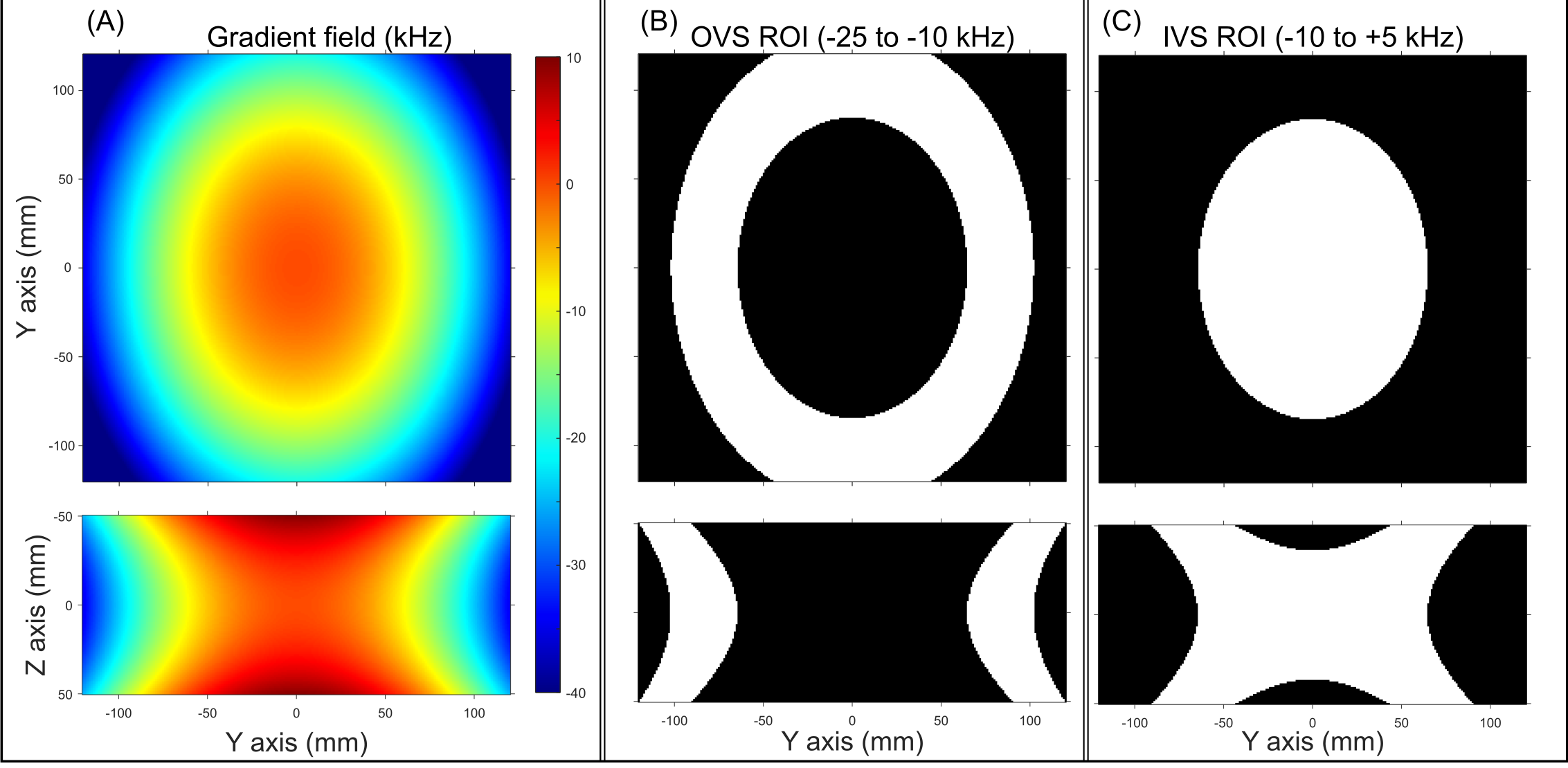
The total gradient field Gtotal(x,y,z) utilizing ECLIPSE and system gradients to produce an elliptical ROI can be described as:Gtotal(x,y,z) = GZ2·(2z2–(x2+y2))+GX2Y2·(x2-y2)+GXY·(xy)+GX·(x)+GY·(y)+GZ·(z).
Here GZ2, GX2Y2, and GXY are quadratic magnetic field amplitudes in Hz/mm2, and GX, GY, and GZ are linear magnetic field amplitudes in Hz/mm. The ratio between GZ2 and GX2Y2 amplitudes determine the axial elliptical shape (Figure 1 A-B), and GX, GY, and GZ amplitudes allow translation of the ellipse along the three primary axes, as previously described1-2. An example of outer volume elliptical ring selection (used for ECLIPSE-OVS) or inner volume elliptical selection (used for ECLIPSE-IVS) is illustrated in Figure 1 C-D, and E-F respectively.
Methods
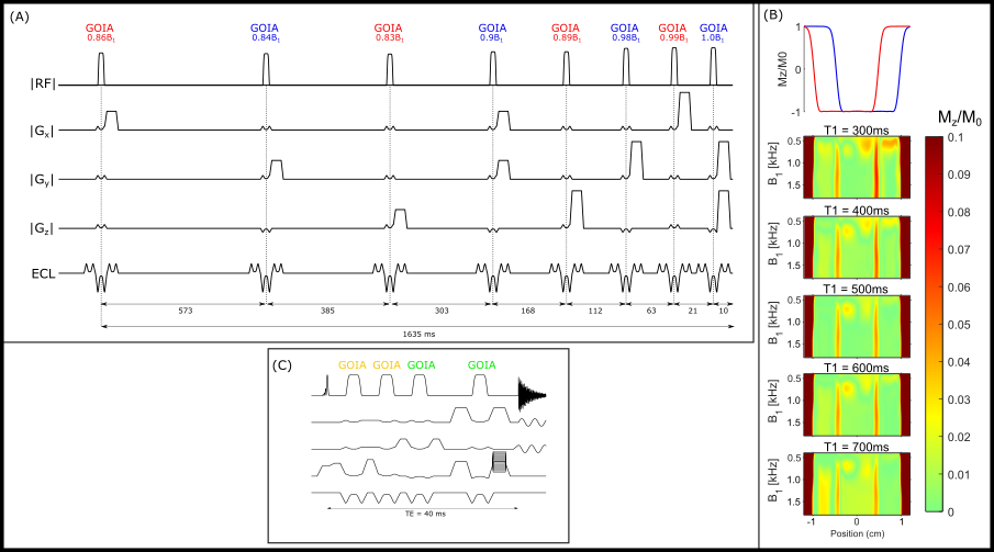
A two-slab ECLIPSE-OVS method executed with two-sets-of-four RF pulses was developed utilizing a 3 ms GOIA-WURST (12-4,7) pulse3-4 (BW = 15 kHz) (Figure 2A). A two-slab ECLIPSE-OVS method creates three unique ROIs in which 1) spins are only perturbed by one OVS module or, 2) spins are perturbed by both OVS modules. To maintain a high level of lipid suppression, OVS pulse angles and inter-pulse delays were optimized simultaneously over all three regions, in addition to covering T1 species spanning 300 – 700 ms, and a B1+ variation of ± 65%. The two-slab ECLIPSE-OVS method can be supplemented with a two-slab ECLIPSE IVS method (Fig. 2C) to achieve near-complete 3D coverage of the human brain. The two-slab ECLIPSE IVS method closely resembles a sLASER method, whereby adiabatic refocusing pulses 1A-1B select one ECLIPSE ROI, and 2A-2B select a second ECLIPSE ROI. ROI placement and brain coverage was evaluated in vivo with the two-slab ECLISPE-OVS method on two volunteers. Whole brain coverage achievable with a four-slab ECLIPSE method was evaluated in simulation using acquired anatomical images from a volunteer.All MR experiments were performed on a 4 T 94 cm Medspec scanner (Bruker corporation. Ettlingen, Germany) with actively shielded gradients capable of switching 30 mT/m in 1150 µs. The ECLIPSE system1 is a home-built, unshielded gradient insert consisting of Z2, X2Y2, and XY second order spherical harmonic magnetic fields, controlled by a home-built multi-channel gradient controller5.
Results
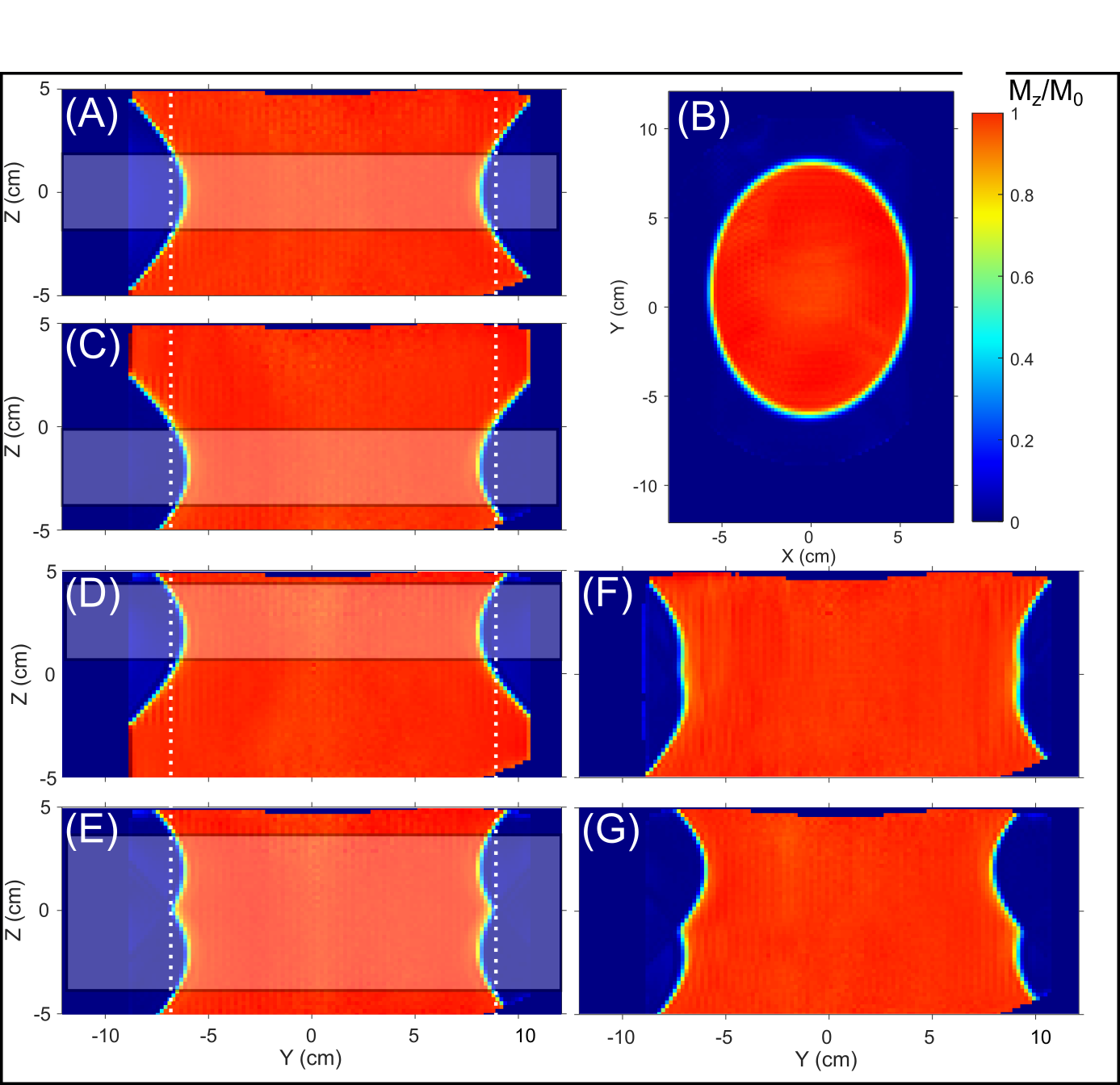
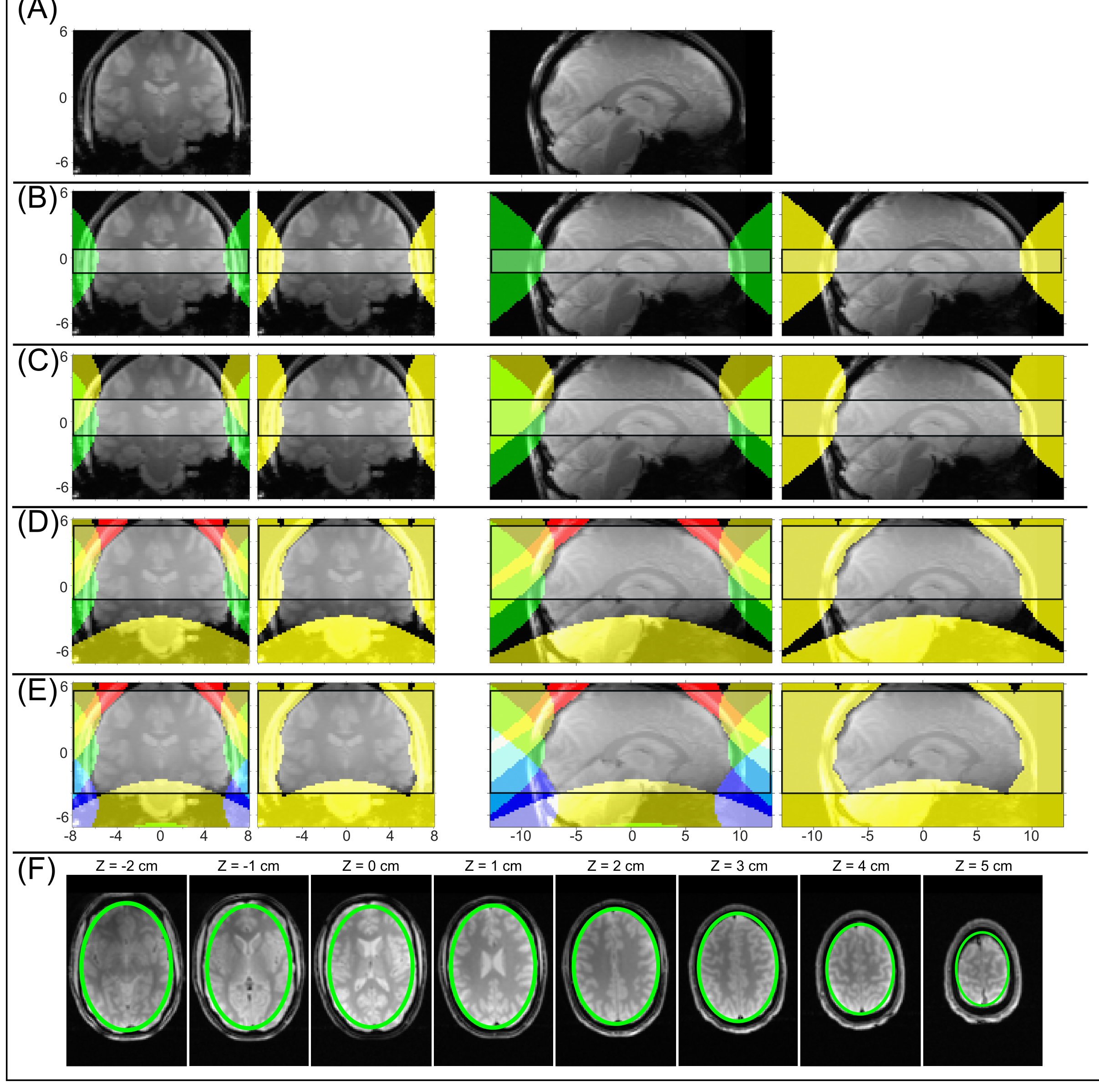
The optimized two-slab ECLIPSE-OVS method (Figure 2A) provides a 107-fold in mean lipid suppression in simulation, over the optimized T1 and B1 span, accounting for lipid suppression in all three regions described above (Figure 2B). The transition zone of each RF pulse, however results in a narrow strip with notably poorer signal suppression, down to 10-fold. Combining one OVS and one IVS module for two-slab suppression can mitigate the reduced suppression in spatial areas of overlap. The two-slab ECLIPSE-OVS method results in ~ 1.9-fold increase in time-averaged power, relative to a single slab ECLIPSE-OVS method previously reported2.Figure 3 illustrates experimental ECLIPSE-OVS ROI profiles in the Y-Z plane for a single slab OVS placed at Z = 0 (A). (B) and (C) illustrate identical ROI’s to (A) with the ± 2 cm Z-offsets. The ROI generated when both slab 1 and 2 OVS in (B) and (C) are simultaneously turned on with the two-slab ECLIPSE OVS method is shown in (D), demonstrating improved coverage in the Z-direction and effective signal suppression throughout the cumulative ROI.
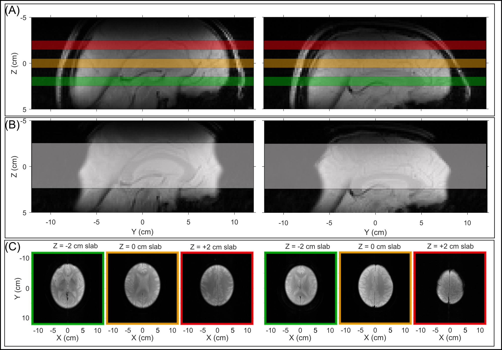
Figure 4 illustrates examples of MRI’s obtained with the two-slab ECLIPSE-OVS method applied on two volunteers. In both cases the OVS ROI were placed to achieve 5 cm of Z coverage.
Figure 5 illustrates brain coverage in simulation with up to four ECLIPSE ROI’s, by combining the two-slab ECLIPSE-OVS and two-slab ECLIPSE-IVS components. For the illustrated example, the four scenarios provide up to 2.6 cm, 3.6 cm, 7 cm, and 9.4 cm of coverage in terms of lipid suppression in the Z-direction, respectively. The four-slab ECLIPSE method allows 94% brain coverage over an 8 cm span in the Z direction. The inherent curvature of the ECLIPSE ROI in the Z direction can be exploited to adhere closely to the head shape, as demonstrated by the red and blue ROI’s in the four-slab method illustrated in Figure 5 D.
Discussion
Experimental implementation of the two-slab ECLIPSE IVS method is currently underway. The combination of the two-slab ECLIPSE IVS and two-slab ECLIPSE OVS method is expected to require a sequence TR = 4000 ms to meet time-averaged power requirements. However, a 3D MRSI data acquisition with a concentric ring readout6 would allow whole brain acquisitions at 1 x 1 x 1 cm3 nominal resolution in ~ 20 min.Acknowledgements
This research was supported by NIH grant R01- EB014861.References
[1] de Graaf RA, Brown PB, De Feyter HM, McIntyre S, Nixon TW. Elliptical localization with pulsed second-order fields (ECLIPSE) for robust lipid suppression in proton MRSI. NMR in biomedicine 2018;31(9):e3949.
[2] Kumaragamage C, De Feyter HM, Brown P, McIntyre S, Nixon TW, de Graaf RA. ECLIPSE utilizing gradient-modulated offset-independent adiabaticity (GOIA) pulses for highly selective human brain proton MRSI. NMR in Biomedicine 2020; 34:e4415.
[3] Tannus A, Garwood M. Adiabatic pulses. NMR in Biomedicine 1997;10(8):423–434.
[4] Kumaragamage C, Coppoli A, Brown P, McIntyre S, Nixon T, De Feyter H, Mason G, and de Graaf R. Short-TE ECLIPSE for Macromolecular-Nulled MRSI in the Human Brain. Proceedings of the Anuual Joinnt Meeting ISMRM, Virtual-2021; 0287.
[5] Nixon TW, McIntyre S, de Graaf RA. The design and implementation of a 64 channel arbitrary gradient waveform controller. Proc Int Soc Magn Reson Med. 2017;25:969.
[6] Emir UE, Burns B, Chiew M, Jezzard P, Thomas MA. Non-water-suppressed short-echo-time magnetic resonance spectroscopic imaging using a concentric ring k-space trajectory. NMR in Biomedicine 20016; e3714.
Figures