3283
SENSE-like reconstruction of multi-average body DWI to remove motion-induced signal loss: application in liver DWI1Department of Diagnostic and Interventional Radiology, Technical University of Munich, Munich, Germany, 2Philips Healthcare, Best, Netherlands, 3Philips GmbH Market DACH, Hamburg, Germany
Synopsis
Motion-induced signal loss and phase maps were generated and used in a SENSE-like reconstruction routine to solve for a single DWI from a multi-average liver diffusion-weighted imaging experiment. The proposed reconstruction yields homogeneous liver signal and possibly improves diagnostic values.
Purpose
Diffusion-weighted imaging is nowadays used in routine body oncological imaging to detect lesions [1]. However, body DWI remains strongly challenged by motion effects that induce different types of artifacts including the motion-induced signal loss typically observed in the left liver lobe [2, 3]. With respiratory gating, the motion-induced signal loss in the left liver lobe is caused primarily by cardiac motion [2]. To mitigate the signal loss problem, multiple averages of the same b-value and diffusion-encoding direction are typically acquired and averaged. The conventional sum-of-squares (SOS) averaging method disregards the difference in the quality of the averages and treat all averages equally. As a result, significant liver signal inhomogeneity can still be observed. A previous work has tried to learn with a network the quality (weights) of different averages and perform the weighted averaging [4]. In this work, complex motion-induced signal loss and phase maps were generated and used in a SENSE-like reconstruction routine to solve for a single diffusion-weighted image (DWI) from a multi-average liver diffusion-weighted imaging experiment.Methods
Data acquisition: One volunteer and two patients were scanned on a 3T whole-body scanner (Ingenia Elition, Philips Healthcare) using the 16-channel torso coil and the built-in-table 12-channel posterior coil. A spin echo diffusion-weighted sequence in combination with a single-shot EPI readout was employed. Patient data were acquired with the following parameters: FOV = 420 x 370 mm2, voxel size = 3 x 3 x 4 mm3, 60 slices with 0.4 mm gap, b-values = 0, 50, 300, 600 s/mm2 with the number of averages of 2, 1, 2, 6, respectively, 3 diffusion encoding directions (b-value > 0). For volunteer data, 10 slices with 6 mm gap, b-values = 0, 200, 400, 600 s/mm2 with the number of averages of 4, 5, 6, 8, respectively were acquired. All scans were respiratory triggered with a fixed trigger delay of 200 ms.Image reconstruction: Individual averages for each b-value and diffusion direction were reconstructed offline. The complex motion-induced signal loss and phase (MISLP) maps were determined as
$$S_{(k,d,b)}(r) = \frac{I^{lr}_{(k,d,b)}(r)}{\max_{k}|I^{lr}_{(k,d,b)}(r)|}$$
where $$$I^{lr}_{(k,d,b)}$$$ is the complex low resolution DWI at average $$$k$$$, diffusion direction $$$d$$$, and b-value $$$b$$$. Further smoothing of the magnitude of the MISLP maps were achieved by employing a 2D median filter of an empirical width of 8 x 8 voxels.
Let $$$m$$$ be the signal-loss-free DWI reconstructed from the multi-average data, similar to a SENSE problem with reduction factor of 1, we have
$$\begin{pmatrix}I_{(k_1, b, d)}(r) \\
I_{(k_2, b, d)}(r) \\
\vdots \\
I_{(N_k, b, d)}(r)
\end{pmatrix} = \begin{pmatrix}S_{(k_1, b, d)}(r) \\ S_{(k_2, b, d)}(r) \\ \vdots \\S_{(N_k, b, d)} \end{pmatrix}m(r) $$
where $$$N_k$$$ is the number of averages for direction $$$d$$$ and b-value $$$b$$$, $$$I_{(k, d, b)}$$$ is the high resolution DWI. The solution for the above equation is
$$m(r) = \frac{\sum_k {S^*}_{(k,d,b)}(r)I_{(k,d,b)}(r)}{\sum_k|S_{(k,d,b)}(r)|^2}.$$
Results
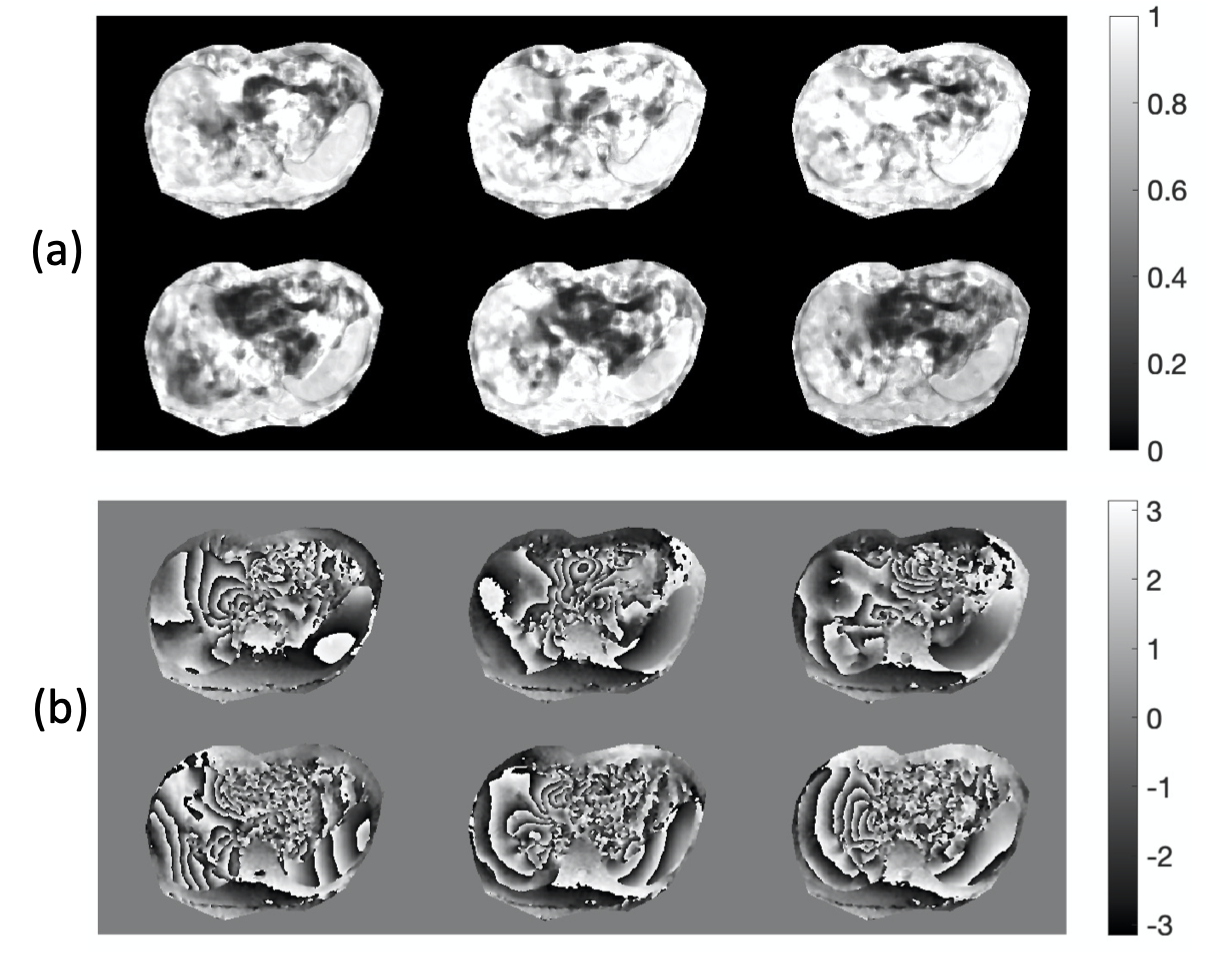
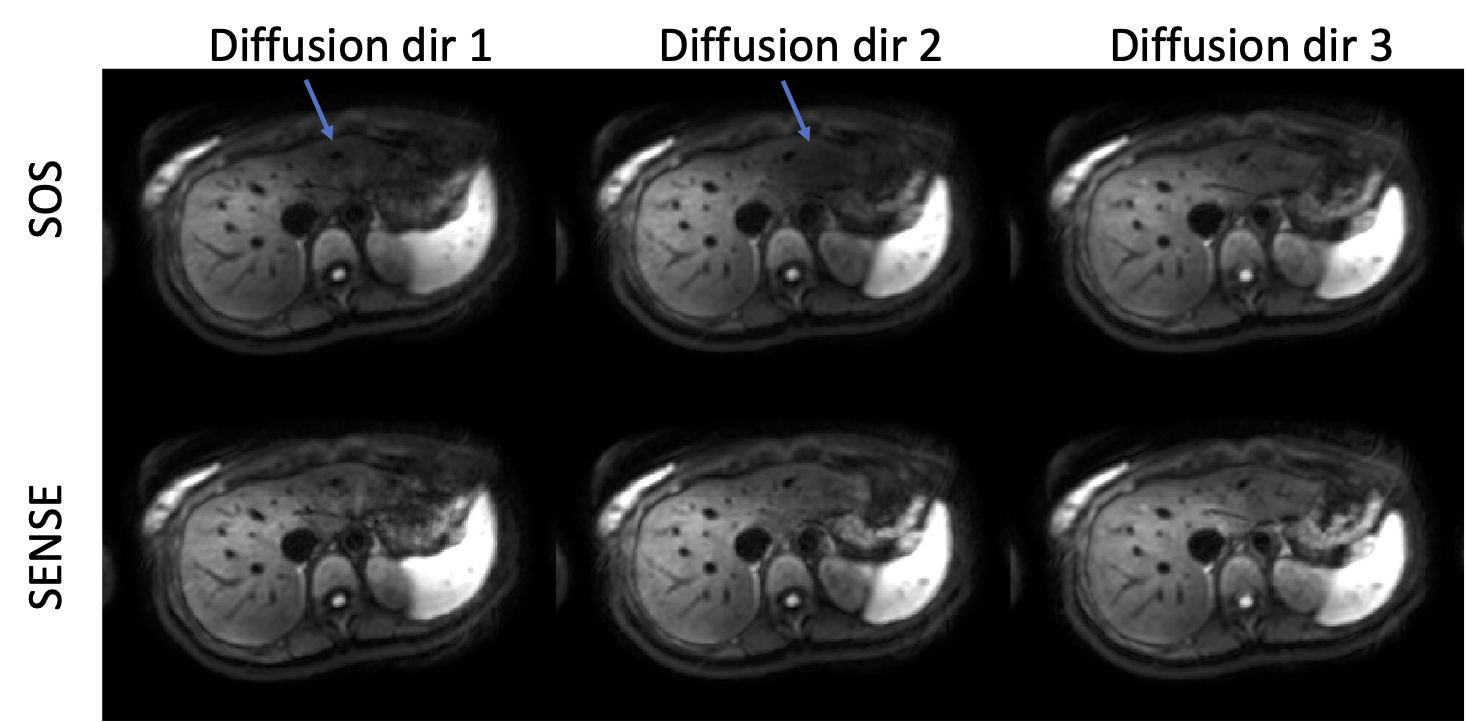
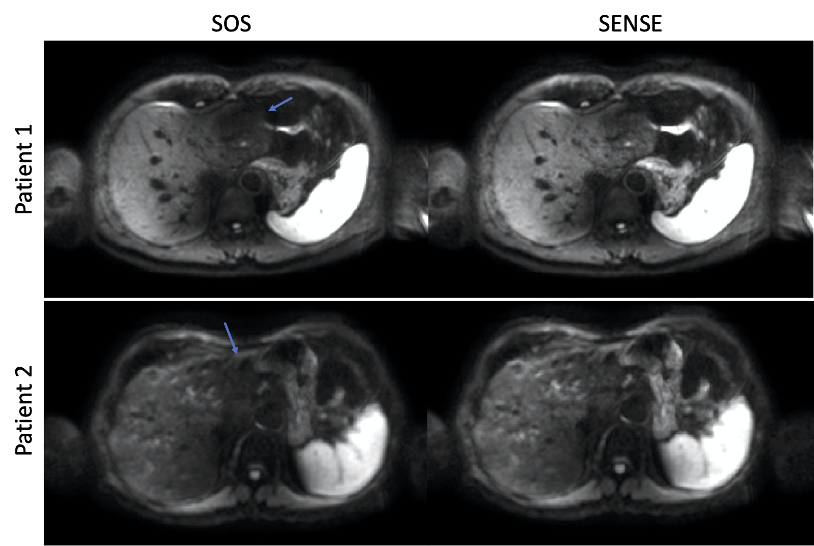
Figure 1 shows examples of the MISLP maps from 6 averages in one volunteer slice. Areas of signal loss especially in the left liver lobe and their different appearance across averages were captured (Figure 1a). Rapidly varying motion-induced phases that are different from average to average can also be observed (Figure 1a). Figure 2 shows the DWIs from three diffusion encoding directions with SENSE-like reconstruction (SENSE-like) and SOS averaging. The SENSE-like results remove the observed signal loss in comparison the SOS results, yielding more homogeneous liver signal. Average DWI across three diffusion encoding directions are presented in Figure 3 for two patients. Similar to the volunteer data, SENSE-like reconstruction results significantly reduce the appearance of signal loss, giving better visualization of anatomies and pathologies (lesion on the left lobe of patient 1).Discussion
A methodology was proposed formulating the averaging of the DWIs as reconstruction problem with SENSE at R=1 and treating the motion induced signal loss maps as sensitivity maps. The proposed SENSE-like reconstruction of the multi-averages DWIs mitigated motion-induced signal loss compared to the conventional SOS averaging method. The proposed SENSE-like reconstruction of multi-average DWI data yields more homogeneous liver signal and should be tested whether it improves diagnostic image quality in a larger clinical DWI dataset.Acknowledgements
The present work was supported by Philips Healthcare.References
1. Lewis S, Dyvorne H, Cui Y, Taouli B. Diffusion-weighted imaging of the liver: techniques and applications. Magn Reson Imaging Clin N Am 2014;22(3):373-395.
2. Kwee TC, Takahara T, Niwa T, Ivancevic MK, Herigault G, Van Cauteren M, Luijten PR. Influence of cardiac motion on diffusion-weighted magnetic resonance imaging of the liver. MAGMA 2009;22(5):319-325.29.
3. Taouli B, Koh DM. Diffusion-weighted MR imaging of the liver. Radiology 2010;254(1):47-66.
4. Gadjimuradov F, Benkert T, Nickel MD, Maier A. Deep Learning-based Adaptive Image Combination for Signal-Dropout Suppression in Liver DWI. Proc. Intl. Soc. Mag. Reson. Med. 29 (2021):0534
Figures