3271
Effect of Inhaled Oxygen Concentration on 129Xe Chemical Shift of Red Blood Cells in Rat Brain1Hospital for Sick Children, Toronto, ON, Canada, 2University of Toronto, Toronto, ON, Canada, 3University of Pennsylvania, Philadelphia, PA, United States
Synopsis
Hyperpolarized 129Xe RBC chemical shift in the rat brain was measured to be higher during hypoxic ventilation than during normoxic ventilation.
Methods: All protocols were approved by the Animal Care Committee of the Hospital for Sick Children. MR spectra were acquired on a clinical 3T scanner (Prisma, Siemens, Erlangen, Germany) using a 1.7cm radius, 5.7cm long transmit/receive birdcage coil (Morris Instruments, Ottawa, Canada) centred on the rat brain. 5 female Sprague-Dawley rats (290 ± 30g, Taconic Biosciences, Rensselaer, NY) were ventilated for 10 minutes without xenon at an FiO2 of either 22% (i.e. normoxia) or 14% (i.e. hypoxia) as previously described.10 After 10 minutes, 129Xe gas was added to the ventilation mix, while the FiO2 was maintained. After 10 seconds, 10 FIDs were acquired using the following parameters: α = 90°, TR = 1 s, bandwidth = 15 kHz, and 2048 spectral points. The transmit and receive frequencies were centred on the RBC chemical shift, 210ppm from the gas frequency. The averaged FIDs were filtered in the time domain with a 10Hz exponential line broadening function and fit to 6 exponentials, (5 dissolved resonances and 1 gas resonance) using an open-source MATLAB toolkit.11 After Fourier transformation, the gas peak was used as the reference for estimating the RBC and the grey matter chemical shifts.
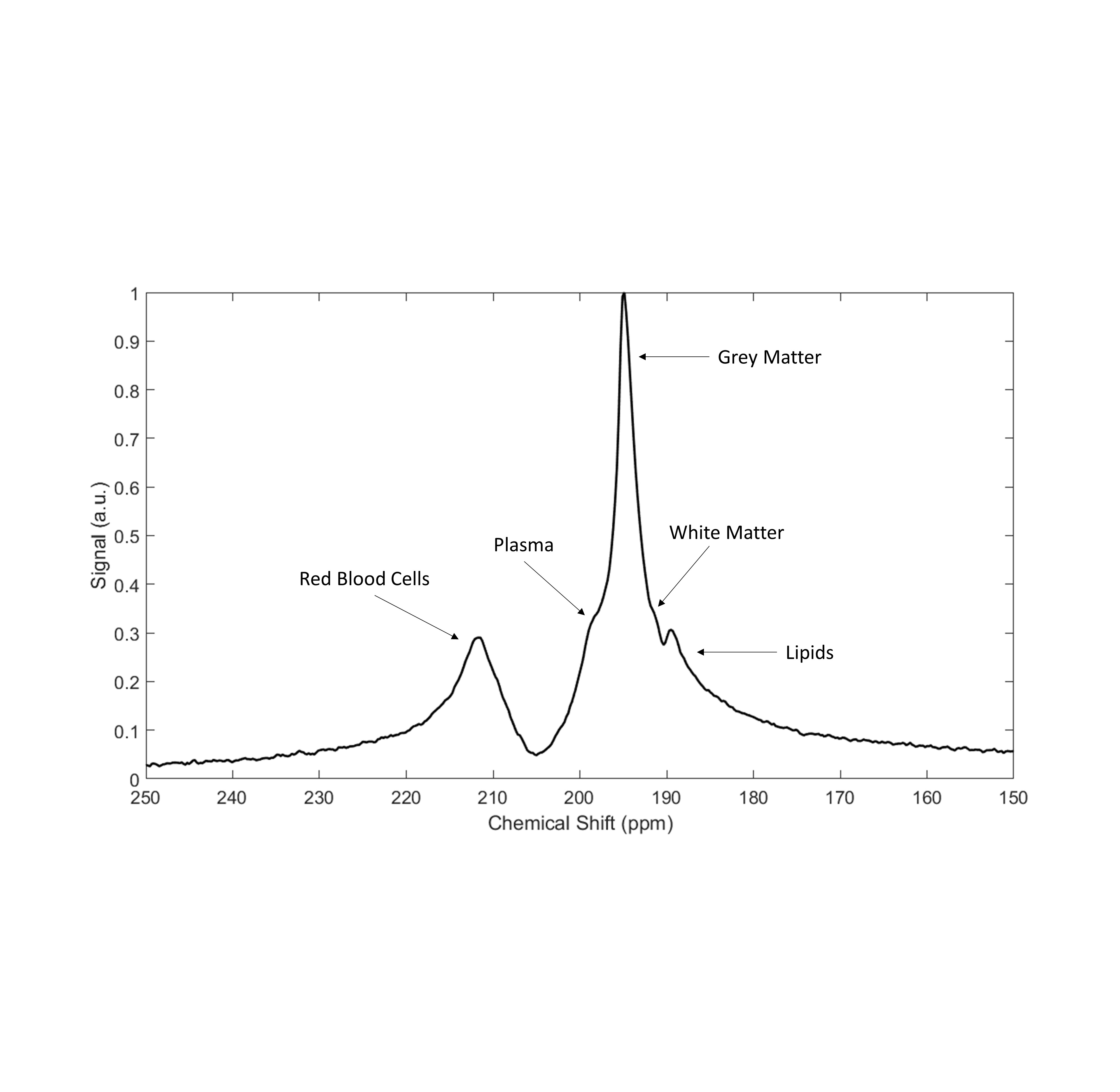
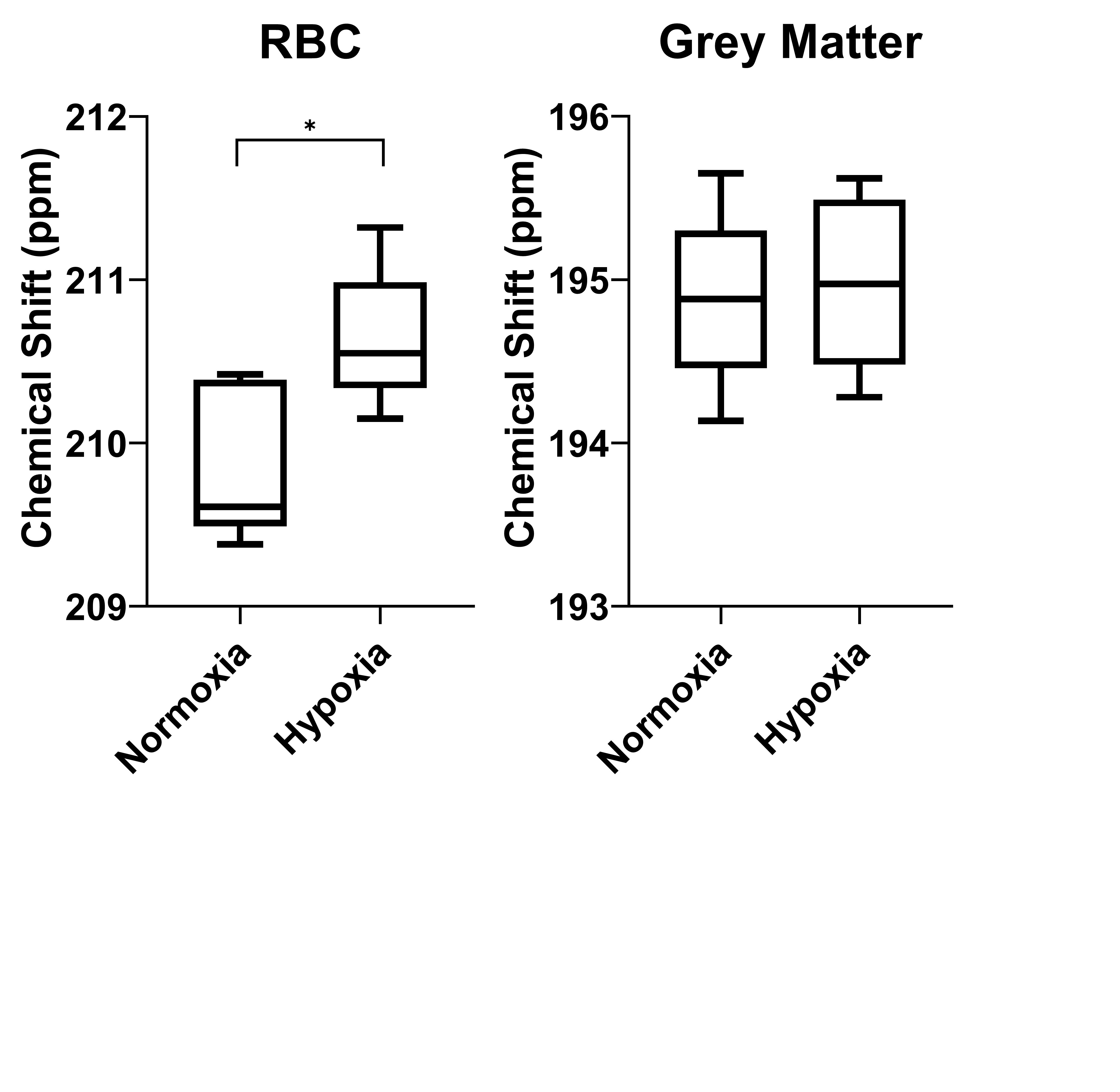
Results: Figure 1 shows a representative 129Xe spectrum acquired in the rat brain and figure 2 demonstrates the effect of hypoxia on the RBC and grey matter chemical shifts. The RBC chemical shift increased during hypoxia (FiO2 = 14%) by 0.77ppm ± 0.42 (p = 0.015) while no effect was seen for the grey matter chemical shift.
Discussion: The increase in RBC chemical shift at 14% O2 is very similar to the increase measured in the lungs at the same FiO2 (0.79ppm ± 0.34).6 As the change in 129Xe RBC chemical shift is principally dependent on conformational changes of the haemoglobin molecule,3 it may not be surprising that the effect is independent of the organ. These results also reinforce that the relationship between oxygenation and RBC chemical shift in rats is opposite to that observed in humans, where RBC chemical shift has been observed to decrease with hypoxia.4,5 In vivo measurement of cerebral blood oxygenation (SO2) in rats could be an important tool for evaluating the progression and treatment of pre-clinical models of brain diseases such as stroke or brain tumors. While the results presented here provide evidence of the relationship between hypoxia and RBC chemical shift in the rat brain, it does not provide an empirical relationship between SO2 and chemical shift. SO2 is directly dependent on FiO2 but there are conflating physiological effects that create variability in the relationship between SO2 and FiO2. Therefore, future experimental setup should be prescriptive of SO2 instead of FiO2. Alternatively, an empirical relationship between SO2 and chemical shift could be developed through in vitro experiments as was previously done for human blood.4,5 An empirical equation relating SO2 to RBC chemical shift in vivo is further complicated by local changes in bulk magnetic susceptibility that affect the regionally dispersed RBC chemical shift but not the gas reference frequency. This is evidenced by the difference in RBC chemical shift at normoxia measured in the brain (209.9ppm ± 0.48) and the lungs (210.7ppm ± 0.1).6 To control for this, the chemical shift should be measured regionally (using CSI or single voxel spectroscopy) and measured relative to a local reference frequency (e.g. using 1H spectroscopy).12 Conclusions: The RBC chemical shift increased during hypoxia (FiO2 = 14%) by 0.77ppm ± 0.42. This relationship is the same as previously reported for the rat lungs. This provides the groundwork for future experiments that measure the effect of disease on RBC chemical shift in the brain.
Acknowledgements
This work was supported by grant funding from NSERC Discovery Grant (RGPIN-2015-03832). Y.F. was financially supported by a Queen Elizabeth II Graduate Scholarship in Science and Technology.References
1. Rao M, Stewart NJ, Norquay G, Griffiths PD, Wild JM. High resolution spectroscopy and chemical shift imaging of hyperpolarized 129Xe dissolved in the human brain in vivo at 1.5 tesla. Magn Reson Med. 2016;75(6):2227-2234. doi:10.1002/mrm.26241
2. Kershaw J, Nakamura K, Kondoh Y, Wakai A, Suzuki N, Kanno I. Confirming the existence of five peaks in 129Xe rat head spectra. Magn Reson Med. 2007;57(4):791-797. doi:10.1002/mrm.21186
3. Norquay G, Wolber J, Wild JM. Chapter 20. 129Xe Chemical Shift and Spin–Lattice Relaxation Dependences on Blood Oxygenation. In: Hyperpolarized Xenon-129 Magnetic Resonance: Concepts, Production, Techniques and Applications. The Royal Society of Chemistry; 2015:365-391. doi:10.1039/9781782628378-00365
4. Wolber J, Cherubini A, Leach MO, Bifone A. Hyperpolarized 129Xe NMR as a probe for blood oxygenation. Magn Reson Med. 2000;43(4):491-496. doi:10.1002/(SICI)1522-2594(200004)43:4<491::AID-MRM1>3.0.CO;2-6
5. Norquay G, Leung G, Stewart NJ, Wolber J, Wild JM. 129Xe chemical shift in human blood and pulmonary blood oxygenation measurement in humans using hyperpolarized 129Xe NMR. Magn Reson Med. 2016;77(4):1399-1408. doi:10.1002/mrm.26225
6. Friedlander Y, Zanette B, Lindenmaier AA, et al. Effect of inhaled oxygen concentration on 129Xe chemical shift of red blood cells in rat lungs. Magn Reson Med. 2021;86(3):1187-1193. doi:10.1002/mrm.28801
7. Friedlander Y, Zanette B, Lindenmaier A, et al. Chemical shift of 129Xe dissolved in red blood cells: Application to a rat model of bronchopulmonary dysplasia. Magn Reson Med. 2020;84(1):52-60. doi:10.1002/mrm.28121
8. Bier EA, Robertson SH, Schrank GM, et al. A protocol for quantifying cardiogenic oscillations in dynamic 129Xe gas exchange spectroscopy: The effects of idiopathic pulmonary fibrosis. NMR Biomed. 2019;32(1):e4029. doi:10.1002/nbm.4029
9. Mummy DG, Bier EA, Wang Z, et al. Hyperpolarized 129Xe MRI and Spectroscopy of Gas-Exchange Abnormalities in Nonspecific Interstitial Pneumonia. Radiology. July 2021:204149. doi:10.1148/radiol.2021204149
10. Friedlander Y, Zanette B, Stirrat E, Couch M, Kassner A, Santyr G. Dependence of the Chemical Shift of 129Xe Dissolved in Red Blood Cells on Transit Time from the Lung Gas Exchange Region in Rats. In: ISMRM 26th Annual Meeting. ; 2018:512. doi:10.1002/mrm.24992.4.
11. Robertson SH, Virgincar RS, Bier EA, et al. Uncovering a third dissolved-phase 129Xe resonance in the human lung: Quantifying spectroscopic features in healthy subjects and patients with idiopathic pulmonary fibrosis. Magn Reson Med. 2017;78(4):1306-1315. doi:10.1002/mrm.26533
12. Antonacci MA, Zhang L, Burant A, McCallister D, Branca RT. Simple and robust referencing system enables identification of dissolved-phase xenon spectral frequencies. Magn Reson Med. 2018;80(2). doi:10.1002/mrm.27042
Figures