3270
A Radial Flow Cell Perfusion System for Hyperpolarized 13C NMR Metabolic Studies at Low Oxygen Levels1Cancer Biology, University of Pennsylvania, Philadelphia, PA, United States, 2Radiology, University of Pennsylvania, Philadelphia, PA, United States
Synopsis
We have developed new methods for studying cultured cancer cell metabolism with hyperpolarized 13C magnetic resonance spectroscopy (HP 13C MRS) at low oxygen concentrations. Cells grown on the surfaces of microcarriers inside the spectrometer were perfused radially (rather than axially) in a modified NMR tube at controlled oxygen levels. Computational and experimental results demonstrated that cell mass oxygen profiles with radial flow were much more uniform than with conventional axial flow. The metabolism of HP [1-13C]pyruvate was markedly different between the two flow configurations, demonstrating the importance of avoiding large oxygen gradients in cell perfusion experiments.
INTRODUCTION
Hyperpolarized 13C magnetic resonance spectroscopy (HP 13C MRS) is a unique technology that is well suited for the determination of metabolic flux in vivo [1-3]. Many hyperpolarized 13C compounds have been examined as metabolic tracers [4] but none have been shown to be specific indicators of hypoxia as occurs in solid cancers [4]. The primary goal of this work was to develop cell culture methods that allow the examination of HP 13C substrate metabolism at low, controlled oxygen levels. Fixed beds of microcarriers are commonly used for cell culture NMR studies. The flow of medium through the cell mass has historically been axial (parallel to the long axis of the NMR tube). However, direct measurement of perfusate oxygen levels have shown that the oxygen gradient across the cell mass can be very large [5]. With large gradients, the quantitative study of hypoxia is not possible. To potentially improve oxygen delivery, we examined changing the direction of perfusion from axial to radial. Because radial flow exposes a larger surface area of the bed for perfusion (approximately 3-fold) the total volumetric flow rate of perfusate can be significantly increased (also 3-fold), without increasing the linear velocity or shear at the microcarrier surface.METHODS
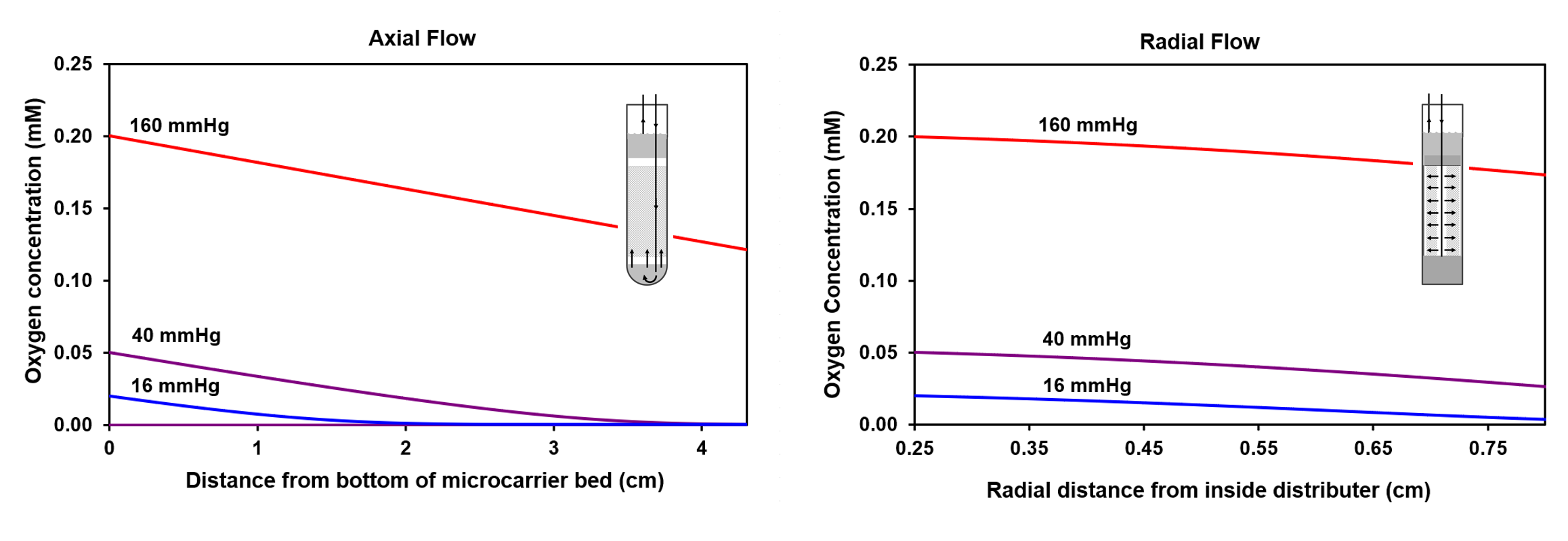
Rat hepatocellular carcinoma (HCC) cells were immobilized on non-porous microcarriers [5]. For axial flow, the microcarriers were held between two porous high-density polyethylene (HDPE) filters in a standard screwcap 20-mm NMR tube. For radial flow, the microcarriers were held between two cylindrical HDPE filters (inner: 1.6 mm I.D. × 5 mm O.D.; outer: 16 mm I.D. × 17.4 mm O.D.), inside an 18.4 mm I.D. x 20.0mm O.D. NMR tube. Medium flowed down an inner polyetherimide tube, across the inner HDPE cylinder, radially across the cell mass, and out of the NMR tube through the space between the outer HDPE cylinder and the NMR tube. The entire perfusion apparatus was constructed of low oxygen permeability materials to enable accurate oxygen measurements with polarographic probes [6]. HP injections were conducted during continuous medium perfusion to maintain steady state oxygen levels. Oxygen was removed from the aqueous HP substrates by counter-current helium stripping [6]. NMR spectra were acquired with an Oxford 9.4 Tesla 8.9cm bore magnet (Oxford Instruments) interfaced to a 400 MHz Varian DirectDrive Console. A 20-mm liquids probe (Varian) was used to acquire 31P (to determine cell mass levels) and 13C spectra. HP 13C spectra were acquired with a single 12° pulse, a repetition time of 3 s, 8192 points, and a spectral width of 25,000 Hz.RESULTS
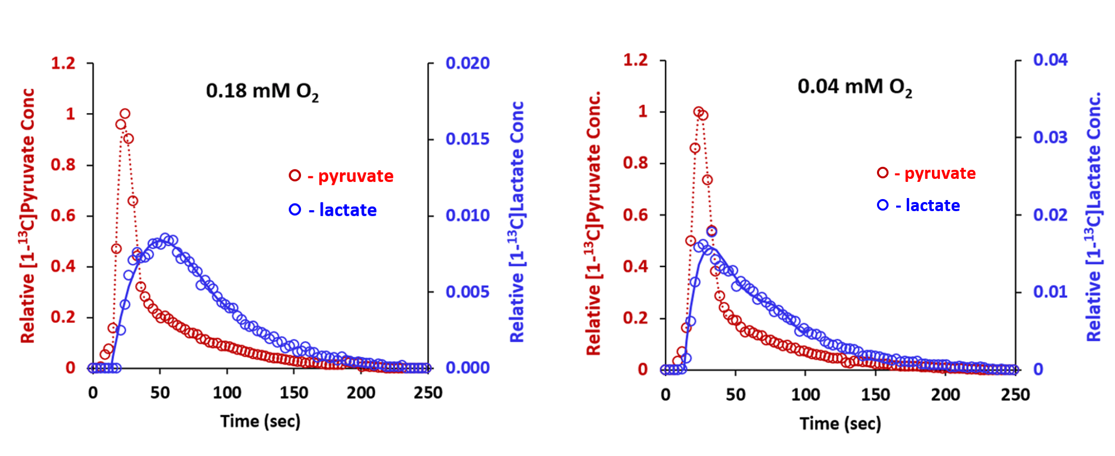
Oxygen concentration profiles through the cell mass were calculated as described previously [6] and the results are shown in Figure 1. At the lowest inlet oxygen concentrations, oxygen is completely depleted with axial flow before the perfusate reaches the end of the cell mass. With radial flow, oxygen transport is markedly improved and the oxygen gradients are much smaller. These results are consistent with our experimental polarographic probe measurements.For axial, injections with [1-13C]pyruvate were conducted at high (0.18 mM) and low (0.04 mM) inlet oxygen levels (Figure 2). Both [1-13C]lactate and [1-13C]alanine were formed. The time to peak intensity for lactate was 27 seconds after the time to peak intensity for [1-13C]pyruvate (TPL = difference between the two peak times). At 0.04 mM inlet oxygen, lactate formation was more rapid; the TPL was only 8 seconds. These results are consistent with the increased rate of lactate formation observed with offline analysis of the extracellular medium (not shown).
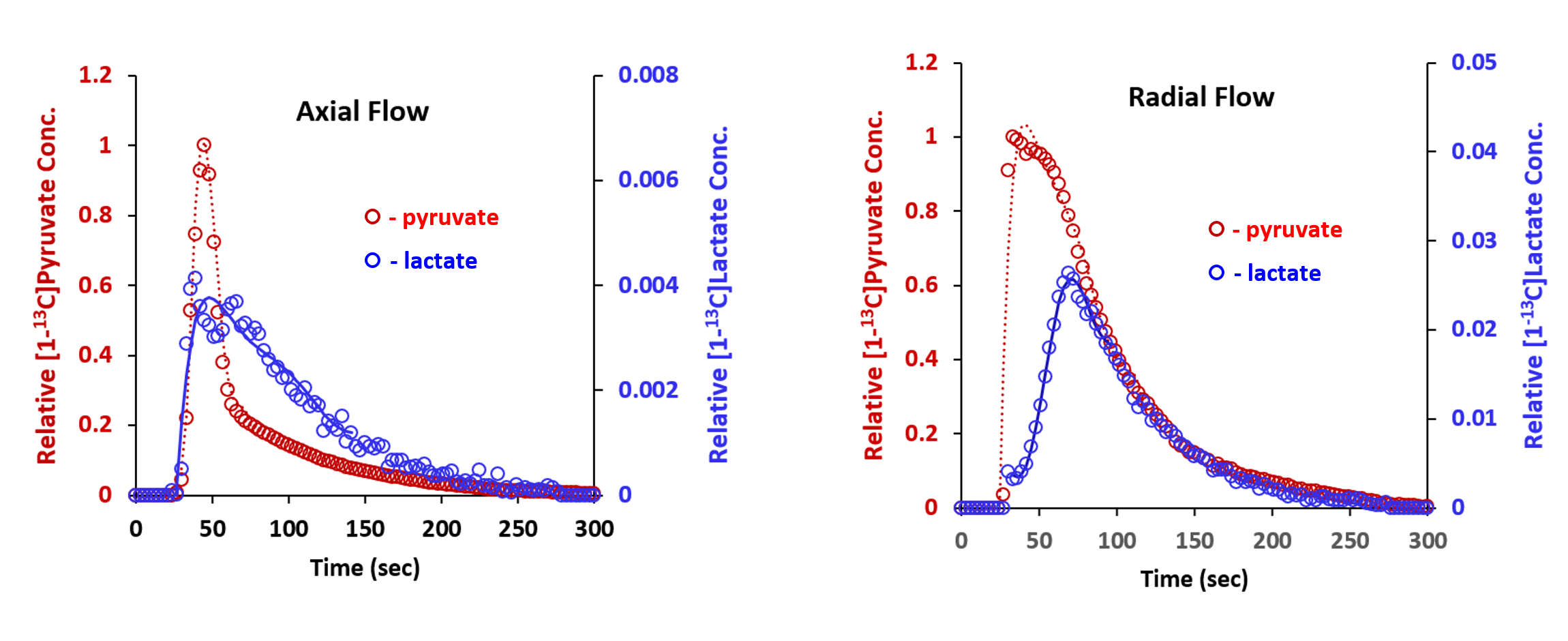
HP [1-13C]pyruvate injections were conducted for inlet oxygen concentrations of 0.06 mM with both flow schemes (Figure 3). The results show that TPL with axial flow (3 s) was much shorter than it was for radial flow (37 s), indicating the presence of very low oxygen levels (and hence large gradients) with axial flow. For the more homogeneous radial flow, the results are very similar to those observed under normoxia (Figure 2).
DISCUSSION
The calculations and the experimental data both demonstrate that radial flow significantly improves the transport of oxygen and consequently reduces the oxygen gradient size. It thereby allows NMR spectroscopy measurements to be made under more well-defined conditions. It is very important to note that if oxygen gradients were assumed to be negligible with axial flow, the axial flow results could be incorrectly interpreted to indicate that pyruvate metabolism at with 0.06 mM was markedly different from that at 0.18 mM.Even more homogenous oxygen profiles would be possible with cell lines that have lower oxygen consumption rates. Other cancer cell lines that have been examined with cell perfusion NMR have oxygen consumption rates that are one-fifth to one-tenth that for HR-2 cells [6]. In addition, further gains in oxygen homogeneity would be possible with a larger inner distributer. For example, the microcarrer bed dimensions could be changed to 9 mm I.D. and 20 mm O.D. to allow an approximate halving of the oxygen gradient. This would require a 25-mm NMR tube/probe and a slight loss of SNR/spin.
CONCLUSIONS
Radial flow perfusion reduces oxygen gradients with immobilized cells and could be very useful for identifying HP 13C NMR substrates that are specific indicators of hypoxia.Acknowledgements
Financial support from this work was provided by grants of TPFG: NIH DP5OD021391, Radiological Society of North America, NIBIB T32 EB00431112, NCRR UL1RR024134, and NCATS UL1TR000003.References
[1] Golman K, Zandt RI, Lerche M, et al. Metabolic Imaging by Hyperpolarized 13C Magnetic Resonance Imaging for in Vivo Tumor Diagnosis, Cancer Res. 2006, 66(22), 10855–10860.
[2] Day SE, Kettunen MI, Gallagher FA, et al. Nat. Med. 2007, 13(11), 1382–1387.
[3] Nelson SJ, Kurhanewicz J, Vigneron DB, et al. Metabolic Imaging of Patients with Prostate Cancer Using Hyperpolarized [1-13C]Pyruvate. Sci. Transl. Med. 2013, 5, 198ra108.
[4] Chaumeil MM, Najac C, Ronen SM, Studies of Metabolism Using 13C MRS of Hyperpolarized Probes. Methods Enzymol. 2015, 561, 1–71.
[5] Mancuso A, Beardsley NJ, Wehrli S, et al. Real-Time Detection of 13C NMR Labeling Kinetics in Perfused EMT6 Mouse Mammary Tumor Cells and BetaHC9 Mouse Insulinomas. Biotechnol. Bioeng. 2004, 87(7), 835–848.
[6] Mancuso A, Pourfathi M, Kiefer RM, et. al., Radial Flow Perfusion Enables Real-Time Profiling of Cellular Metabolism at Low Oxygen Levels with HP 13C NMR. Metabolites 2021, 11(9), 576.
Figures