3266
Correlation between MRI texture analysis and histo-pathology of Alzheimer’s disease: evaluation in the hippocampus of the Tgf344-AD rat model.1Magnetic Resonance Imaging Core Facility, IDIBAPS, Barcelona, Spain, 2CIBER-BBN, Barcelona, Spain, 3Universitat de Barcelona, Barcelona, Spain, 4Laboratory of Surgical Neuroanatomy, Universitat de Barcelona, Barcelona, Spain, 5Institute of Neurosciences, Universitat de Barcelona, Barcelona, Spain
Synopsis
β-amyloid plaques and neuroinflammation, two of the pathological processes associated to Alzehimer's disease (AD) are not visible by conventional MRI. However, its presence may induce image changes that can be evaluated using texture analysis. We evaluated the potential of texture analysis of conventional MRI to characterize these changes. T2-weighted MRI of a transgenic rat model of AD were acquired and texture measures in hippocampus were correlated with inmunohistochemical quantification of plaques and microglia, showing significant correlation. Coherently, significant differences in the texture measures were found between transgenic and control animals, pointing to the potential of texture analysis as AD biomarker.
INTRODUCTION
β-amyloid plaque deposition and neuroinflammation are two processes that have been related to Alzheimer’s disease (AD)1. However, its early detection based on non-invasive methods such as magnetic resonance imaging (MRI) is still challenging. Recently, the ability of texture features to help AD diagnosis or prognosis has been evaluated, either by themselves or as part of the features considered by radiomics algorithms2,3. The hypothesis underlying the use of image texture is that deposition of plaques or other processes may induce changes in image intensity and therefore it will have an effect in the spatial variation in voxel intensity levels2. However, to the best of our knowledge, the correlation between AD pathological hallmarks and texture metrics has not been assessed. In this work, we evaluated the correlation between MRI texture and histologic features in the hippocampus of a transgenic rat model of AD (TgF344-AD)4; as well as the potential of these features to discriminate between transgenic and control animals.METHODS
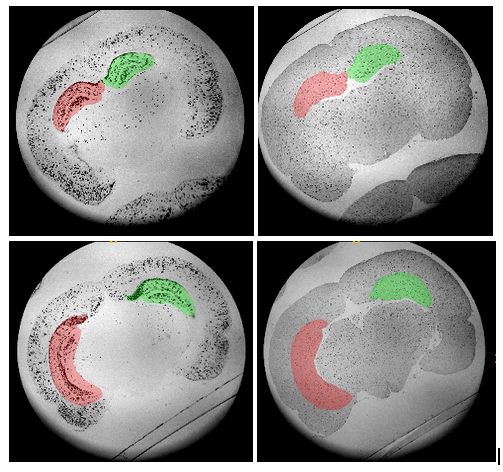
MRI acquisition was performed in a cohort of 25 TgF344-AD animals (12 females/13 males) and their 26 control littermates (12 females/14 males) every 4 months from 3 to 19 months of age. Structural T2-weighted images were acquired using a RARE sequence with effective echo time TE = 35.3 ms, repetition time TR = 6,000 ms and RARE factor = 8, voxel size = 0.12 × 0.12 mm2, 40 slices, slice thickness = 0.8 mm, and field of view FoV = 30 × 30 × 32 mm3. Together with this, the brains of four transgenic animals were excised and fixed and ex-vivo MRI acquisition performed with the same acquisition protocol.Immunohistochemical analysis were performed in fixed brains to visualize β-amyloid protein and microglia, with ab4 and Iba1 antibodies respectively. These were quantified in four hippocampal regions of each brain (anterior right, anterior left, posterior right and posterior left) as shown in Figure 1.
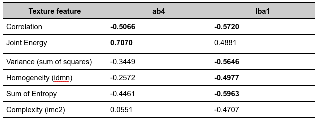
GLCM (gray-level co occurrence matrices) was considered for texture analyses and six features were evaluated: correlation, joint energy, variance, homogeneity (inverse difference normalized, idn), sum of entropy, and complexity (informational measure of correlation 2, imc2) using pyradiomics5. Hippocampus was automatically segmented in the T2-w images based on a rat brain atlas using diffeomorphic elastic registration as described in6 and texture analysis was performed in this region. In the ex-vivo images, after automatic hippocampus segmentation, the MR slices corresponding to the histological sections were visually identified to establish the correspondence between them, so 4 regions equivalent to those identified in histological sections were identified in each MRI volume. Texture measures of these regions were computed and their value was correlated with ab4 and Iba1 quantification. Kruskal-wallis tests were performed between transgenic and control groups to evaluate statistical differences in texture metrics between genotypes.
RESULTS
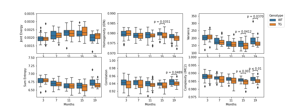
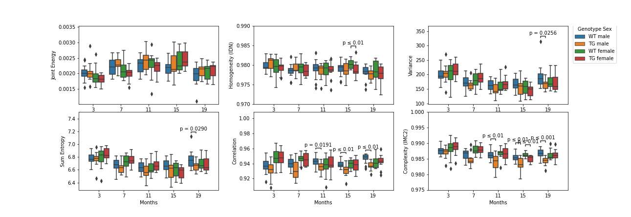
Table 1 shows the correlation between the evaluated texture metrics and ab4, iba1. Strong correlation was observed between histological and texture properties, namely, ab4 was significantly correlated with joint energy and correlation measures; while iba1 showed significant (anti)correlation with correlation, variance, homogeneity and entropy.As shown in Figure 2, group-wise comparison showed significantly lower values of homogeneity at 15 months of age, entropy and IMC2 at 15 and 19 months and correlation at 19 months. Since a significant effect of sex was observed, we evaluated separately the effect of genotype in each sex (Figure 3). In transgenic females only a significant decrease was observed in homogeneity and IMC2 with respect to wild-type at 15 months of age, measures that were shown to be correlated with iba1 but not with β-amyloid load. More changes were observed in the transgenic males with respect to controls, including decreased variance and entropy at 18 months, while correlation and iMC2 were significantly reduced from 11 months on.
DISCUSSION
Texture analysis is a promising technique in the research for AD early biomarkers. Not only is altered in transgenic animals with respect to controls, but it is also strongly correlated with pathological hallmarks in AD. Therefore it could be indirectly a marker of the presence of β-amyloid plaques and/or neuroinflammation. Further investigation in bigger histological data-set will be required to confirm these findings. To note, while in females significant differences were observed in properties significantly correlated with iba1 staining at 15 months of age, in transgenic males, differences were observed starting at 11 months and in measures correlated with both beta-amyloid load and iba1. To mention, while in human population, about two thirds of persons diagnosed with AD dementia are women, our results point to stronger pathological effect in males in this transgenic model7. Further investigation must be performed to elucidate this sex-related differences in this model and identify if different processes or different progression are related to the disease in each sex.CONCLUSION
Texture measures derived from conventional MRI acquisitions significantly differ between TgF344-AD and wild-type animals suggesting its potential as a biomarker of the disease. The correlation between texture and histological measures such as beta-amyloid load and neuroinflammation points to its ability to characterize the underlying pathological process.Acknowledgements
This work has been funded by the project PI18/00893, integrated in the Plan Nacional I+D+I and co-funded by ISCIII-Subdirección General de Evaluación and European Regional Development Fund (ERDF) "A way to make Europe" and Secretaria d’Universitats i Recerca del Departament d’Empresa I Coneixement de la Generalitat de Catalunya AGAUR 2017 SGR 01003. CIBER-BBN is an initiative financed by the Instituto de Salud Carlos III with assistance from the European Regional Development Fund. We are indebted to the Experimental MRI 7T Unit of the IDIBAPS.References
[1] DeTure, M.A., Dickson, D.W. The neuropathological diagnosis of Alzheimer’s disease. Mol Neurodegeneration 14, 32 (2019).
[2] Jia-Hui C. et al. Magnetic Resonance Texture Analysis in Alzheimer's disease,Academic Radiology, 27(12):1774-1783, (2020)
[3] Feng Q, Ding Z. MRI Radiomics Classification and Prediction in Alzheimer's Disease and Mild Cognitive Impairment: A Review. Curr Alzheimer Res. 17(3):297-309 (2020)
[4] Cohen RM, et al. A transgenic Alzheimer rat with plaques, tau pathology, behavioral impairment, oligomeric aβ, and frank neuronal loss. J Neurosci. 33(15):6245-56. (2013)
[5] Griethuysen, J.J.M., et al. Computational radiomics system to decode the radiographic phenotype. Cancer Research, 77(21), e104–e107 (2017)
[6] Mielke MM. Sex and Gender Differences in Alzheimer's Disease Dementia. Psychiatr Times. 35(11):14-17 (2018)
[6] Muñoz-Moreno E, et al. Brain connectivity during Alzheimer's disease progression and its cognitive impact in a transgenic rat model. Netw Neurosci.4(2):397-415 (2020)
Figures