3261
The Contribution of Increased Choroid Plexus Volume in the Alzheimer’s Disease1College of Biomedical Engineering & Instrument, Zhejiang University, Hangzhou, China, 2Psychology, Beijing Normal University, Beijing, China, 3Biomedical Engineering, University of Virginia, Charlottesville, VA, United States, 4Department of Computer Science, State University of New York at Binghamton, Binghamton, NY, United States, 5Radiology, Beth Israel Deaconess Medical Center and Harvard Medical School, Boston, MA, United States
Synopsis
Alzheimer’s disease (AD) may be caused by the dysfunction of glymphatic system, in which anatomic changes of the choroid plexus may be associated with reduced CSF production. Our previous work showed significantly increased choroid plexus volume in AD patients compared to healthy controls. However, contributions of the lateral ventricle volume were not investigated. Here, the lateral ventricle and choroid plexus were segmented using two cascaded deep learning models and the volumes were measured on 733 subjects. Increased lateral ventricle volume was found in AD patients, similar to previous studies. More importantly, choroid plexus volume showed a unique contribution to AD.
Introduction
The glymphatic system and cerebrospinal fluid (CSF) provide drainage of Amyloid-beta and their malfunction can be associated with Alzheimer’s disease (AD) [1]. The choroid plexus located in the lateral ventricle secretes the CSF, and therefore its anatomic changes were assumed to be associated with AD. Tadayon et al. investigated the assocations between the choroid plexus volume and CSF proteins [2]. In our previous research [3], the choroid plexus volume was found to be significantly larger in AD patients compared to healthy controls. However, brain atrophy and enlarged lateral ventricles are associated with AD [4]. A possible cause of enlarged choroid plexus in AD patients could be more unfolded choroid plexus in the larger lateral ventricle of AD patients. To test this hypothesis, we segmented the lateral ventricle and choroid plexus using two cascaded deep learning models. Then the volumes were quantified and their contributions to the AD were investigated statistically.Methods
733 subjects were selected from the Alzheimer’s Disease Neuroimaging Initiative (ADNI) database based on the 3D MPRAGE sequence and 3T Siemens scanners. It included 409 healthy controls (73.3 ± 7.5 years old, 40.1% male), 188 mild cognitive impairment (MCI) patients (75.0 ± 8.5 years old, 54.8% male), and 136 AD patients (75.5 ± 8.3 years old, 54.4% male).T1W images were resampled to a spatial resolution of 1 mm3 isotropic. Skull stripping was achieved using the deepbrain toolbox [5] and morphological corrections. Then, a 160 × 200 × 160 volume was cropped according to the brain center of each subject.
Lateral ventricle segmentation and the choroid plexus segmentation models were implemented based on a modified 3D U-Net structure [6, 7]. Residual connection and PReLU activation function were adopted in the convolution block. Dice loss was used to update models’ parameters. Lateral ventricle segmentation model was trained and validated on 35 subjects from the MICCAI 2012 challenge dataset [8] with further in-house manual correction. Another dataset of manually labeled 50 ADNI subjects was used for the choroid plexus segmentation model. With the well-trained models, the lateral ventricle segmentation was deployed on the ADNI dataset first, then the ventricle patch was cropped (96×96×80) as the input of the choroid plexus segmentation model.
The lateral ventricle and choroid plexus volume of each subject were calculated as the number of voxels in the segmentation results. Their contributions were calculated statistically using SPSS 26. First Shapiro-Wilk test and Levene test were used to identify the normality and homogeneity of variance for lateral ventricle volume, which were prerequisites for choosing a reasonable method, then a nonparametric Kruskal-Wallis test was used to compare the medians of lateral ventricle volume of the healthy controls, MCI and AD groups. Linear correlation between the lateral ventricle and choroid plexus volume was checked using the Pearson correlation coefficient. Furthermore, the ventricle volume and choroid plexus volume were normalized using z-score. Then, an ordinal multi-class logistic regression was conducted to examine the choroid plexus contribution by controlling the lateral ventricle volume effects.
Results
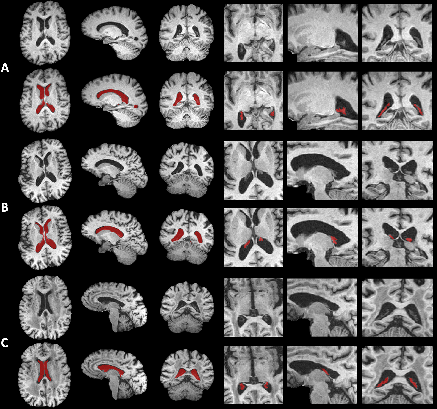
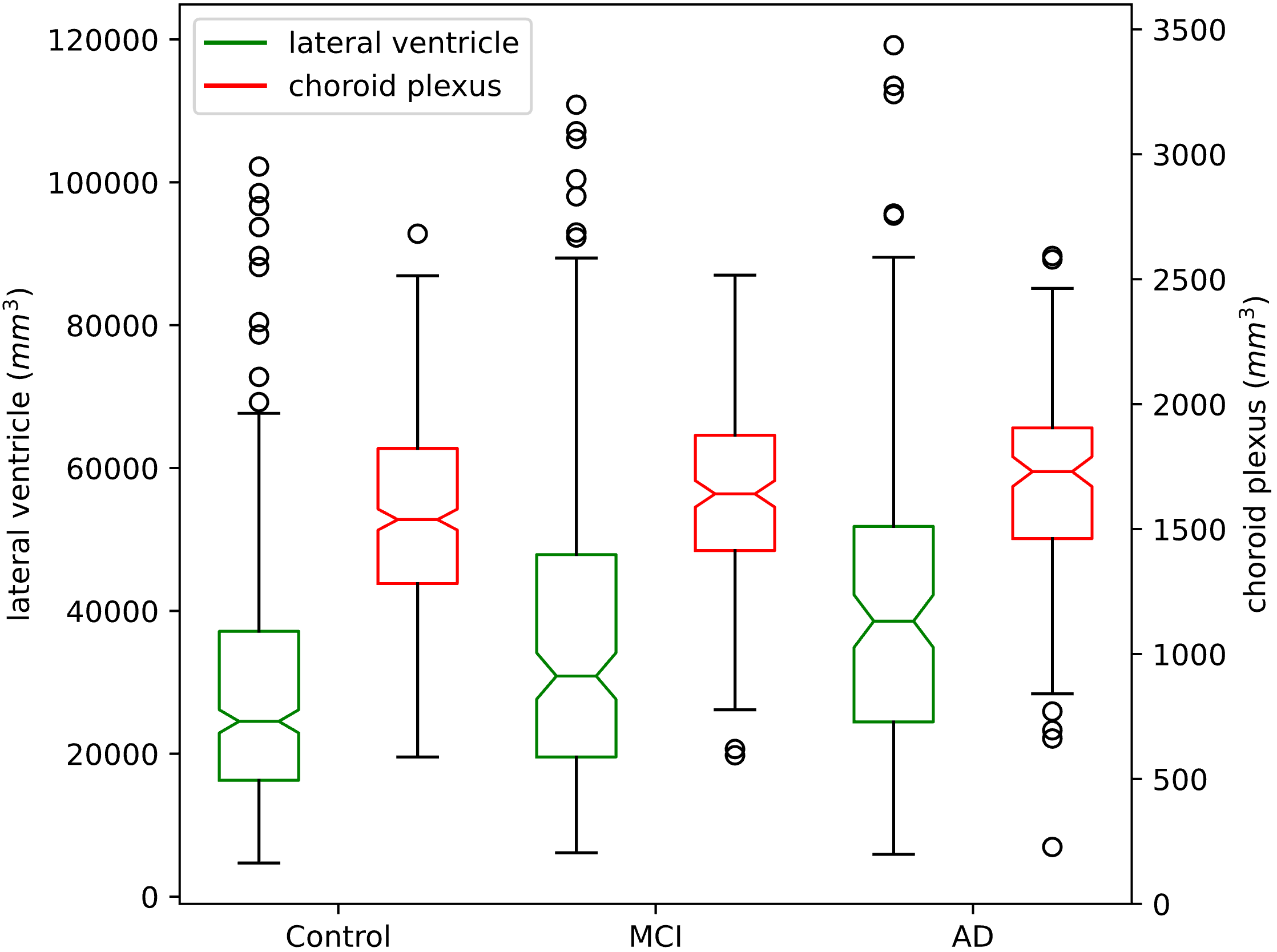
Segmentation results of deep learning models are shown in Figure 1. The proposed methods accurately segmented the regions of interest. The Dice coefficients were 0.854 (five-fold cross-validation) in the lateral ventricle and 0.727 in the choroid plexus regions.The lateral ventricle volumes of AD patients, MCI patients, and healthy volunteers were 41,447 ± 22,395mm3, 36,047 ± 21,616mm3 and 28,946 ± 17,139mm3. The overall distribution of the lateral ventricle volume and choroid plexus volume is shown in Figure 2. Results of the Shapiro-Wilk test (p<0.05 in three groups) and Levene test (p<0.05) suggest that the lateral ventricle volume of each group with homogeneity of variance follows the non-normal distribution. Therefore, a nonparametric Kruskal-Wallis test was appropriate for the comparison. The Kruskal-Wallis test shows significant differences among the three groups (H=45.64, p<0.001), and p values of post hoc multiple comparisons are less than 0.05.
The choroid plexus volume was increased in the AD patients compared to the healthy controls, as shown in Figure 2 and our previous work [2]. The Pearson correlation coefficient between the lateral ventricle and choroid plexus volumes was 0.343, which showed a moderate correlation between them. In ordinal multi-class logistic regression, the standardized regression coefficients for lateral ventricle volume and choroid plexus volume were 0.418 and 0.161, respectively (p<0.05). It indicated that the volume change of the choroid plexus had a unique impact on AD.
Discussion
In this study, we quantified the lateral ventricle and choroid plexus volume by leveraging deep-learning segmentation models. The lateral ventricle and choroid plexus volume of healthy volunteers, MCI patients, and AD patients increased sequentially and significantly. In the ordinal multi-class logistic regression, choroid plexus volume showed a unique contribution to the risk of Alzheimer’s disease. This finding demonstrated the increased choroid plexus volume was associated with AD which may provide a novel biomarker.Acknowledgements
This work is supported in part by the National Institute of Mental Health through R01MH080729, the Alzheimer’s Association through AARF-18-566347, the Fundamental Research Funds for the Central Universities, MOE Frontier Science Center for Brain Science & Brain-Machine Integration, Zhejiang University.References
[1] Iliff JJ, Wang M, Liao Y, Plogg BA, Peng W, Gundersen GA, Benveniste H, Vates GE, Deane R, Goldman SA, Nagelhus EA. A paravascular pathway facilitates CSF flow through the brain parenchyma and the clearance of interstitial solutes, including amyloid β. Science translational medicine. 2012 Aug 15;4(147):147ra111.
[2] Tadayon E, Pascual-Leone A, Press D, Santarnecchi E, Alzheimer's Disease Neuroimaging Initiative. Choroid plexus volume is associated with levels of CSF proteins: relevance for Alzheimer's and Parkinson's disease. Neurobiology of aging. 2020 May 1;89:108-17.
[3] Zhao L, Feng X, Hu Y, Wu D, Meyer CH, Alsop DC. Changes of Choroid Plexus Volume in Alzheimer’s Patients. In Proceedings of the International Society for Magnetic Resonance in Medicine 2021(2374).
[4] Nestor SM, Rupsingh R, Borrie M, Smith M, Accomazzi V, Wells JL, Fogarty J, Bartha R, Alzheimer's Disease Neuroimaging Initiative. Ventricular enlargement as a possible measure of Alzheimer's disease progression validated using the Alzheimer's disease neuroimaging initiative database. Brain. 2008 Sep 1;131(9):2443-54.
[5] Deep Learning tools for brain medical images. https://github.com/rockstreamguy/deepbrain
[6] Çiçek Ö, Abdulkadir A, Lienkamp SS, Brox T, Ronneberger O. 3D U-Net: learning dense volumetric segmentation from sparse annotation. In International conference on medical image computing and computer-assisted intervention 2016 Oct 17 (pp. 424-432). Springer, Cham.
[7] Zhao L, Feng X, Meyer CH, Alsop DC. Choroid plexus segmentation using optimized 3D U-Net. In 2020 IEEE 17th International Symposium on Biomedical Imaging (ISBI) 2020 Apr 3 (pp. 381-384). IEEE.
[8] http://www.neuromorphometrics.com/2012_MICCAI_Challenge_Data.html
Figures