3242
Development of a Benchtop 7T cryogen-free animal MRI system1Institute of Electrical Engineering, Chinese Academy of Sciences, Beijing, China, 2USC Stevens Neuroimaging and Informatics Institute, Keck School of Medicine, University of Southern California, Los Angeles, CA, United States, 3School of Information Technology and Electrical Engineering, The University of Queensland, Brisbane, Australia
Synopsis
A small-size, light-weight, cryogen-free 7T animal MRI system is being developed in IEE CAS. The magnet system has been completed and acceptable magnetic field strength and homogeneity indices have been achieved. The gradient assembly with passive shimming is going into the final assembly stage and the RF coil is ready for tunning. The integration of the whole system will be very soon and a mouse experiment will be conducted then. It is expected the ultimate product will largely boost the scientific research activities that are unapproachable to advanced MRI scanners.
Introduction
High-field MRI has significant advantages in terms of signal to noise ratio (SNR) and resolution, which can not only image the microscopic structure of biological tissue, but also explore the cognitive function, and even conduct metabolic detection of the living body [1]. However, existing high-field MRI systems usually have a bulk volume with a vast amount of liquid helium stored, having a high fabrication and maintenance cost beyond the budget of many MRI users. In this work, a small-size, light-weight, 7T cryogen-free animal MRI scanner is under development in the Institute of Electrical Engineering, Chinese Academy of Sciences (IEE CAS). Until now, the fabrication of the superconducting magnet of this Benchtop 7T system has been completed, the gradient coil is in the final assembly stage, and the RF coil is ready for tuning. We report here on progress details of the hardware system development.Method
In designing the actively-shielded superconducting magnet of the Benchtop 7T MRI system, we adopted a radial current density classification scheme by using different wire gauges on the primary long solenoid coils [2]. Based on the magnetic field of the primary coils, several adjusting coils were necessary to elevate the central field strength further and offset the unwanted inhomogeneous magnetic field components simultaneously, and shielding coils were used to constrain the 5-Gauss line range. The magnet was wound with NbTi superconducting wires on aluminium alloy bobbins to guarantee a large heat transfer coefficient. Oxygen-free copper was used as the conduction tape connected to a pulse tube cryocooler [3]. The magnet coils were hoisted in a helium-free cryogenic vessel and operated in a dry mode. Inside the warm bore of the magnet, a thin gradient assembly will be installed. The three-axis gradient coils were all unshielded to reduce the total thickness, where a hollow z gradient coil also plays a role of cooling pipe, and the shim trays were placed in the inner wall of the coil bobbin. A relatively large available space will be reserved to install the RF coil and then use for the imaging detection.Results
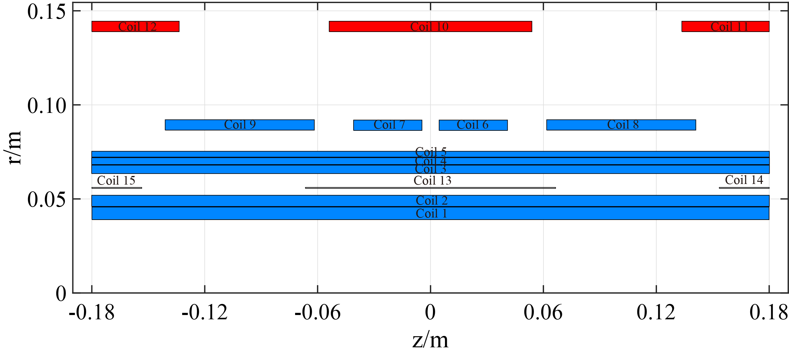
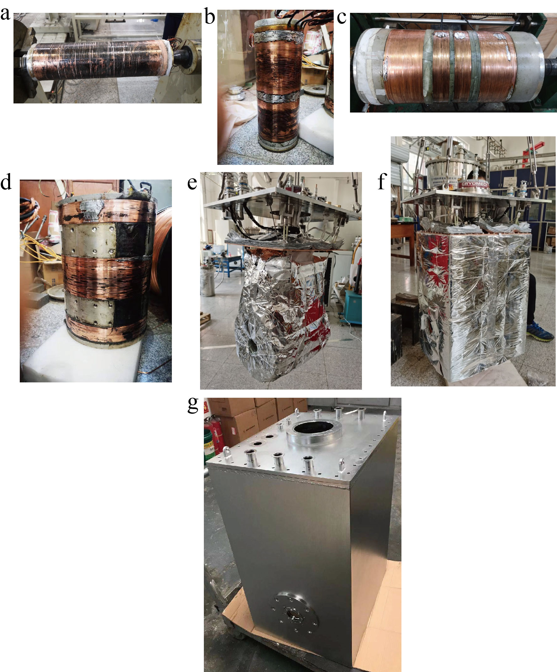
The coils structure of the main magnet is illustrated in Fig. 1, which includes five primary coils 1-5, four adjusting coils 6-9, three shielding coils 10-12, and three electromagnetic interference screening (EIS) coils 13-15 [4]. The fabricated magnet coils, heat conduction structure, cold shield and vacuum container are shown in Fig. 2. The magnet was dedicatedly protected with thermal insulation, and one pulse tube cryocooler was directed and connected to the magnet endplate.The final magnet system was tested, as shown in Fig. 3, and specified magnet strength and homogeneity were achieved. The magnet has a dimension with a length of 635mm, width 490mm, and height 883mm, the horizontal warm bore diameter is 54mm with 30mm DSV, and the total mass of the magnet is 255kg. The temperature of the magnet coils was cooled to less than 4K in a steady state.
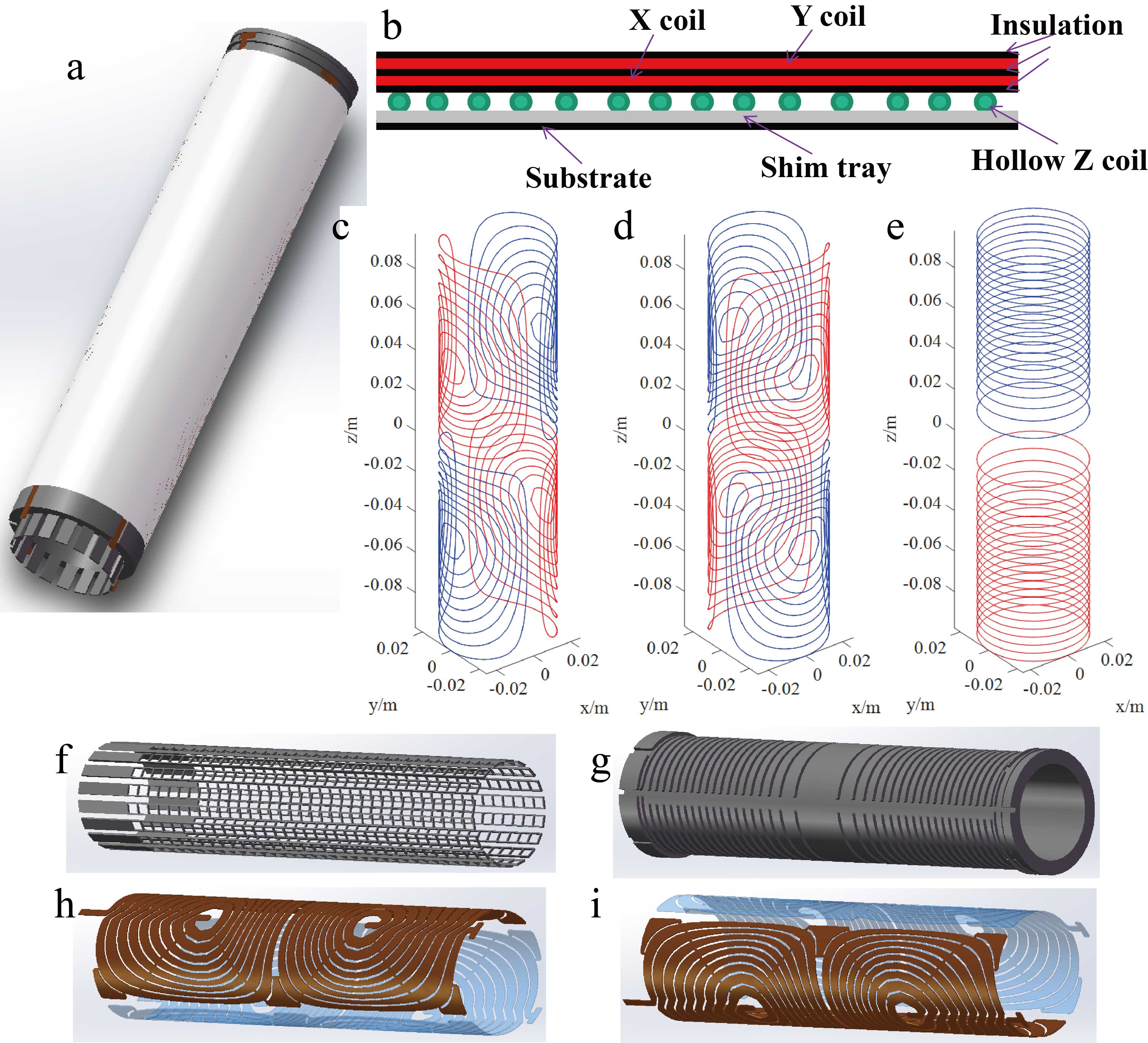
The gradient assembly design is illustrated in Fig. 4, where the integral scheme is presented in Fig. 4(a), the layout of the assembly structure is diagrammed in Fig. 4(b), the designed coil patterns are plotted in Fig. 4(c-e). The engineering components of the assembly are shown in Fig. 4(f-i). A passive shim tray with 14 strips in the circumferential direction and 34 in the axial direction was embeded in the substrate. The transverse gradient coils will be cut out on the copper plates, and the axial gradient coil was wound with a hollow copper pipe. The outer diameter of the gradient assembly is 53mm, leaving a 1-mm assembling gap to the magnet warm bore, and the inner diameter is 40.68mm.
The RF coil adopts a birdcage structure with eight legs, and there are six capacitors at one end ring and eight capacitors at the other. The capacitors have an identical capacitance of 23 pF and contribute a resonance frequency 299MHz.
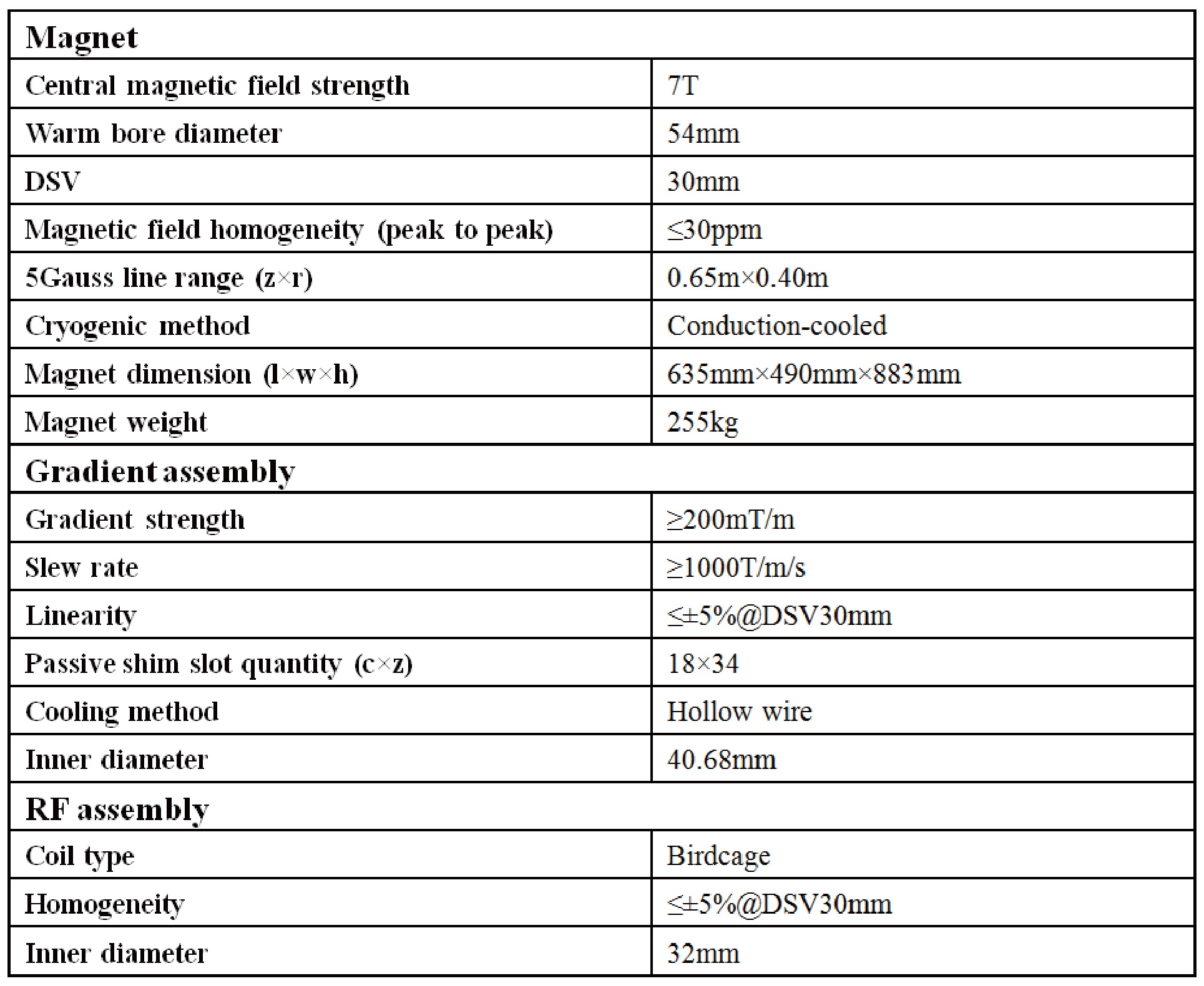
The main parameters of the Benchtop 7T cryogen-free animal MRI system are listed in Table I.
Discussions
Since the gradient coils were not actively shielded to enlarge the available detection bore size, we will be very careful of using a fast scanning sequence. The relatively slow sequences are workable based on our previous experience due to weak eddy current, and the follow-up study on the image correction method under fast imaging is essential to support more sequence requirements. Alternatively, we will also consider the actively shielded gradient coil design.Conclusion
A Benchtop 7T cryogen-free animal MRI system is under development at IEE CAS, whose clear detection bore diameter is around 32mm, which is feasible for the imaging of small-size organisms, such as mice. Significant merits of the system include cryogen-free operation, small magnet size, light weight, etc., which result in very high performance to cost ratio. The ultimate product is expected to boost a wide range of scientific research programs that require ultra-high-field MRI technology.Acknowledgements
This work is funded by CAS Pioneer Hundred Talents Program (Grant No. Y8402A1C31) and National Key Research and Development Program of China (Grant No. 2020YFF01014702).References
[1] R. Warner, "Ultra-high field magnets for whole-body MRI," Superconductor Science and Technology, vol. 29, p. 094006, 2016.
[2] Y. Wang, Q. Wang, H. Wang, S. Chen, X. Hu, Y. Liu, et al., "Actively-shielded ultrahigh field MRI/NMR superconducting magnet design," Superconductor Science and Technology, 2021 (online).
[3] A. Rybakov, A. Bagdinov, E. Demikhov, E. Kostrov, V. Lysenko, N. Piskunov, et al., "1.5 T Cryogen Free Superconducting Magnet for Dedicated MRI," IEEE Transactions on Applied Superconductivity, vol. 26, pp. 1-3, 2016.
[4] G. Gabrielse, J. Tan, P. Clateman, L. A. Orozco, S. L. Rolston, C. H. Tseng, et al., "A superconducting solenoid system which cancels fluctuations in the ambient magnetic field," Journal of Magnetic Resonance, vol. 91, pp. 564-572, 1991.
Figures