3241
Design of a 5-channel local B0 shimming coil for rat brain imaging at 3T1Paul C. Lauterbur Research Center for Biomedical Imaging, Shenzhen Institutes of Advanced Technology, Chinese Academy of Sciences, Shenzhen, China, 2Key Laboratory for Magnetic Resonance and Multimodality Imaging of Guangdong Province, Shenzhen, China, 3Department of Biomedical Engineering, State University of New York at Buffalo, New York, NY, United States
Synopsis
To improve B0 magnetic field homogeneity and minimize interferences on RF coils, local shimming coils with a few channel number can be applied. In this study, we designed and constructed a 5-channel local B0 shimming coil for the rat brain. There was a marginal SNR loss within 5% after converting the local shimming coil into a 3-channel RF coil. The reduction of B0 inhomogeneity for the rat brain with respect to the basic set was about 34%. A large portion of the distortion in EPI images was recovered and some image artifacts were eliminated after using the local shimming coil.
Introduction
In MRI, the B0 magnetic field inhomogeneity caused by the variation of magnetic susceptibility distribution of tissue may introduce geometric distortion, signal dropout warping artifacts, incomplete fat suppression, or other issues related to frequency optimized excitation, particularly in echo-plane imaging (EPI) 1. To mitigate the effect, the B0 shimming coils can be applied. However, in some realizations, there has a 10-15% signal-to-noise ratio (SNR) loss 2. To minimize the coupling between B0 shimming and RF received coils, local shimming coils with a few channel number can be applied 3. In this work, a 5-channel local shimming coil was designed and constructed in combination with a 3-channnel RF received coil at a 3 T Siemens TrioTim MRI system. SNR of the RF coil and B0 shimming performance were evaluated to demonstrate the feasibility of this local shimming coil designed with a few channel number.Methods
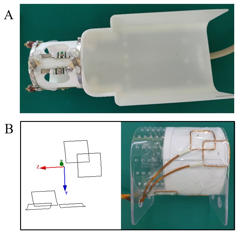
B0 maps of the rat brains were measured with a 2D multi-echo GRE sequence by using the RF received coil, as shown in Figure 1A. The parameters were as followings: TE=[3.68 … 13.44] ms, echo train length=5, echo spacing=2.44 ms, TR=300 ms, FA=10o, resolution=0.5×0.5×1.5 mm3, slices=24, BW=500 Hz/pixel. Brain masks were created from the magnitude images and spatial phase unwrapping was applied to the B0 maps 4. Only 9 slices contained the rat brain. Five rat brain B0 maps were used for optimization, and three rat brain B0 maps were used for validation of the optimization outcome.The local shimming coil was positioned on a cylinder with a diameter of 70 mm. All the shimming loop elements had an identical square shape. The B0 shimming relevant z-component of the magnetic field generated by the square shimming loops on the cylinder can be derived analytically by the biot-savart law. Optimization were performed with four degrees of freedom, including current of each shimming loops, side length of the square shimming loops, angular coordinate (θ) and axial coordinate (Z) or height of the center of shimming loops in the cylinder surface. The current was constrained to [-2 2] A, and the lower and upper bounds for the side length of the shimming loops were 20 mm and 50 mm, respectively. The shimming loops should lay between –π and +π, and the height of the center of shimming loops were constrained to [-25 25] mm. Before optimization, the initial current was 0.1 A, and the initial side length was 30 mm. The initial shimming loops were distributed uniformly on the surface of the cylinder. Particle swarm optimization method was implemented to find the minimum standard deviation (SD) of the B0 maps of the five rat brains. The performance of the optimized local shimming coil was evaluated and compared with the basis set. After optimization, the local shimming coil was constructed, as shown in Figure 1B.
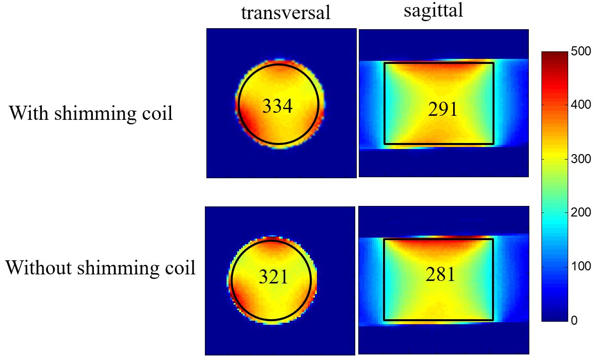
A 2D GRE sequence was used for the SNR measurements in the transversal and sagittal planes with a cylinder phantom. The parameters were as followings: TR/TE =300/10 ms, FA=30o, resolution=0.5×0.5×3 mm3, BW=200 Hz/pixel. The SNR maps were calculated by using the root sum-of-squares method 5. The B0 shimming performances was evaluated by animal studies, including B0 maps and EPI images evaluations. The EPI images were acquired with following parameters: resolution=1.1×1.1×1.5 mm3, FA=90°, TR/TE=2000/29 ms, slices=24, measurements=100, BW=1324 Hz/Px.
Results
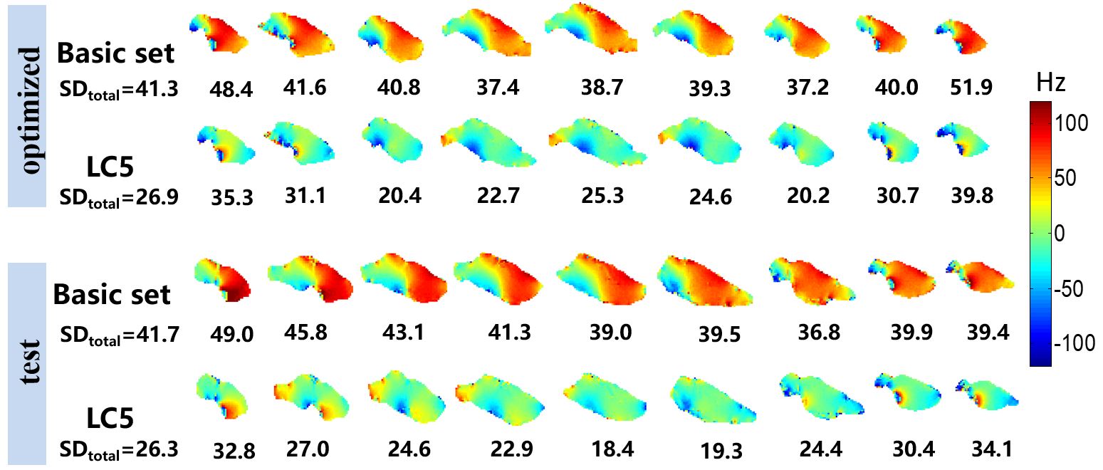
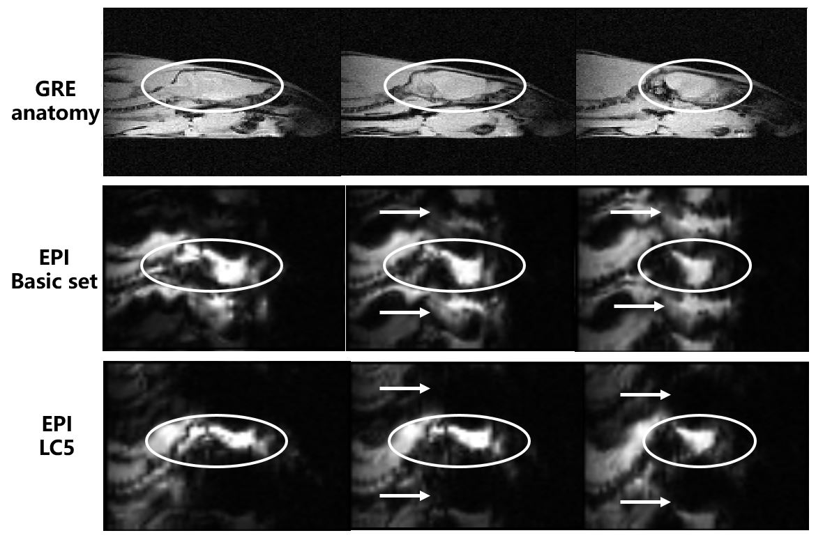
Figure 2 depicts the shimming performance of two representative rat brain B0 maps in simulations, one for optimized and the other for test. In comparison to the basic set, SD of the total rat brain B0 maps after shimming decreased by 35% and 37%, respectively for optimized and test in simulations. Figure 3 displays SNR measurements with the shimming coil and without shimming coil. These results show a marginal SNR loss within 5%. Figure 4 shows the B0 shimming performance of the 5-channel local shimming coil in animal study. The inhomogeneity of the rat brain B0 maps was reduced by 34% after shimming, which was consistent with the simulation results. Figure 5 illustrates the impact of the local B0 shimming on correction of geometric distortion in EPI images.Three representative slices were shown. Anatomical images obtained by using GRE sequence are used as undistorted reference images. From the images, it can be known that a large portion of the distortion was recovered and some image artifacts were eliminated after local shimming, as depicted by the circles and the arrows, respectively.Discussions and Conclusions
The decreased SD for the whole mouse brains was 31% using a 48-chnanel multi-coil compared with the basic set in a 9.4 T MRI system as reported in Juchem et al 6. The results achieved in this study show a comparability with this result.A 5-channel local shimming coil was designed and constructed for rat brain imaging. By using the 5-channel local shimming coil, the SNR loss was marginal within 5% and B0 field inhomogeneity was reduced by 34%. EPI images with less distortion and less artifacts can be obtained. It is anticipated a variety of applications that are sensitive to B0 inhomogeneity can benefit from the local shimming coils.
Acknowledgements
This work was supported in part by NSFC under Grant No. 61801466; the Strategic Priority Research Program of Chinese Academy of Sciences (Grant No. XDB25000000); National Key R&D Program of China, 2021YFE0204400; city grant RCYX20200714114735123; city grant ZDKJ20190204003 and ZDKJ20190204004.References
1. Stockmann JP, Wald LL. In Vivo B0 Field Shimming Methods for MRI at 7 T. Neuroimage, 2018, 168: 71-87.
2. Stockmann J P, Witzel T, Keil B, Polimeni J R, Mareyam A, LaPierre C, Setsompop K, Wald LL. A 32-channel combined RF and B0 shim array for 3T brain imaging. Magnetic resonance in medicine, 2016, 75(1): 441-451.
3. Zhou J, Stockmann JP, Arango N, Witzel T, Scheffler K, Wald LL, Lin FH. An orthogonal shim coil for 3T brain imaging. Magnetic Resonance in Medicine, 2020, 83(4):1499-1511.
4. Abdul-Rahman HS, Gdeisat MA, Burton DR, Lalor MJ, Lilley F, Moore CJ. Fast and robust three-dimensional best path phase unwrapping algorithm. Applied Optics. 2007, 46: 6623–6635.
5. Roemer P B, Edelstein W A, Hayes C E, Souza S P, Mueller O M. The NMR phased array. Magnetic resonance in medicine, 1990, 16(2): 192-225.
6. Juchem C, Brown PB, Nixon TW, McIntyre S, Rothman DL, de Graaf RA. Multicoil shimming of the mouse brain. Magnetic resonance in medicine. 2011; 66(3):893-900.
Figures