3238
Bore integrated coil for whole-body imaging at 7 Tesla based on patch antennas
Georgiy Solomakha1, Svetlana Egorova1, Alena Shchelokova1, Egor Kretov1, and Stanislav Glybovski1
1Department of Physics and Engineering, ITMO University, Saint Petersburg, Russian Federation
1Department of Physics and Engineering, ITMO University, Saint Petersburg, Russian Federation
Synopsis
This work proposes a novel transmit radiofrequency coil integrated into the scanner bore for whole-body MRI for 7 Tesla MRI based on patch antennas. Numerical studies with a homogeneous phantom demonstrated 4.5-fold higher transmit efficiency in the phantom center compared to the state-of-the-art bore integrated stripline array.
Synopsys
This work proposes a novel transmit radiofrequency coil integrated into the scanner bore for whole-body MRI for 7 Tesla MRI based on patch antennas. Numerical studies with a homogeneous phantom demonstrated 4.5-fold higher transmit efficiency in the phantom center than the state-of-the-art bore integrated stripline array.Purpose
To develop and characterize numerically novel and efficient bore-integrated coil based on patch antenna array for 7 Tesla whole-body MRI.Introduction
Ultra-high field (field strength higher than 7 Tesla) imaging is an extensively developing field. Recently UHF MRI-scanner was approved for limited clinical use for head and extremity imaging with static CP-mode excitation [1]. For the body imaging at such high field strengths, volume coils integrated to RF-shield of MR-scanner became inefficient because of strongly inhomogeneous RF-magnetic field in the human body due to interference effects [2]. To obtain a homogenous field in the desired region, local transmit arrays combined with the so-called RF-shimming procedure [3]. A recently different approach based on eight stripline coils integrated into the bore of the 7 T scanner was introduced [3]. Such design allows to reduce SAR and optimize receive and transmit coils separately but suffers from the low Tx-efficiency. This work presents a new bore integrated array for a 7 T MRI of the human body. The proposed body array elements demonstrate 4.48 times better B1 in the desired region than the bore integrated stripline array [3]. Patch-antennas are well known in the microwave frequency range as low profile and robust solutions with a simple design that can efficiently operate near the conducting surfaces. Patch antenna consists of the metallic half-wavelength sheet (patch) with a rectangular shape above the electric shield. The operation principle of the patch antenna is based on the excitation of TE10 eigenmode in the volume under the patch. This resonant mode excites two linear equivalent magnetic currents at the edges of the patch. Such currents act as two in-phase slot antennas. Patch antennas were already proposed for MRI application as CP-polarized forward view antenna for the head [4] or small animal imaging [5]. Here, we describe and demonstrate in simulations a new bore integrated array for a 7 T MRI of the human body based on patch antennas that allow increasing Tx-efficiency 4.48 times compared to the state-of-the-art stripline array.Methods
The design of the proposed bore-integrated array is presented in Figure 1. It consists of an octagonal-shaped bore with 680 mm diameter and eight patch antennas with air substrate. In planar configuration, patch antennas with aligned radiating slots are well decoupled. When these eight antennas are placed in the bore environment, coupling between them becomes significant. Passive magnetic resonators were inserted between non-radiating slots of the patch antennas. The passive magnetic resonator consisted of two loops connected with two capacitors. The total length of the patch was chosen 460 mm to be resonant at the 298 MHz frequency inside the bore. The width of each element was chosen at 200 mm to fit the eight-channel configuration. A bore integrated stripline array was used as a reference coil array. For numerical simulation, the finite-element method implemented in CST Microwave Studio 2021 software was used. Decoupling and resonant frequency of patches were adjusted by changing the length of the magnetic resonator.Results
Figure 2 demonstrates the distribution of B1+ for two circular polarized modes, CP1 (45 deg phase lag between element) and CP2 (90 deg phase lag between element), both for patch and stripline arrays in the central axial plane of the elliptical phantom. CP1 mode of both arrays gives maximum field in the center. The same field distribution in the sagittal plane is depicted in Figure 3. The power balance of numerical simulation, B1+ value in the center of the homogenous phantom, local SAR-maxima value, and SAR-efficiency for the phantom center presented in Table 1Conclusion
We developed a novel concept of a bore-integrated coil for 7 Т body imaging. We can conclude from the presented results that the proposed coil increases Tx-efficiency 4.47 times compared to the stripline array. . However, the SAR efficiency of the proposed and reference was almost the same. Measured S11 for the reference and proposed array lower than -20 dB. The coupling between neighbor elements of the patch array was -3 dB. With an additional decoupling resonator, it was improved to the -15 dB level. In the future, we plan to perform a numerical evaluation with a voxel model for SAR performance evaluation and on-bench and MRI measurements of the prosed coil. After successful evaluation proposed design could be used as a prototype for the first bore-integrated transmit coil for clinical 7 Tesla MRI.Acknowledgements
This work was supported by the Russian Science Foundation (Project 21-19-00707).References
- Ladd, M. E., Bachert, P., Meyerspeer, M., Moser, E., Nagel, A. M., Norris, D. G., ... & Zaiss, M. (2018). Pros and cons of ultra-high-field MRI/MRS for human application. Progress in nuclear magnetic resonance spectroscopy, 109, 1-50.
- Vaughan, J. T., Snyder, C. J., DelaBarre, L. J., Bolan, P. J., Tian, J., Bolinger, L., ... & Ugurbil, K. (2009). Whole‐body imaging at 7T: preliminary results. Magnetic Resonance in Medicine, 61(1), 244-248.
- Orzada, S., Bitz, A. K., Johst, S., Gratz, M., Völker, M. N., Kraff, O., ... & Ladd, M. E. (2017). Analysis of an integrated 8-channel Tx/Rx body array for use as a body coil in 7-Tesla MRI. Frontiers in Physics, 5, 17.
- Shajan, G., Hoffmann, J., Balla, D. Z., Deelchand, D. K., Scheffler, K., & Pohmann, R. (2012). Rat brain MRI at 16.4 T using a capacitively tunable patch antenna in combination with a receive array. NMR in Biomedicine, 25(10), 1170-1176.
- Hoffmann, J., Shajan, G., Budde, J., Scheffler, K., & Pohmann, R. (2013). Human brain imaging at 9.4 T using a tunable patch antenna for transmission. Magnetic Resonance in Medicine, 69(5), 1494-1500.
Figures
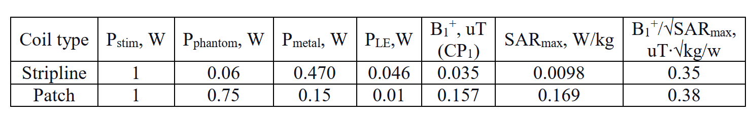
Table 1. Power
balance of investigated bore-integrated arrays.
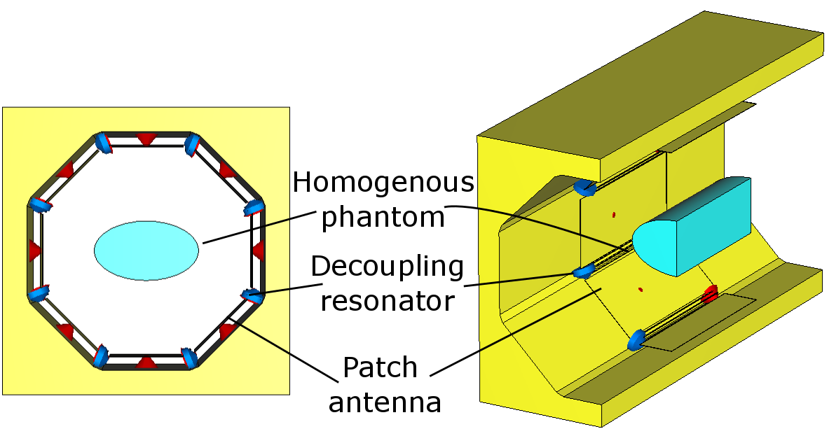
Figure 1.
Proposed design of bore integrated patch array.
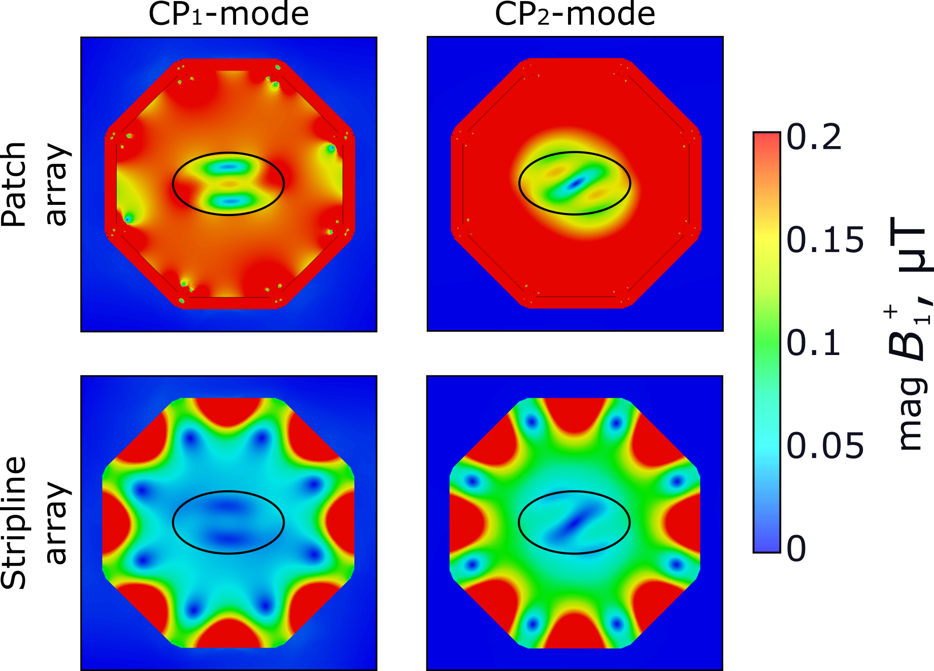
Figure 2. Results of B1+ simulation of reference and proposed arrays
(central axial plane)
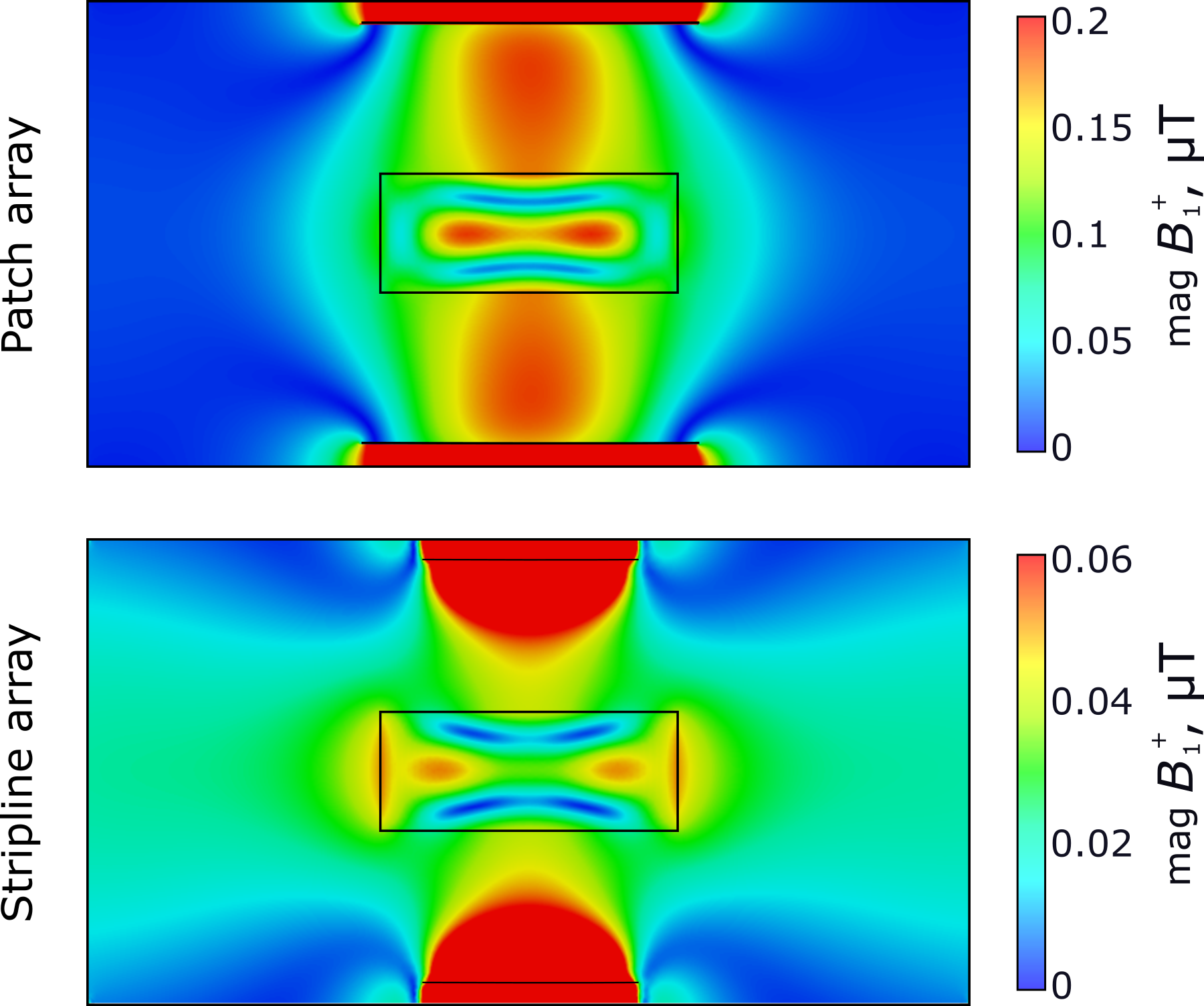
Figure 3. Results of B1+ simulation of reference and proposed arrays
(central sagittal plane)
DOI: https://doi.org/10.58530/2022/3238