3235
Multichannel MRI receive coil based on artificial material1School of Physics and Engineering, ITMO University, Saint-Petersburg, Russian Federation
Synopsis
Metamaterials and metasurfaces, in particular, are gaining increasing popularity in recent years. One of the novel applications of metasurfaces are receive coils for magnetic resonance imaging. The major drawback of using such structures is the lack of parallel MRI capabilities due to the single-channel acquisition regime. In this work, we will demonstrate that it is possible to design a multichannel metasurface.
Synopsis
Metamaterials and metasurfaces, in particular, are gaining increasing popularity in recent years. One of the novel applications of metasurfaces is receive coils for magnetic resonance imaging. The major drawback of using such structures is the lack of parallel MRI capabilities due to the single-channel acquisition regime. In this work, we will demonstrate that it is possible to design a multichannel metasurface.Main findings
In this work, we show the concept of an artificial dielectric-based metasurface for MRI with multichannel reception. It provides a higher signal-to-noise ratio in contrast with the classical loop coil.Introduction
Nowadays, most MRI coils are phased arrays based on loop coil1. While being sufficiently cheap, durable, and practical, loop coils are not flawless. Their most relevant shortcoming in the context of our work would be the necessity to balance the field penetration depth, decoupling, and signal-to-noise ratio2. Moreover, a single loop has only one sensitivity profile. Phased arrays are introduced to achieve a higher region of interest coverage while maintaining the same SNR. An alternative approach is to use metasurfaces3. They often show higher SNR values than traditional coils. In particular, our proposed device has two sensitivity profiles on a single frequency and shows higher SNR than loop coils of comparable dimensions.Methods
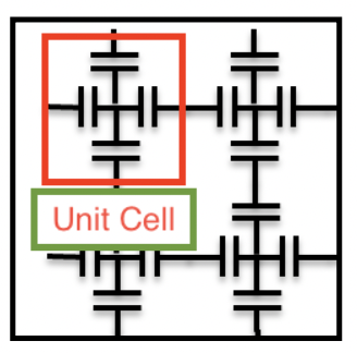
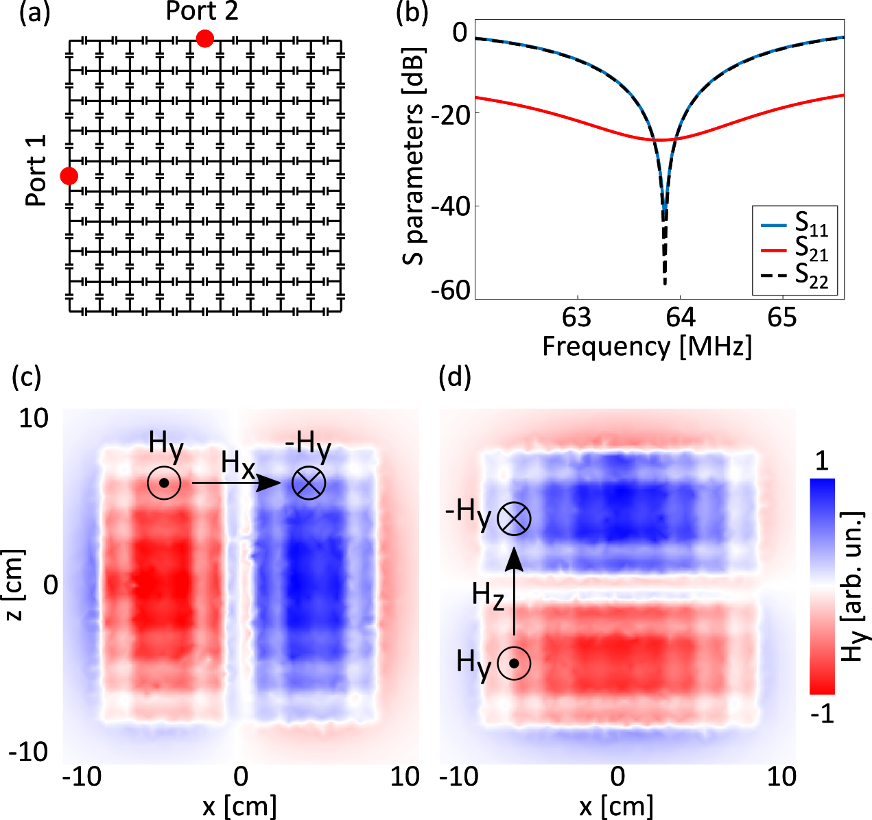
Our proposed device is based on the so-called artificial dielectric. Recently, it has been shown that such metasurface may be used in clinical MRI as a substitute for expensive and heavy dielectric pads4. It is possible because of the high index of refraction that this structure possesses. In the context of this work, we use the term «refractive index» as a radio-frequency wave compression factor in media. We propose using the theory of Fabry-Perot resonances in order to obtain multiple modes on the same frequency. We start with a unit cell geometry, provided in fig. 1. Then, we carefully pick the dimension, capacitance, and number of unit cells on each axis to achieve a predetermined phase shift along this direction. In our case, it is 2π and π. In terms of Fabry-Perot resonances, we have a mode (m,p) = (2,1). Since we are investigating a square sample, it is helpful to look at the symmetries of the structure. If we rotate the metasurface by 90 degrees along its normal vector, we get the same metasurface. This fact implies that the second mode has order (1,2). In practice, we use these two modes for independent reception channels. It is possible because of good implicit decoupling between ports (see fig.2). The feeding port positions and overall design is shown in Fig 2. a. All numerical calculations were carried out in CST Microwave Studio. We used a dielectric cubic phantom with ε = 72 and σ= 0.7 S/m as a load for our numerical simulations (imitating a human body). The dimension of the cube is slightly greater than the side of the metasurface.Results
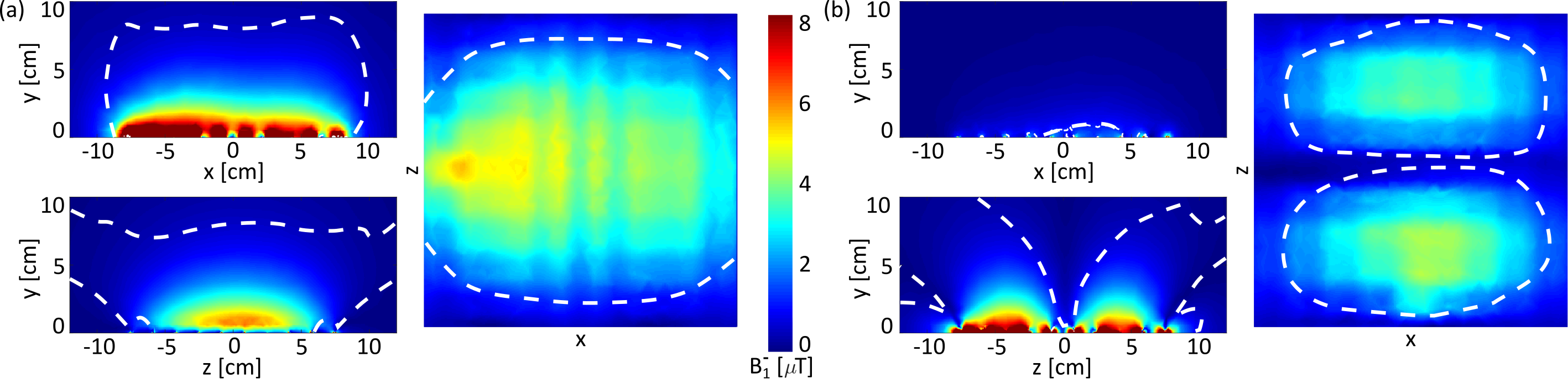
We performed numerical simulations as well as analytical calculations of field distributions and scattering parameters of the proposed metasurface. In fig.2. b we can see that S21 between two ports is approximately - 25 dB. This means that two ports are effectively decoupled. S11=S22 ≈ -55 dB. Due to high decoupling, it is possible to use these two modes as independent receive channels for MRI. Signal sensitivity increase compared to loop coil is evident from fig.3, which demonstrates receive B1- field.Discussion and Conclusion
Higher SNR typically means the higher diagnostic quality of the images. Moreover, the implicit decoupling of modes is beneficial for the applications of parallel MRI techniques. The proposed device is simple and highly scalable. With adjustments to capacitances and dimensions of the unit cell, the device may be implemented in 1- 3 Tesla or higher field machines. The proposed analytical approach to the description of the unit cells based on the Fabry-Perot resonances is universal; we use it to investigate anisotropic cells that can be used to obtain more than two resonant field modes on the same frequency. Future development of this design will make it possible for additional channels to be initiated in parallel MRI techniques.Acknowledgements
This work was supported by the Russian Science Foundation (Project No.21-79-30038).References
1. Ohliger, Michael A., and Daniel K. Sodickson. An introduction to coil array design for parallel MRI, NMR in Biomedicine: An International Journal Devoted to the Development and Application of Magnetic Resonance In vivo 2006; 19.3:300-315.
2. Lee, Ray F., Randy O. Giaquinto, and Christopher J. Hardy. Coupling and decoupling theory and its application to the MRI phased array. Magnetic Resonance in Medicine: An Official Journal of the International Society for Magnetic Resonance in Medicine 2002; 48.1: 203-213.
3. E. Stoja, S. Konstantin, D. Philipp, R. N. Wilke, D. Betancourt, T. Bertuch, J. Jenne, R. Umathum, and M. Gunther, Improving magnetic resonance imaging with smart and thin metasurfaces, Scientific Reports 11, 2021.
4. V. Vorobyev, A. Shchelokova, A. Efimtcev, J. D. Baena, R. Abdeddaim, P. Belov, I. Melchakova, and S. Glybovski, Improving homogeneity in abdominal imaging at 3 T with light, flexible, and compact metasurface, Magnetic Resonance in Medicine 2021.
Figures