3233
A novel three-channel endorectal coil for prostate magnetic resonance imaging at 3T1Lauterbur Research Center for Biomedical Imaging, Shenzhen Institutes of Advanced Technology, Chinese Academy of Sciences, Shenzhen, Guangdong, 518055, China, Shenzhen, China, 2Department of Biomedical Engineering, State University of New York at Buffalo, NY, United States., Buffalo, NY, United Stats, Buffalo, NY, United States, 3Union Hospital, Tongji Medical College, Huazhong University of Science and Technology, Wuhan, China
Synopsis
We designed and fabricated a novel three-channel endorectal prostate coil and validated its feasibility of high-resolution magnetic resonance imaging (MRI) at 3T. A commercial 3T surface coil was used for comparison. Both phantom imaging and in-vivo prostate imaging show that the 3-channel endorectal coil provides an improved SNR than the external surface coil. Prostate images at resolution of 0.47 mm × 0.47 mm × 4.0 mm are acquired using the 3-channel endorectal coil to demonstrate the performance. With the improved SNR and multichannel design, the proposed endorectal coil could enable parallel imaging based fast imaging in prostate imaging studies.
INTRODUCTION
Signal-to-noise ratio (SNR) is the key parameter of RF coils for MRI. A high SNR means clear imaging, high resolution, and potentially fast imaging speed. Since the signal intensity decreases rapidly with the increase of distance to a coil, it is difficult to obtain a high SNR for prostate imaging with external surface coils. To address this problem, endorectal coils were proposed and demonstrated the SNR superiority compared with external surface coils1,2,3. In this work, a novel three-channel endorectal coil for prostate MRI was designed and manufactured, which could further boost SNR and also enable fast imaging acquisition through parallel imaging. in vitro study as well as in vivo study were performed to validate the performance.METHOD
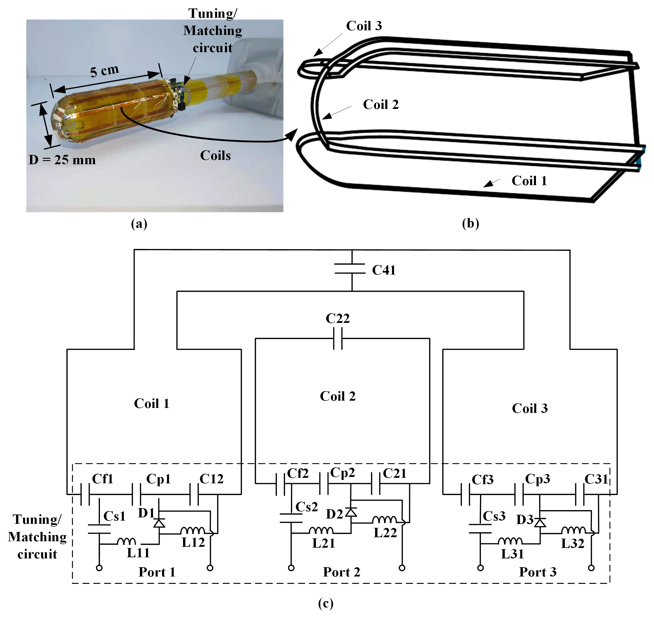
A structural part as shown in figure 1 (a) was made with PVC material. The structural part is a cylinder with a diameter of 25 mm and a length of 5 cm, a part of which was cut off. The D-shaped cross-section ensures that the coil imaging area is sufficient while making the coil thin enough to be inserted into the rectum smoothly. As shown in figure 1 (a), the coil 1 to 3 are arranged overlappingly on the cylinder in sequence, and coil 1 and coil 3 are decoupled by means of a capacitor. The corresponding circuit schematic diagram is shown in figure 1 (b).Of the in vitro study, a 12 cm-diameter phantom with a 3 cm-diameter hole in the center and filled with 0.9% normal saline was imaged to compare the SNR of the endorectal coil and the surface coil. A fast- spin-echo (FSE) sequence with TR = 2895 ms, TE = 86.08 ms, FOV = 240 × 240 mm2, matrix = 512 × 512, slice thickness = 4 mm, and NEX =1 was performed on a 3T MRI scanner (uMR 790, Shanghai United Imaging Healthcare, Shanghai, China).Of the in vivo study, an endorectal coil was inserted into a beagle’s rectum to image its prostate. The beagle was lying on his back on the hospital bed, and the same sequence as that performed in the in vitro study was performed. As a comparison, a 12-channel commercial surface coil was covered on the abdomen of the beagle for imaging.RESULTS AND DISCUSSION
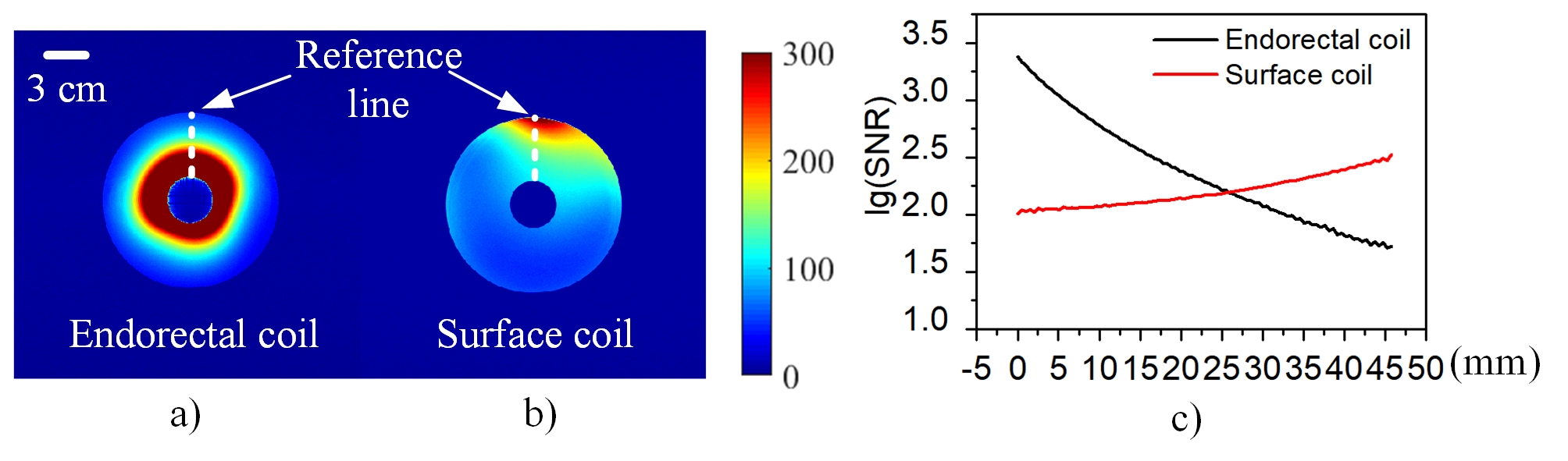
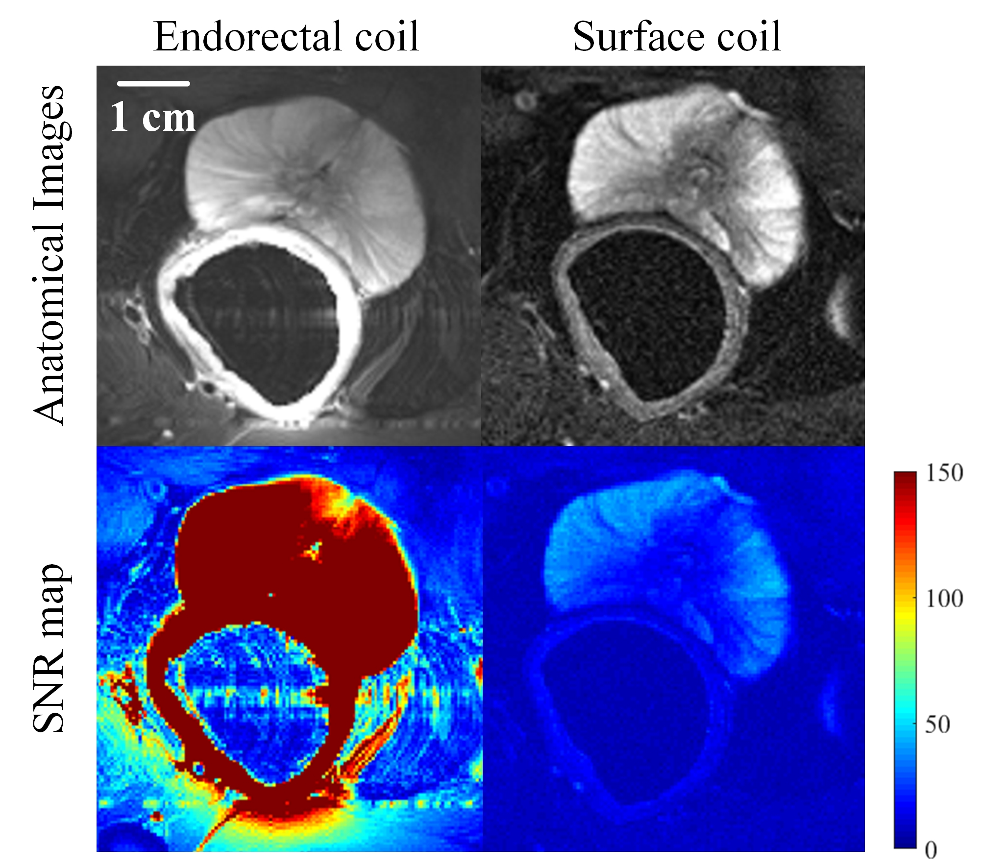
Of the in vitro study, we select a reference line as shown in Fig.2. a), then plot the change of the SNRs along the reference line in Fig.2 b). The SNR of the endorectal coil (black line) is higher than the SNR of the surface coil (red line) in the area where the penetration depth of the rectal coil is less than 25 mm.Of the in vivo study, prostate imaging was acquired with endorectal coil and surface coil (Figure 3). The image obtained by the endorectal coil shows richer details and lower noise than that obtained with the surface coil. As expected, the SNR of the endorectal coil is significantly higher than that of the surface coil in the prostate area.CONCLUSION
In this work, we designed and manufactured a novel three-channel endorectal coil array for prostate MR imaging at 3T. The results demonstrate a higher SNR in a penetration depth of 25 mm than a commercial external surface coil. Of the in vivo study in a beagle dog, the proposed endorectal coil array showed much improved image quality and SNR superiority compared with the external surface coil at 3T.Acknowledgements
This work was supported in part by the National Key Scientific Instrument Development Project (Grant No. 81927807), city grant RCYX20200714114735123, National Key R&D Program of China, 2021YFE0204400, Strategic Priority Research Program of the Chinese Academy of Sciences (Grant No. XDB25000000), Guangdong Province grants 2018B030333001, and Youth Innovation Promotion Association of CAS No. 2017415References
1. Rata, Mihaela, et al. "Endoluminal MR‐guided ultrasonic applicator embedding cylindrical phased‐array transducers and opposed‐solenoid detection coil." Magnetic resonance in medicine 73.1 (2015): 417-426.
2. Arteaga de Castro, C. S., et al. "Improving SNR and B1 transmit field for an endorectal coil in 7 T MRI and MRS of prostate cancer." Magnetic resonance in medicine 68.1 (2012): 311-318.
3. Metzger G J, van de Moortele P F, Akgun C, et al. Performance of external and internal coil configurations for prostate investigations at 7 T[J]. Magnetic resonance in medicine, 2010, 64(6): 1625-1639.
Figures