3229
A dual-nuclei 1H/23Na knee coil for high-resolution 1H and 23Na MR imaging at 7T1Shenzhen Institutes of Advanced Technology,Chinese Academy of Sciences, Shenzhen, China
Synopsis
An ideal MRI study for early osteoarthritis should provide both structure imaging and functional imaging, which can show early morphologic degenerative changes and physiology changes of the cartilage. Most of previous dual-nuclei coils pushed the limits of non-proton nuclei sensitivity at the expense of 1H performance. To get high performance imaging for both nuclei, a novel dual-nuclei 1H/23Na coil array was developed. The bench test results show that all coils were sufficiently matched and decoupled, and the interference between sodium coils and proton coils was nearly negligible, thus can be expected to get high-resolution 1H imaging and quantitative 23Na imaging.
Introduction
The main histopathological features of osteoarthritis is articular cartilage degeneration.The progressive loss of normal cartilage structure and function, leads to the clinical syndrome of osteoarthritis 1,2. Quantitative sodium MRI can be used to measure glycosaminoglycan (GAG) content in knee cartilage, as a means of detection and assessment of the degree of biochemical degradation in early osteoarthritis 3,4. An ideal MRI study for early osteoarthritis should provide both structure imaging and functional imaging, which can show early morphologic degenerative changes and physiology changes of the cartilage 5.Most of previous dual-nuclei coils pushed the limits of non-proton nuclei sensitivity at the expense of 1H performance whilst providing less than optimal performance (SNR and/or field-of-view) of 1H imaging 3,6,7. The purpose of this study was to construct a dual-nuclei 1H/23Na knee coil, which can get sufficiently matching and isolation between coil elements of all coil arrays, and can provide adequate field of view of 1H imaging. The coil configurations were optimized for either high-resolution 1H imaging and quantitative 23Na imaging.
Method
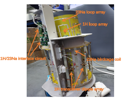
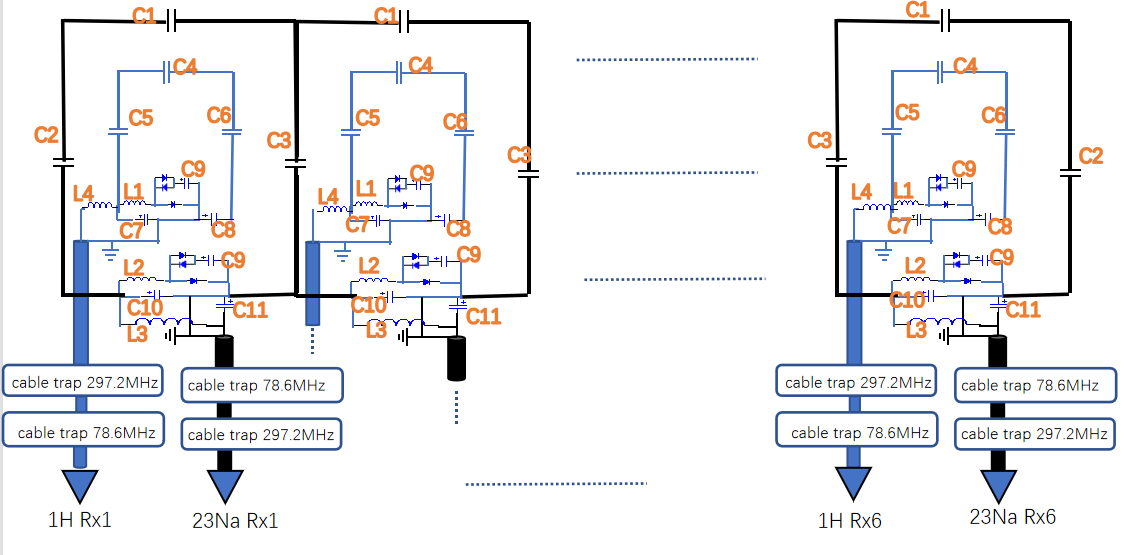
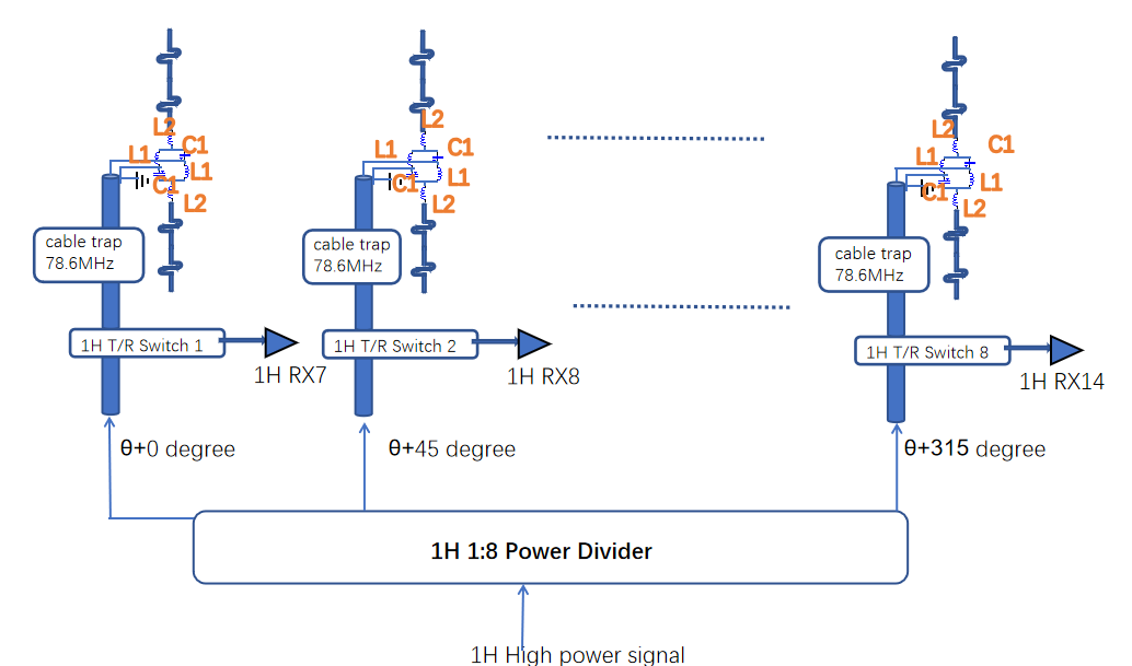
The dual-nuclei 1H/23Na knee array consisted of a combination of four RF coils arranged in three layers (Fig 1): the innermost layer consisted a six-channel sodium loop array and a six-channel proton loop array nested within the sodium array, which were built on a flexible substrate to minimize coil-to-tissue distance for high receive performance (Fig 2). A 27cm-diameter high-pass birdcage coil which has 16 rungs was built for homogeneous sodium transmission on the outmost layer. Between these two layers, eight transceiver dipole antennas were mounted symmetrically on a 20.6cm-diamater tube for proton transmission and receive (Fig 3), which was to improve the SNR performance and the field-of-view along the direction of the main magnetic field of proton imaging. The coil former was divided into anterior and posterior sections for patient entry.To evaluate the proton signal degradation caused by the sodium coils, the ratio of unloaded to loaded Q-factors of the proton loop coils were measured with and without the sodium coils present. And vice versa, the ratio of unloaded to loaded Q-factors of the sodium loop coils was measured with and without the proton coils present to evaluate the sodium signal degradation caused by the proton coils.
Results and Discussion
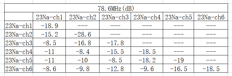
All the coil elements of proton coils were sufficiently matched and decoupled. Table 1 shows the reflection coefficient of dipole antennas and loop coils are no larger than -17.6dB and -18.4dB at 297.2MHz, respectively. Sufficient inter-element decoupling of the dipole array and the loop array were achieved, with the minimum isolation are -11.5dB and -13.2dB at 297.2 MHz, respectively. And the isolation between dipole antennas and loop coils are less than -14.2dB at 297.2MHz.Table 2 shows the reflection coefficient of sodium loop coils are less than -17.8 dB at 78.6MHz, and the isolation between neighboring coils are less than -15.2 dB at 78.6MHz. The next-nearest neighbors were coupled by less than -8.5 dB, and extra 25dB isolation can be provided by preamplifier decoupling.
The Q-factors (unloaded/loaded) of the six-loop sodium array with/without the proton coils at 78.6MHz were 161.7 ± 16 / 41.6 ± 4 and 171 ± 15 / 40 ± 5, respectively. And the ratios of the unloaded to loaded Q-factors of the proton loop coils at 297.2MHz were 2.67 ±0.2 and 2.52 ± 0.18 with/without the sodium coils.
Conclusion
In this study, a novel dual-nuclei 1H/23Na coil array was developed. This coil could provide adequate field of view of proton imaging along the direction of the main magnetic field. The bench test results show that all coils were sufficiently matched and decoupled, and the interference between sodium coils and proton coils was nearly negligible, thus can be expected to get high-resolution 1H imaging and quantitative 23Na imaging.Future investigation will focus on phantom and clinical study on a 7T MR system.Acknowledgements
No acknowledgement found.References
1.Joseh A. Buckwalter, Henry J.Mankin,..et al. Articular cartilage and osteoarthitics. Instr Course Lect.2005.
2.Luyten FP,Denti M,Filardo G,...et al. Definition and classification of early osteoarthfitis of the knee[J].Knee Surg Sports Traumatol Arthrosc 20(3):401—406
3.Ryan Brown, Karthik Lakshmanan, ...et al. A Flexible Nested Sodium and Proton Coil Array with Wideband Matching for Knee Cartilage MRI at 3T. Magnetic Resonance in Medicine 2015.
4. Henning Madry,Frank P. Luyten,Andrea Facchini. Biological aspects of early osteoarthritis. Knee Surg Sports Traumatol Arthrosc 20:407–422.
5.Garry E. Gold, Christina A. Chen, ...et al. Recent Advances in MRI of Articular Cartilage. Musculoskeletal Imaging • Review. AJR:193, September 2009.
6.Mirkes, C., Shajan, G., Chadzynski, G., ...et al. 31P CSI of the human brain in healthy subjects and tumor patients at 9.4 T with a three-layered multi-nuclear coil: initial results. Magnetic Resonance Materials in Physics, Biology and Medicine, 29(3), 579–589.
7.Graham C. Wiggins, Ryan Brown and Karthik Lakshmanan. High-performance radio frequency coils for 23Na MRI: brain and musculoskeletal applications. NMR Biomed. 2016; 29:96–106
Figures