3228
Prostate imaging with Integrated Multi-modal Antenna with coupled Radiating dipoles (I-MARS) at 7T1School of Information Technology and Electrical Engineering, The University of Queensland, Brisbane, Australia, 2Siemens Healthineers, Brisbane, Australia, 3School of Human Movement and Nutrition Sciences, The University of Queensland, Brisbane, Australia, 4The Center for Magnetic Resonance in Biology and Medicine, Aix-Marseille University, Marseille, France
Synopsis
Prostate imaging is challenging at 7T due to its small size seated deeply inside the pelvic region. As such, both transmit receive performance of the RF coils are heavily challenged. We have recently developed a new paddle-shape Integrated Multi-modal Antenna with coupled Radiating structures (I-MARS) allowing an ensemble of eight elements to be placed tightly together among each other and closely to the imaging region. In the current work, we demonstrate optimized work-flow and in vivo imaging results using such an 8-element paddle I-MARS coil with an upgraded MAGNETOM 7T Plus system.
Introduction
The prostate is a walnut-size gland located deep inside the pelvic cavity. At 7T, MRI of the prostate is challenging due to dielectric effects caused by the shortened RF wavelength. Furthermore, high RF power is required for effectively calibrating the B1 field in deep areas, resulting in elevated SAR and limitations in regards to obtainable of sequence parameters. Recently, we have developed a novel RF coil system called Integrated Multi-modal Antenna with coupled Radiating Structure (I-MARS) 1 for 7T imaging. These initial pTx I-MARS coils, designed with a meander architecture, have high efficiency which is especially important for imaging of deep structures in the human body. In this work, we present the design and application of a new, narrower paddle I-MARS coil which allows an ensemble of eight coils to be placed close together in parallel over an imaging region of interest with the capacity to provide higher B1 field efficiency 2. In the current work, the new paddle I-MARS coils were used on the recently upgraded 7T MRI scanner (Magnetom to Magnetom Plus) located at The University of Queensland. This upgrade entailed an RF chain upgraded from 1 kW/channel to 2 kW/channel with an inline B1 shimming capability, facilitating more streamlined workflow. In the current work, an 8-element paddle I-MARS coil was used to acquire high quality prostate images with such a hardware configuration.Methods
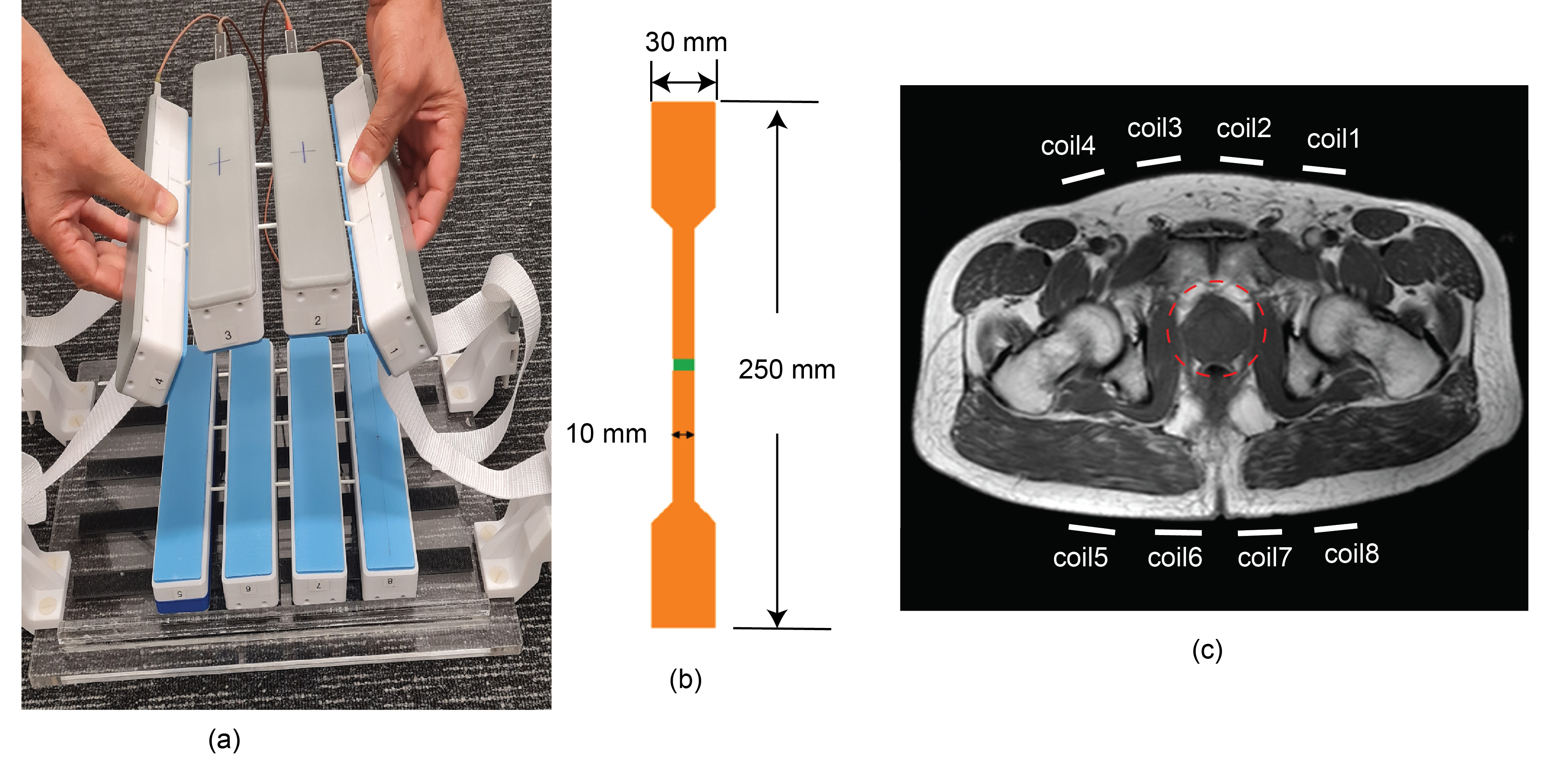
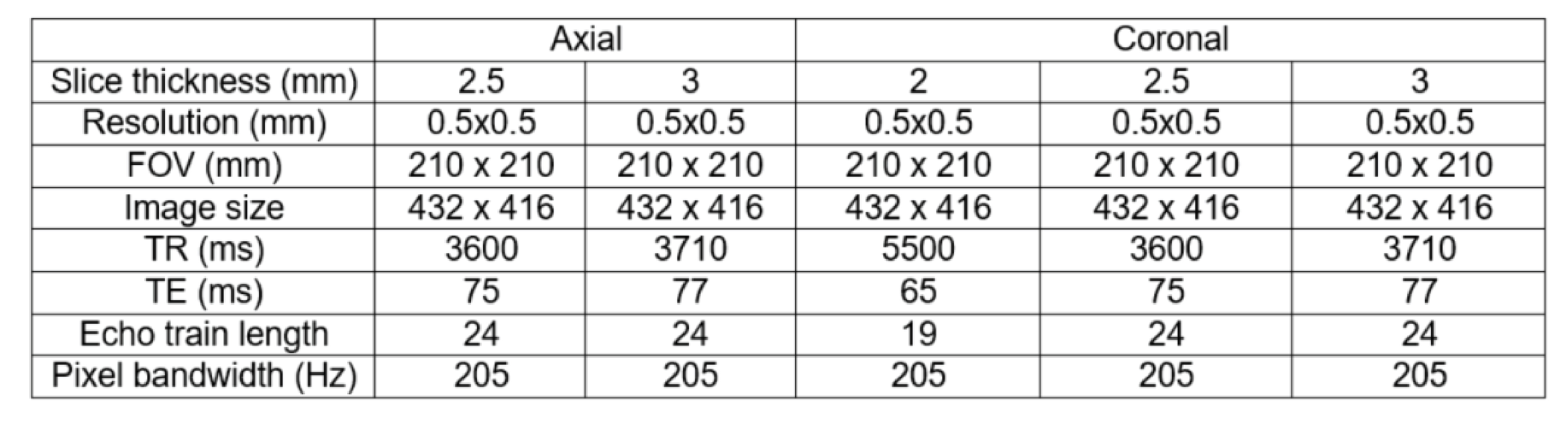
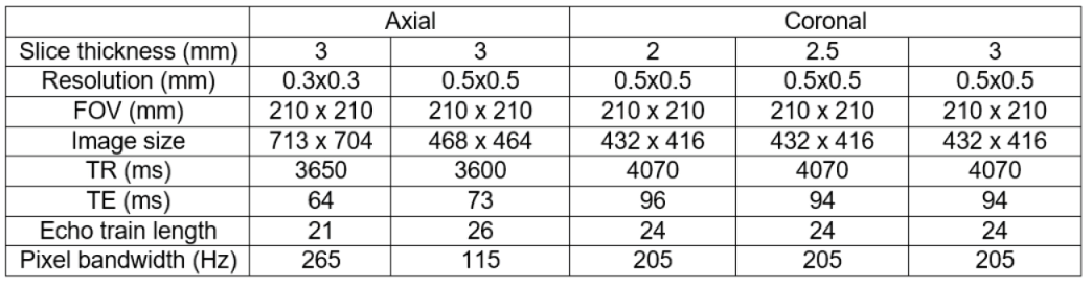
The 8-element paddle I-MARS coil was used to acquire axial and coronal T2-weighted turbo spin echo (TSE) images of the prostate in healthy volunteers (volunteer 1 was 57-years-old, 84 kg, 178 cm; volunteer 2 was 36-year-old, 88 kg, 181 cm) on an investigational whole-body 7T scanner (Siemens Healthcare, Erlangen, Germany) located at the Centre for Advanced Imaging, The University of Queensland. Detailed sequence parameters for the T2W TSE images are listed in Table I and Table II. Before the RF chain was upgraded to 2 kW/channel, the phase-only B1 shimming was normally applied due to insufficient power 3. In contrast, the inline B1 shimming has the capacity to provide adequate B1 field strength for fast 180˚ refocusing, optimizing both the amplitude and phase of each channel. Moreover, increased RF power enables increased penetration depth, which is beneficial for imaging deep structures such as the prostate. While performing B1 shimming at 7T, the management of local SAR10g is also critical 4. Therefore, Virtual Observation Points (VOP)5 were calculated, based on which the vendor-supplied inline monitored is constantly performed. The VOPs were determined using the Finite-Difference Time-Domain (FDTD)6 based electromagnetic simulation software Sim4Life (ZMT, Zurich, Switzerland) and the Duke human model from the Virtual Family 7.As shown in Figure 1(a), the 8-element array consists of a 4-element anterior array and a 4-element posterior array. The posterior array is velcro-attached to a dedicated base that is locked to the patient bed of the MRI system. The anterior array is then placed on top of the patient over the pelvic region and again firmly secured to the MRI gantry by straps and clips. An individual paddle I-MARS coil element is shown in Figure 1 (b). The patient-coil setup is shown in Figure 1 (c), where it can be seen that all coil elements can be placed closely together in parallel anterior and posterior configurations over the region the prostate benefitting from the narrow width of the paddle coil elements.
Results
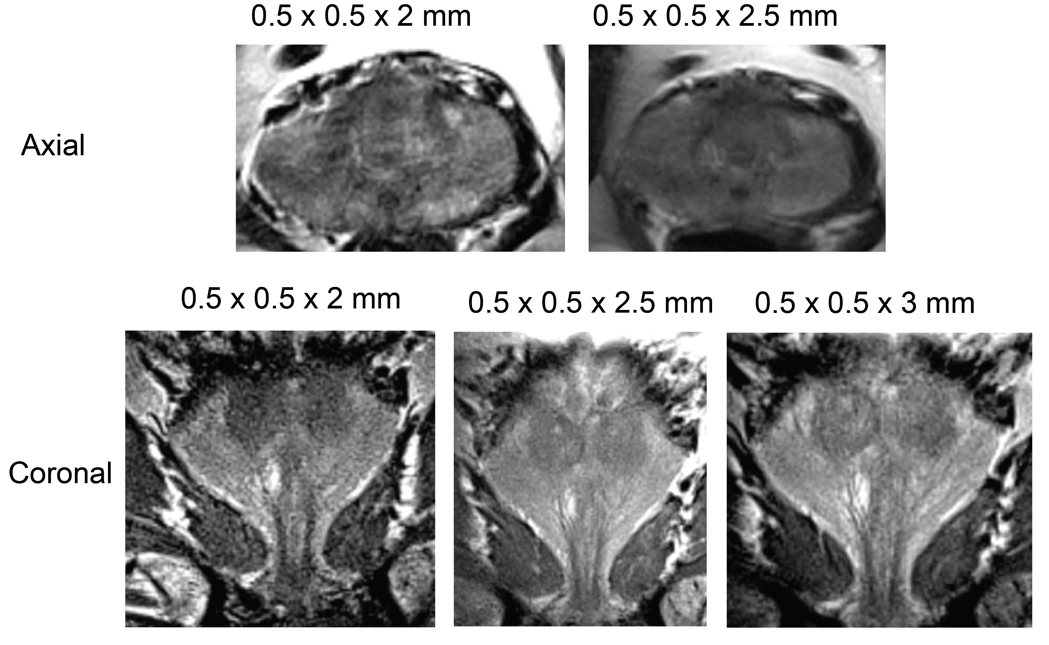
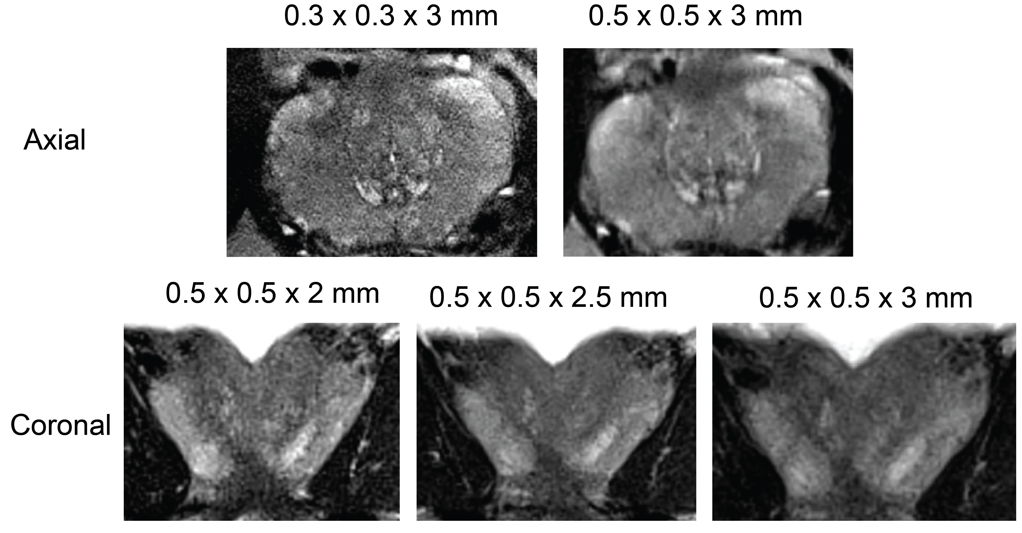
The paddle IMARS coil successfully acquired coronal and axial prostate images on the 7T Magnetom Plus system using a T2-weighted TSE sequence, a typical sequence used in clinical imaging to evaluate the zonal anatomy of the prostate. In the axial images of both volunteers (Figure 2 & 3), which were acquired with a range of slice thicknesses (2mm, 2.5mm, 3mm) and in-plane resolutions (0.3, 0.5mm) for preliminary testing, the peripheral zone can be differentiated from the transition zone and central zones in all cases. In the high resolution (0.3 x 0.3 x 3 mm) axial images acquired from the second volunteer, the boundary between the posterior medial peripheral zone and posterior transition zone is well visualized in comparison with the less clear boundary between the anterior peripheral zone and anterior transition zone. However, these structures are better characterized in the axial images with a slightly lower resolution (0.5 x 0.5 x 3 mm). In the coronal images acquired from the 57-year-old volunteer, the chosen images display the peripheral zone, transition zone and central zone; the centrally located prostatic urethra and superior seminal vesicles are well seen.Conclusion
In this work, we have demonstrated the capability of newly developed paddle I-MAR coils to acquire high-quality, high-resolution MR images of the prostate at 7T. In future work, optimization of the T2w TSE sequence to enhance contrast and SNR characteristics will be undertaken in conjunction with a multiparametric imaging protocol of the prostate, including the acquisition of DWI images at 7T. To exploit the potential clinical uses of these specialized coils, we plan to carry out a more detailed evaluation of the performance of the I-MARS coil for imaging of the prostate at 7T, particularly with patients having biopsy-proven prostate cancer.Acknowledgements
The authors wish to thank ZMT Zurich MedTech for providing the simulation software package Sim4Life.References
1. Destruel A, Jin J, Weber E, et al. Integrated Multi-modal Antenna with coupled Radiating Structures (I-MARS) for 7T pTx body MRI. IEEE Transactions on Medical Imaging. 2021:1-1.
2. Aurelien Destruel, Ewald Weber, Mingyan Li, Jin Jin, Craig Engstrom, Feng Liu, and Stuart Crozier. A novel type of radiofrequency antenna for multi-regional 7T MRI. 2021 ISMRM & SMRT Virtual Conference; 2021.
3. Mingyan Li, Aurelien Destruel, Ewald Weber, Aiman Al-Najjar, Shekhar Chandra, Feng Liu, Craig Engstrom, and Stuart Crozier. A Comparative Study on High Resolution Prostate Imaging at 7T and 3T MRI. 2020 ISMRM & SMRT Virtual Conference; 2020.
4. IEC 60601-2-33:2010 Medical electrical equipment - Part 2-33: Particular requirements for the basic safety and essential performance of magnetic resonance equipment for medical diagnosis. In: International Electrotechnical Commission 2010.
5. Eichfelder G, Gebhardt M. Local specific absorption rate control for parallel transmission by virtual observation points. Magnetic resonance in medicine. 2011;66(5):1468-1476.
6. Yee K. Numerical solution of initial boundary value problems involving maxwell's equations in isotropic media. Antennas and Propagation, IEEE Transactions on. 1966;14(3):302-307.
7. Gosselin M-C, Neufeld E, Moser H, et al. Development of a new generation of high-resolution anatomical models for medical device evaluation: the Virtual Population 3.0. Physics in Medicine and Biology. 2014;59(18):5287-5303.
Figures