3225
A 24-channel ultra-light flexible receiver coil array for human body imaging at 5T1Paul C. Lauterbur Research Center for Biomedical Imaging, Shenzhen Institutes of Advanced Technology, Chinese Academy of Sciences, Shenzhen, China, 2Key Laboratory for Magnetic Resonance and Multimodality Imaging of Guangdong Province, Shenzhen, China, 3Shanghai United Imaging Healthcare, Shanghai, China
Synopsis
To improve signal-to-noise ratio (SNR) and patient comfort, flexible coils need to be made of very light and flexible materials. In this study, a novel 24-channal ultra-light flexible receiver coil array was designed and constructed for human abdominal imaging at a 5T ultra-high field MRI system. The 24-channel ultra-light flexible coil showed much higher SNR and a better parallel acceleration capability compared to a 24-channel conventional receiver coil. High quality human abdominal images were acquired by using the 24-channel ultra-light flexible coil.
Introduction
To improve signal-to-noise ratio (SNR) and parallel acceleration capability, high-density receiver coil array can be applied. In recent years, high-density flexible receiver coils have been developed rapidly because of their ability to fit the imaging subject well to further improve SNR [1-3]. For patient comfort, flexible coils need to be made of very light and flexible materials. Because the flexible coils need to be adapted to patients in different sizes, the coil shape would change, leading to the change of the coupling between coil units. Hence, the flexible coils need to be designed carefully. In this paper, a novel 24-channal ultra-light flexible receiver coil array was designed and constructed for human abdominal imaging at a 5T ultra-high field MRI system. The SNR and parallel acceleration capability of the 24-channel ultra-light flexible coil were evaluated and compared to these of a 24-channel conventional receiver coil. High quality human abdominal images were acquired by using the 24-channel ultra-light flexible coil.Methods
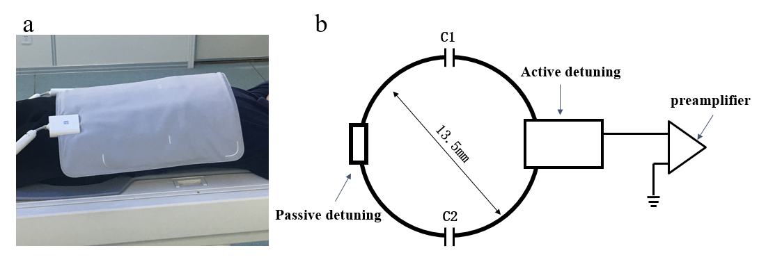
Figure 1 shows the photograph and the of the circuit schematic of the 24-channel ultra-light flexible coil. The passive detuning circuit was designed at the opposite position of the active detuning circuit to further reduce transmit field distortion that was caused by receive array. To diminish the common mode current on the cable shield, cable traps were added on the cables wired within the receive array. Soft waterproof and fireproof materialwas used for the receiver coil package to guarantee the flexibility of the receiver coil and patient safety. The coil was developed and tested on a 5T UIH (Shanghai United Imaging Healthcare, Shanghai, China) MR scanner with volume transmit coil.A 2D density-weighted gradient echo (GRE) sequence was applied for signal acquisitions with a phantom. The parameters were as followings: TR/TE =100/20 ms, flip angle=30o, slice thickness=5mm, matrix=256×256, FOV=500mm×500mm. The noise images were obtained by setting the flip angle to zero. For SNR comparisons, SNR maps were calculated using the sum-of-squares method [4]. For parallel imaging capability evaluation, the inverse g-factor maps were analyzed by using sensitivity encoding (SENSE) reconstructions [5].
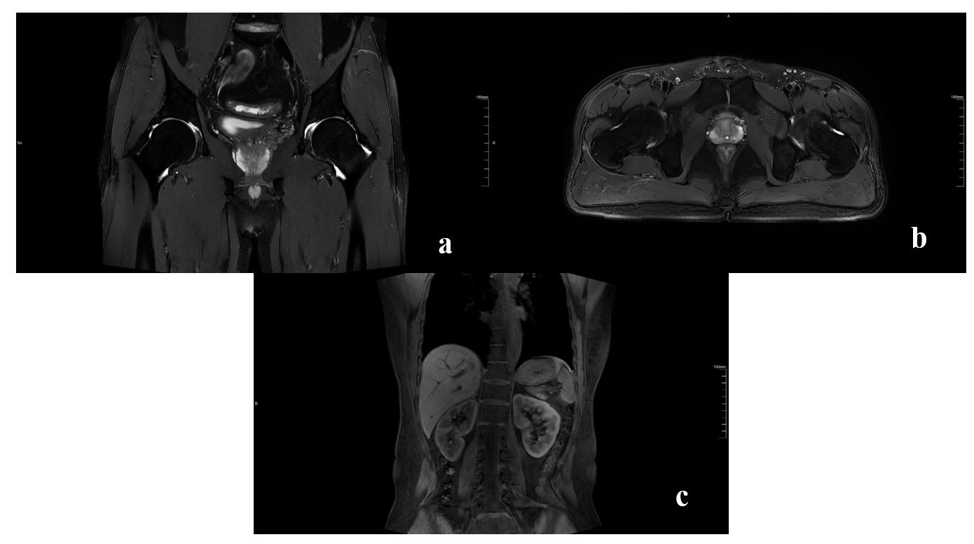
Human abdominal images were acquired using a T2-weighted fast spin echo (FSE) sequence with following parameters: TR/TE=5300/85.12 ms, flip angle=110o, FOV=360mm×300mm, matrix=499×416, reconstructed resolution=0.5×0.5×3mm3, bandwidth=200 Hz/pixel. Additionally, a GRE_quick sequence was applied for the body imaging with following parameters: TR/TE=3.84/1.53 ms, flip angle=25°, FOV=380mm×380mm, matrix=304×304, reconstructed resolution=1.6×1.6×3mm3, bandwidth=600 Hz/pixel.
Results
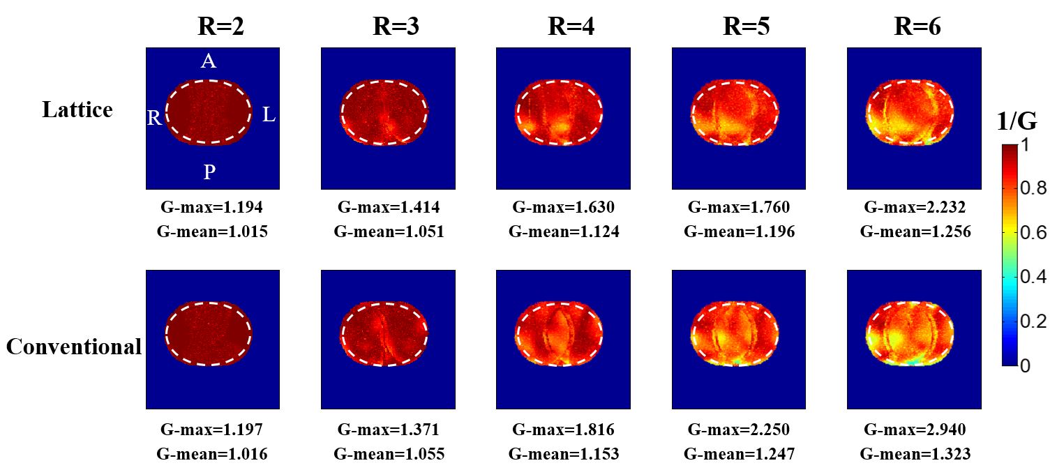
Figure 2 shows phantom SNR maps in the transversal and sagittal planes and SNR profiles in the center. The flexible lattice array demonstrates much higher SNR in the edge region. Figure 3 depicts the inverse g-factor maps in the transverse plane with acceleration factors R from 2 to 6. The results indicate that the parallel imaging capability of the flexible lattice array is better than that of the conventional structure array, particularly at high acceleration factors. Figure 4 displays the human body images using the 24-channel flexible array, which show high quality.Discussions and Conclusions
A novel 24-channal ultra-light flexible receiver coil array was designed and constructed for human abdominal imaging at a 5T ultra-high field MRI system. The 24-channel ultra-light flexible coil showed much higher SNR and a better parallel acceleration capability compared to a 24-channel conventional receiver coil. High quality human abdominal images were acquired by using the 24-channel ultra-light flexible coil. These show great significance in clinical and scientific research applications for human body imaging using the ultra-light flexible coil at 5T.Acknowledgements
This work is supported by the Strategic Priority Research Program of Chinese Academy of Sciences, XDB25000000; National Key R&D Program of China, 2021YFE0204400; Shenzhen city grant, RCYX20200714114735123, ZDKJ20190204003, ZDKJ20190204004.References
[1] Collick BD, Behzadnezhad B, Hurley SA, Mathew NK, Behdad N, Lindsay SA, Robb F, Stormont RS, McMillan AB. Rapid development of application-specific flexible MRI receive coils. Physics in Medicine & Biology. 2020, 65(19): 19NT01.
[2] Vincent JM, Rispoli JV. Conductive thread-based stretchable and flexible radiofrequency coils for magnetic resonance imaging. IEEE Transactions on Biomedical Engineering. 2020, 67(8): 2187-2193.
[3]Chen QY, Xie GX, Luo C, Yang X, Zhu J, Lee J, Su S, Liang D, Zhang XL, Liu X, Li Y, Zheng HR. A dedicated 36-channel receive array for fetal MRI at 3 T. IEEE Transactions on Medical Imaging, 2018, 37(10): 2290-2297.
[4] Roemer PB, Edelstein WA, Hayes CE, Souza SP, Mueller OM. The NMR phased array. Magn Reson Med. 1990, 16(2):192-225.
[5] Pruessmann KP, Weiger M, Scheidegger MB, Boesiger P. SENSE: sensitivity encoding for fast MRI. Magn Reson Med. 1999, 42(5):952-62.
Figures