3223
A quadrature birdcage/48-channel receiver coil assembly for human brain imaging at 5T1Paul C. Lauterbur Research Center for Biomedical Imaging, Shenzhen Institutes of Advanced Technology, Chinese Academy of Sciences, shenzhen, China, 2Key Laboratory for Magnetic Resonance and Multimodality Imaging of Guangdong Province, shenzhen, China, 3Shanghai United Imaging Healthcare, Shanghai, China, shanghai, China, 4Department of Biomedical Engineering, State University of New York at Buffalo, Buffalo, NY, United States
Synopsis
Ultra-high field magnetic resonance imaging (MRI) of human brain with high resolution has been increasing used for clinical and research applications. Due to RF transmit homogeneity and specific absorption issues, clinical use of ultra-high field MRI were limited. In this work, a local quadrature birdcage/48-channel receiver coil assembly was designed and evaluated on a novel whole body 5T MRI scanner. The coil at 5T showed improved SNR, higher parallel acceleration capability and improved detection in vessel wall imaging compared to a 32-channel coil at a 3T commercial scanner.
Introduction
Increasing magnetic field strength can improve signal-to-noise ratio (SNR) and susceptibility contrast to obtain high resolution images. Based on the improvement of the SNR, ultra-high field MRI shows a potential in direct visualization of the vessel wall with high-resolution intracranial vessel wall MR imaging [1]. However, clinical use of ultra-high field MRI were limited due to a number of issues, especially RF transmit homogeneity and specific absorption [2]. Between 3T and 7T, an intermediate magnetic field strength of 5T may provide significant signal-to-noise improvement with less critical RF challenges [3-4]. In this work, we have designed and evaluated a local quadrature birdcage/48-channel receiver head coil array assembly for human brain imaging at 5T (Shanghai United Imaging Healthcare, Shanghai, China). The RF coil assembly was validated by electromagnetic simulations and phantom experimental studies. The signal-to-noise ratio, parallel imaging capability and high-resolution vascular wall imaging with high acceleration factor using the coil at 5T were evaluated and compared to these using a 32-channel coil at a 3T commercial scanner (uMR790, Shanghai United Imaging Healthcare, Shanghai, China).Methods
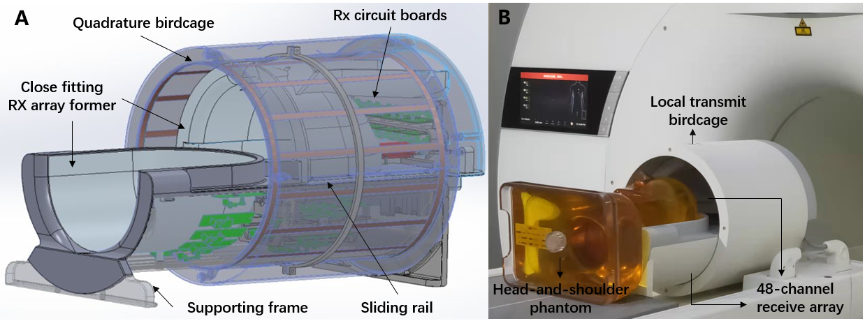
Figure 1 shows photograph of the quadrature birdcage/48-channel receiver coil assembly for 5T. The shielded high-pass birdcage coil was comprised of 16 rungs and was driven in circularly polarized mode. The receiver array was arranged on a close-fitting helmet former (A-P:230mm, L-R:210mm, I-S:266mm), of which 16 elements on the anterior and 32 elements on the posterior. At 3T, a similar size 32-channel receiver coil in combination with the body coil for transmission was used and compared. Before in vivo imaging, the RF coil was validated by electromagnetic (EM) simulation and phantom experimental studies. A head-and-shoulder phantom filled with an aqueous solution of 52.4g polyvinylpyrrolidone (PVP) and 1.15g sodium chloride (NaCl) per 100g of demineralized water was used to mimic the average dielectric properties of brain tissue [5] (conductivity 0.53 S/m and relative permittivity 55.4). The measured B1+ field was characterized using a B1+ mapping sequence. Both the EM simulated and measured B1+ field were obtained and evaluated by normalizing the accepted input power. A 2D density-weighted gradient echo (GRE) sequence was applied for signal acquisitions with the parameters: TR/TE =3000ms/6.5ms, flip angle=300, slice thickness=5mm, matrix=256×256, FOV=200mm×200mm. The noise images were obtained by setting the flip angle to zero. For SNR comparisons, SNR maps were calculated using the sum-of-squares method [6]. For parallel imaging capability evaluation, the inverse g-factor maps were analyzed by using sensitivity encoding (SENSE) reconstructions [7]. Vascular wall images were acquired using a T1-weighted 3D MATRIX (Modulated flip angle technique in refocused imaging with extended echo train) sequence with following parameters: TR/TE=830ms/15ms, flip angle=740, FOV=192mm×232mm, matrix=384×464 (At 3T: TR/TE=800ms/16.2ms, flip angle=750, FOV=180mm×232mm, matrix=360×464), slices per slab=280, Echo train length=40, reconstructed resolution=0.3×0.3×0.3mm3, bandwidth=440 Hz/pixel. uCS(united compressed sensing) acceleration factor=6 and 5.4 at 5T and 3T, respectively.Results
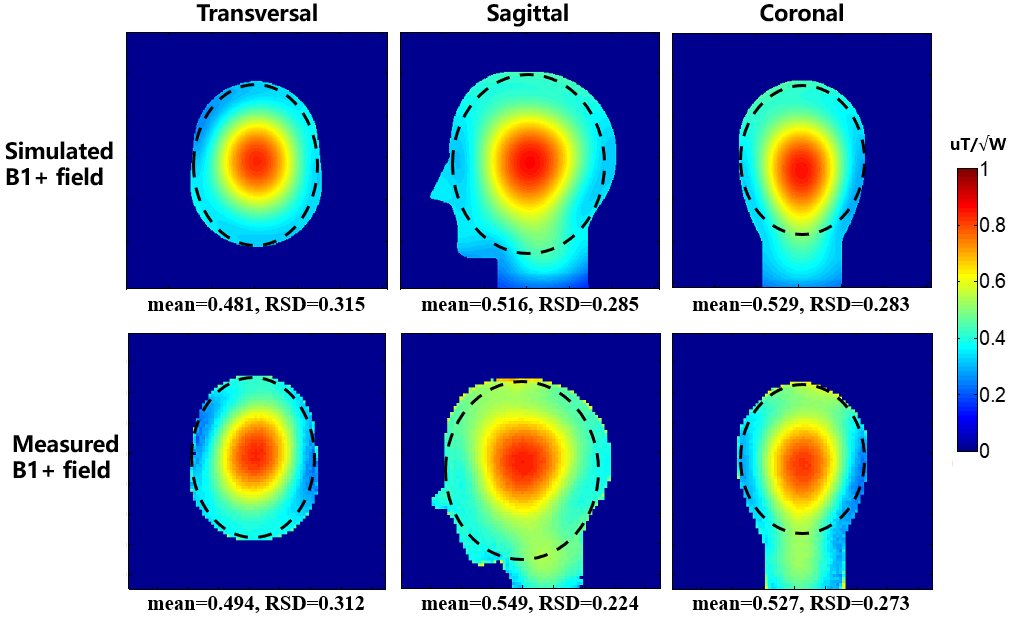
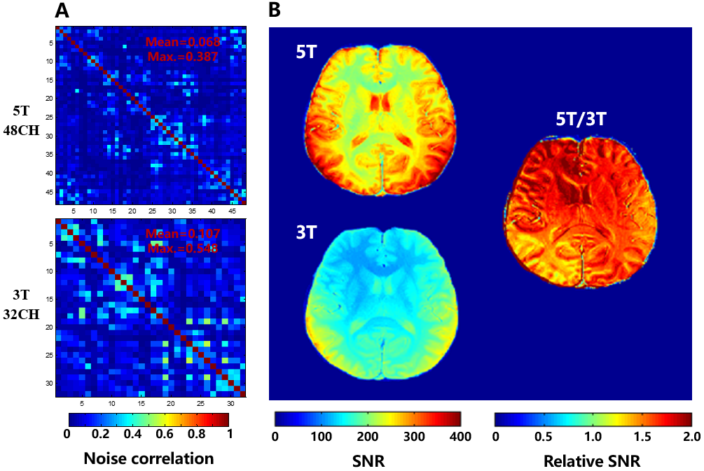
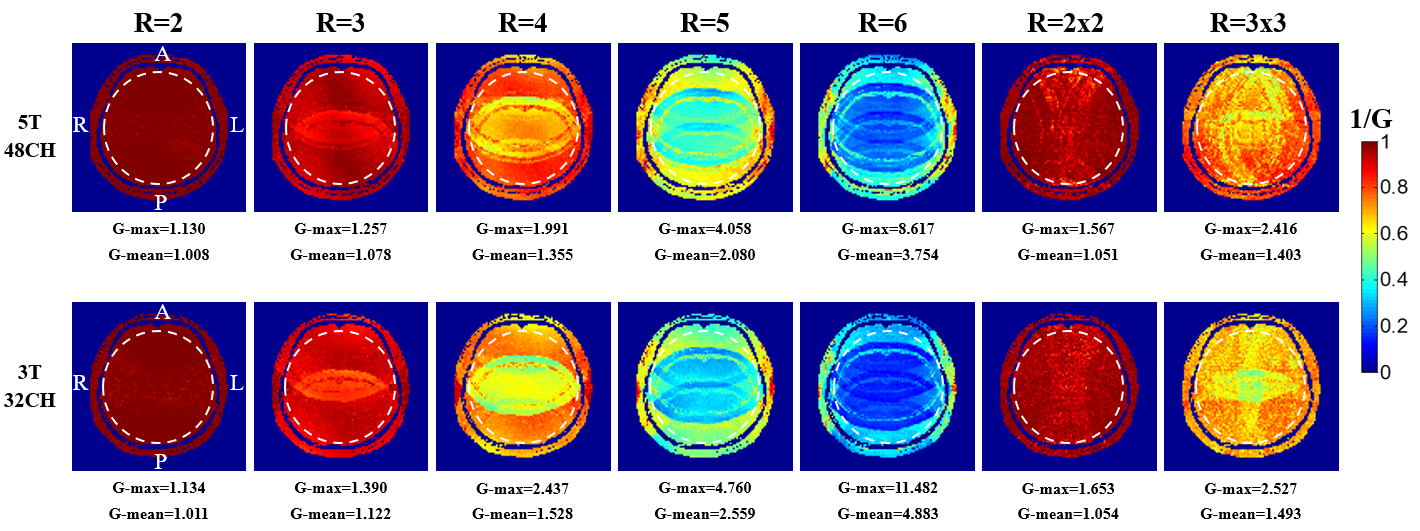
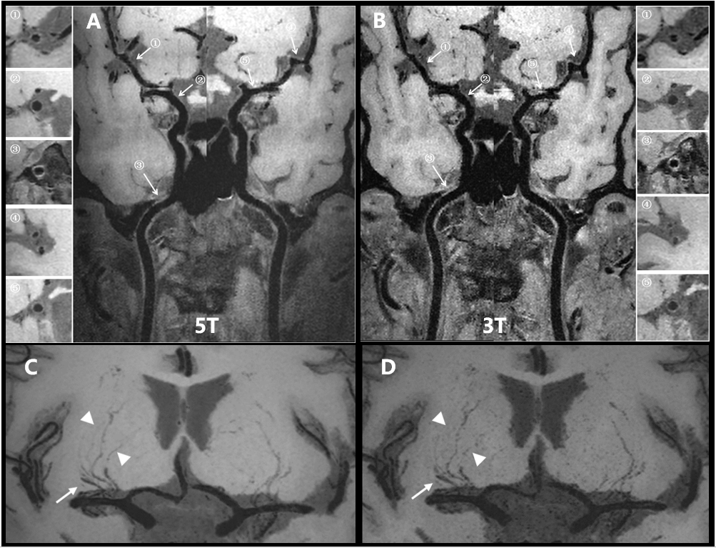
The simulated and measured B1+ field maps at 5T were constructed in the transversal, sagittal, and coronal planes, as shown in Figure 2. The mean and relative standard deviation (RSD) values in the ROI were calculated and depicted in the maps. The measured B1+ field distributions were in good correspondence with simulations. The noise correlation matrix and SNR maps of human brain in the transversal plane acquired at the 5T and 3T MRI scanners are shown in Figure 3. The average SNR over the entire cerebrum at 5T was improved by a factor of 1.6 compared to that at 3T. Known from the relative SNR differences between the SNR maps, it was up to 2 times in some brain regions. Figure 4 depicts the inverse g-factor maps in the transverse plane with acceleration factors R from 2 to 6 and R=2x2, R=3x3. The results indicate that the parallel imaging capability of the coil using at 5T is better than that of the coil using at 3T, particularly at high acceleration factors. Curved multi-planar reformatting images of the left and right intracranial arterial vessel wall acquired at 5T and 3T are shown in Figure 5 A and B. The vessel wall images acquired at 5T show higher SNR than these acquired at 3T. More distal vessels of lenticulostriate arteries (LSAs) can be appreciated at 5 T compared to 3 T (Figure 5 C and D). The delineation of these LSAs allows for visualization of normal vessels as well as the detection of possible pathologies such as arterial dissection and small LSA aneurysms.Discussions/Conclusion
A local quadrature birdcage/48-channel receiver coil assembly for human brain was designed and evaluated at a 5T MRI system. Compared to 3T clinical MRI, the 48-channel receiver head coil at 5T provides significant SNR improvement and higher parallel imaging capability. By using the coil designed at 5T, high-resolution intracranial vessel wall images can be obtained. These show great significance in clinical and scientific research applications for vascular wall imaging using the 5T MRI. Future work includes performance evaluation in high-resolution Time-of-flight MR angiography, susceptibility weighted imaging and functional imaging.Acknowledgements
This work is supported by the Strategic Priority Research Program of Chinese Academy of Sciences, XDB25000000; National Key R&D Program of China, 2021YFE0204400; Shenzhen city grant, RCYX20200714114735123, ZDKJ20190204003, ZDKJ20190204004.References
[1] Mandell DM, Mossa-Basha M, Qiao Y, Hess CP, Hui F, Matouk C, Johnson MH, Daemen MJ, Vossough A, Edjlali M, Saloner D, Ansari SA, Wasserman BA, Mikulis DJ; Vessel Wall Imaging Study Group of the American Society of Neuroradiology. Intracranial Vessel Wall MRI: Principles and Expert Consensus Recommendations of the American Society of Neuroradiology. AJNR Am J Neuroradiol. 2017, 38(2):218-229.
[2] Cao Z, Park J, Cho ZH, Collins CM. Numerical evaluation of image homogeneity, signal-to-noise ratio, and specific absorption rate for human brain imaging at 1.5, 3, 7, 10.5, and 14T in an 8-channel transmit/receive array. J Magn Reson Imaging. 2015, 41(5):1432-9.
[3] Hennig J. Ultra high field MR: useful instruments or toys for the boys? Magn. Reson Mater Phy. 2008; 21: 1-3.
[4] Ye Li et al. In-vivo human brain imaging at 5 T using a 48 channel Tx-Rx array. ISMRM 2021 p1572
[5] Gabriel S, Lau RW, Gabriel C. The dielectric properties of biological tissues: III. Parametric models for the dielectric spectrum of tissues. Phys Med Biol. 1996, 41:2271-2293
[6] Roemer PB, Edelstein WA, Hayes CE, Souza SP, Mueller OM. The NMR phased array. Magn Reson Med. 1990, 16(2):192-225.
[7] Pruessmann KP, Weiger M, Scheidegger MB, Boesiger P. SENSE: sensitivity encoding for fast MRI. Magn Reson Med. 1999, 42(5):952-62.
Figures