3217
Water- fat separated MR Fingerprinting (MRF) with simultaneous B1+ and B0 estimation1State key Laboratory of Modern Optical Science and Engineering, Zhejiang University, Hangzhou, China
Synopsis
A novel water-fat separated MRF approach was proposed to minimize the known biases introduced by the B1+, B0 and water and fat partial volume. By incorporating the water-fat separation approach in the framework, the dictionary size could be greatly reduced with the known B0 and FF map. Multiple maps of parameters including B1+, B0, FF and T1 and T2* of water and fat can be acquired within 14s for one slice.
Introduction
Magnetic resonance fingerprinting (MRF) is an efficient technique to simultaneously provides quantitative maps of different tissue parameters. Considering the inevitable systematic errors, such as RF transmit field (B1+) and B0 field inhomogeneity, a novel MRF sequence has been proposed to enable a simultaneous T1, T2*, B1+ and B0 detection1. However, there is another factor that may cause the inaccuracy of parametric mapping — water and fat partial volume, and the fat characterization is also considered important especially for some fat-related diseases, such as fatty liver. There are two main approaches to solve this problem: (a) Including the fat signal by the multi-component model and matching the temporal signal evolution by an exhausted dictionary approach. This kind of method usually work with the dictionary compression to accelerate the matching2. (b) Incorporating the Dixon method in the sequence to achieve water and fat separation at first, then implementing the independent water and fat MRF matching to avoid the large dictionary matching3,4. Inspired by these approaches, we adapted the existed B0&B1-correction MRF sequence to achieve the fat characteration. The multi-echo signals from Double TR blocks could be used to calculate the Fat Fraction (FF) map and B0 map by three-Dixon method. With the known FF map and B0 map, the dictionary size could also be greatly decreased during the multi-component signal matching. Our proposed framework could enable the T1 and T2* mapping for both water and fat and the reliable B1+ and B0 estimation simultaneously.Methods
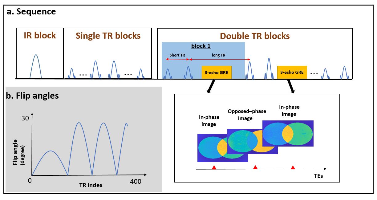
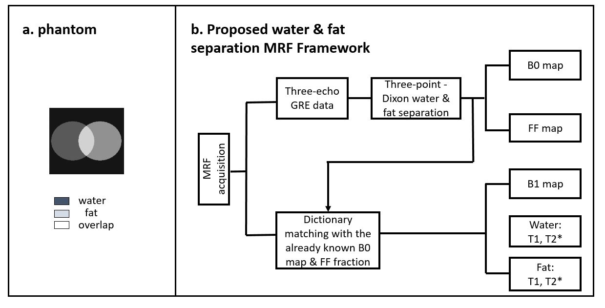
Fig.1a shows the B0&B1-correction MRF sequence we used for our experiments. Basically it is an IR-spoiled GRE sequence with various TRs and TEs. The sequence can be divided into one IR block and multiple double TR block. The IR block is mainly used to efficiently map the T1 parameter and the double TR block that composes of a short TR (12 ms) and a long TR (48 ms) is used to encode the B1+ information. Inside the double TR block, there are four echoes for the B0 and T2* estimation. The first three echoes (TE1/TE2/TE3 = 2.4/13.2/24 ms) will be used as three-echo GRE signal to acquire the in-phase and opposed-phase water and fat images for the Dixon method to calculate the FF map and B0 map. Fig.1b shows the Flip angles of each TR. Spiral trajectory is designed for the sequence with 36 interleaves at 1mm resolution for 220 mm FOV, and the total acquisition time is around 14s/slice.As Fig.2b illustrates, the B0 and FF values estimated from the three echoes will be used as the known parameters to create water-fat dictionary by Bloch simulation. The mixed signal S at echo time TE can be written as:
$$S(TE)=M0[(1-FF)+FF*e^{(i2\pi f_{cs} TE)} ] e^{(i2π∆B_0TE)} $$
Where M0 represents the equilibrium magnetization, $$$ f_{cs}$$$ is the water-fat chemical shift. Noted that we ignore the B1+ and relaxation effects and assume there is only one peak in fat spectrum for a clear illustration. Over multiple excitation pulses, perfect spoiling was assumed for simulations.
Finally, the temporal signal evolutions obtained by the proposed sequence were matched using the created dictionary. We adopted the SVD compression approach to further accelerate the dictionary matching5.
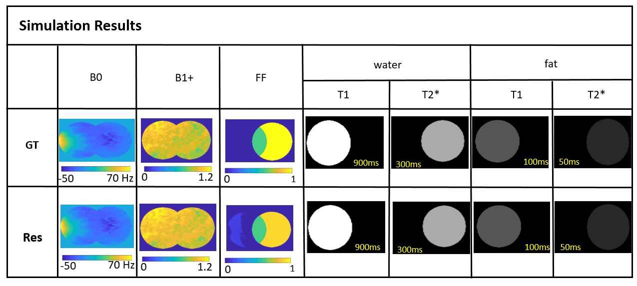
We did the numerical simulations to validate our idea. Fig.2a shows a simple water-fat phantom with an overlapping area between the water and fat circles. Reasonable M0, T1, T2* are set for water and fat, respectively. The T1 (from 200 to 2300 ms with a step size of 10ms), the T2* (from 50 to 600 ms with a step size of 5 ms), the B1+ (from 0 to 1.4 with a step size of 0.1) are adopted for the dictionary creation and temporal signal evolutions.
Results
Fig.3 shows the simulation results of the proposed method. The Ground Truth (GT) and Results (Res) of different parameters (B0, B1+, FF, T1 and T2* of water and fat) are shown in the table. The B0 map and FF map is identical with the real values, and the B1+, T1 and T2* mapping match well with the ground truths. The results further validate the feasibility of our method.Discussion and Conclusion
Simulations suggest that our method could enable the simultaneous mapping of B0, B1+, FF mapping, as well as the T1 and T2* mapping for both water and fat. This preliminary exploration has shown promising results with further real phantom and in-vivo validation. The potential applications of the proposed approach may include the liver and skeletal muscles, where fat plays an important role in the related diseases.Acknowledgements
This work was supported in part by National Key R&D Program of China (No: 2020AAA0109502), by the National Natural Science Foundation of China (No: U1809204, 61525106, 61427807, 61701436), the Fundamental Research Funds for the Central Universities.References
1. H Ye, Q Li, X Cao, et al. MR fingerprinting (MRF) incorporating simultaneous detection of RF transmit field and B0 inhomogeneity. Proceedings of the 26th Annual Meeting of ISMRM, Paris,France,2018.
2. Marty B, Carlier P G. MR fingerprinting for water T1 and fat fraction quantification in fat infiltrated skeletal muscles[J]. Magnetic Resonance in Medicine, 2019, 83(2).
3. Jaubert O, Cruz G, Aurélien Bustin, et al. Water–fat Dixon cardiac magnetic resonance fingerprinting[J]. Magnetic Resonance in Medicine, 2020, 83(6).
4. Koolstra K, Webb A G, Veeger T T J, et al. Water–fat separation in spiral magnetic resonance fingerprinting for high temporal resolution tissue relaxation time quantification in muscle[J]. Magnetic Resonance in Medicine, 2020, 84(2):646-662.
5. D. F. McGivney et al. SVD Compression for Magnetic Resonance Fingerprinting in the Time Domain[J]. IEEE Transactions on Medical Imaging, vol. 33, no. 12, pp. 2311-2322.
Figures