3183
3D T1-weighted sequence of volumetric isotropic turbo spin-echo acquisition with a deep learning constrained Compressed SENSE reconstruction1Department of Radiology, The First Hospital of Jilin University, Changchun,Jilin, China, 2The First Hospital of Jilin University, Changchun,Jilin, China, 3Philips Healthcare,Beijing,China, Beijing, China
Synopsis
Compressed SENSE-AI reconstruction system was introduced with its sufficient noise removal to maintain high image quality while reducing scanning time. Intracranial and carotid vessel wall MR imaging (VW-MRI) are widely available in detecting and characterizing atherosclerosis occurring in the two vascular ranges, however, long scan time had so far limited high resolution VW-MR imaging. In this study, we investigated the impact of Compressed SENSE-AI acceleration factor on the diagnostic quality of magnetic resonance images and compared with standard vessel wall MR imaging protocol.
Introduction
Intracranial and carotid vessel wall MR imaging (VM-MRI) are widely available in detecting and characterizing atherosclerosis occurring in the two vascular ranges1,2. In order to minimize scan time and provide a good vessel wall to lumen contrast for assessing vessel wall characteristics, Compressed-SENSE (CS) has been used in intracranial and carotid VW-MRI jointly3,4. Recently, deep learning-based algorithms have been combined with CS (CS-AI) by learning optimal reconstruction parameters from the data itself and showed superior performance. Thus, the aims of this study were to overcome the drawback of time limitations at VW-MRI and to evaluate the impact of different acceleration factors (AF) for CS-AI on the vessel wall images quality of 3D T1-weighted sequence of volumetric isotropic turbo spin-echo acquisition (VISTA) sequence, and the compared to the existing standard acceleration techniques.Materials and methods
This prospective single center study was approved by the local institutional review board. Written informed consent was obtained from all participants included in the study. From October 1 2021 to October 31 2021, a total of 26 consecutive patients (14 males,12 females, mean age: 63.77, age range: 51–79 years) with suspected intracranial and carotid arterial disease were examined on 3T MR system (Ingenia Elition X, Philips Healthcare).All patients were receiving an optimized 3D T1-VISTA sequence with different AF (6, 8,10 and 12) and reconstruction by CS (CS6,CS8,CS10, CS12) and CS-AI(CS-AI 6, CS-AI 8, CS-AI 10, CS-AI 12).The commercially available SENSE acceleration method were also acquired with AF6 and AF8(S6, S8). Among them, S6 is the commonly used clinical sequence and is shown as reference. Parameters were common to all examinations: voxel size=0.75*0.75*0.75mm3, FOV=200*251mm2, 440 slices, TR=700ms, TE=35ms.The subjective image quality was compared qualitatively between the VISTA CS, VISTA CS-AI and VISTA SENSE. The readers who were blinded to any clinical information evaluated overall image quality on the following 4-point Likert scale: 1 = poor; 2 = fair; 3 = good; 4 = excellent(including degree of noise present, vessel wall characteristics and contrast of the vessel and wall, fat and muscle were scored on a four-point scale).The image quality of all sequences was evaluated objectively by the Signal-to-noise ratio (SNR) and contrast-to-noise ratio (CNR, between vessel, intra-arterial plaques and muscle). Paired t-test was used to compare the SNR, and CNR with the CS, CS-AI and SENSE sequences. The frequency of the scores were tested with the Friedman test and post hoc analysis. P values< 0.05 were considered significant.Results
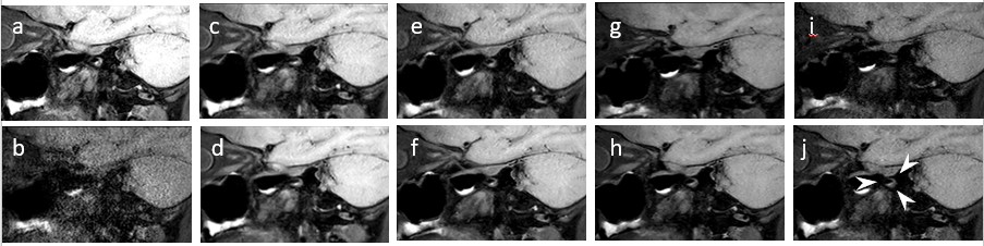
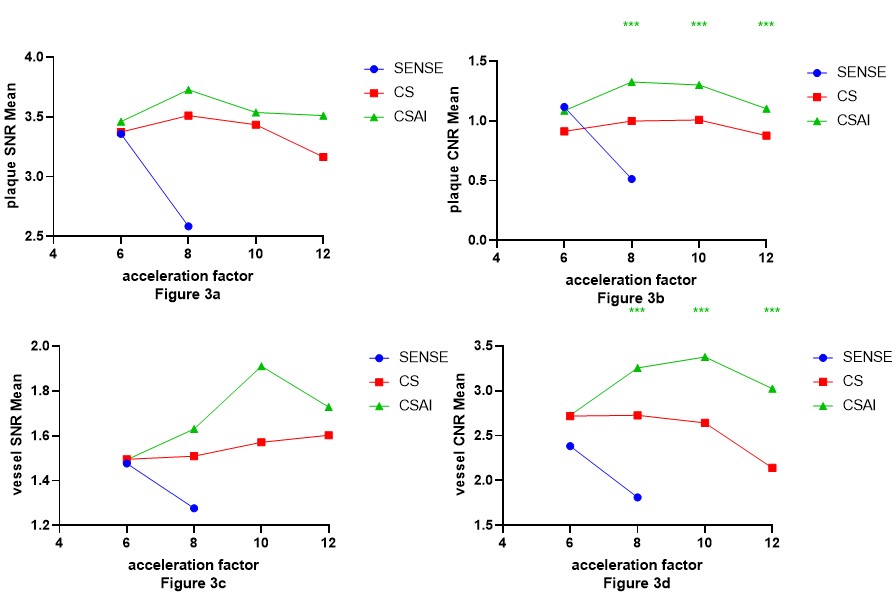
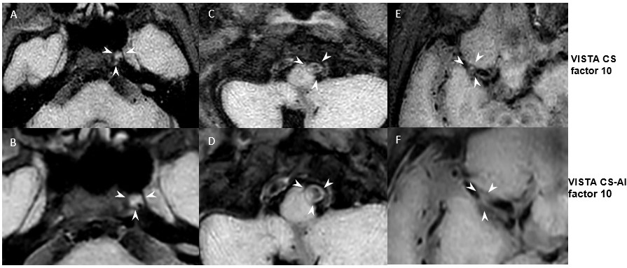
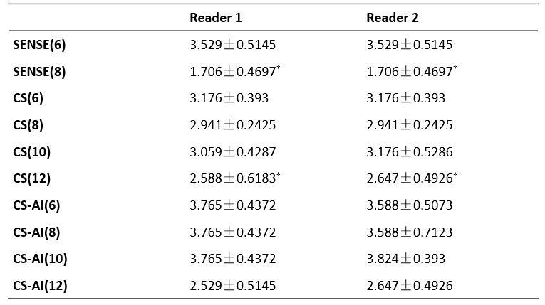
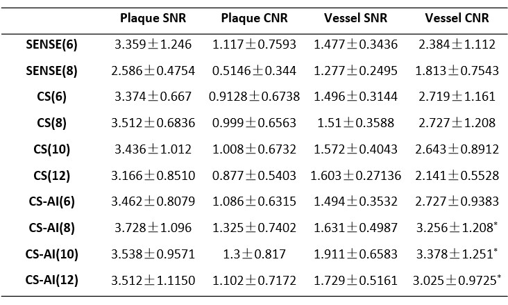
Figure 1 show representative VW-MRI with SENSE, CS and CS-AI acquired with different acceleration factors. Qualitative image scores can be shown in Table 1. The two reviewers had good agreement for image scoring [ICC (95% CI) = 0.76 (0.66, 0.86)]. CS-AI groups showed good image quality comparable to existing scanning sequences (Table 1). Noise enhancement and reduction of the differentiation of low-contrast features are the main artifact observed of the two readers in S8 compared with S6 (reference)(P<0.05). As can be seen in Tabel 2 and Figure 2, no statistically significant difference was found either between the overall the plaque SNR and vessel SNR of CS 6, CS 8, CS 10 or CS 12 accelerated VISTA scan and reference scan (P>0.05). The plaque CNR between plaque and temporal muscle in CS-AI groups is significantly higher than that in CS groups at any acceleration factor, the same on the vessel CNR (P<0.05).Until the AF rises to 10, both the plaque CNR and the vessel CNR for CS-AI groups showed significantly higher than CS groups and SENSE groups (P<0.05). The plaque CNR and the vessel CNR for CS-AI 12 tend to be lower than S6(reference)(P<0.05).Discussion and Conclusion
VISTA-CS can provide sufficient diagnostic quality of accelerated images and normal wall delineation to VISTA SENSE, which is reported in literature5,6. In this study, we observed that VISTA CS-AI provided better quality images in the lumen of both normal and lesion sites without significantly sacrificing image quality. The implementation of higher acceleration factors had good potential. Acceleration factors up to 10 were tested with a good result in producing images of good diagnostic quality relative to reference S6 sequence.(Figure 3) Results obtained with the application of CS-AI are clinically relevant, and this technique has the potential for use in the clinical setting in the evaluation of patients with intracranial and carotid vessel walls and identifying all involved lesions. Further studies and experience are required particularly with regard to establishing the technique aspects in clinical routine. In conclusion, by combining the CS and deep learning based reconstruction enables substantial acceleration of image acquisition while without significantly sacrificing image quality (AF=10 , reduce approximately 50% time).Acknowledgements
No acknowledgement found.References
1.Mandell DM, Mossa-Basha M, Qiao Y, et al. Intracranial Vessel Wall MRI: Principles and Expert Consensus Recommendations of the American Society of Neuroradiology. AJNR Am J Neuroradiol. 2017;38(2):218-229. doi:10.3174/ajnr.A4893
2.Xie Y, Yang Q, Xie G, Pang J, Fan Z, Li D. Improved black-blood imaging using DANTE-SPACE for simultaneous carotid and intracranial vessel wall evaluation. Magn Reson Med. 2016 Jun;75(6):2286-94. doi: 10.1002/mrm.25785. Epub 2015 Jul 8. PMID: 26152900; PMCID: PMC4706507.
3.Jia S, Zhang L, Ren L, Qi Y, Ly J, Zhang N, Li Y, Liu X, Zheng H, Liang D, Chung YC. Joint intracranial and carotid vessel wall imaging in 5 minutes using compressed sensing accelerated DANTE-SPACE. Eur Radiol. 2020 Jan;30(1):119-127. doi: 10.1007/s00330-019-06366-7. Epub 2019 Aug 1. PMID: 31372787.
4.Alexander MD, Yuan C, Rutman A, Tirschwell DL, Palagallo G, Gandhi D, Sekhar LN, Mossa-Basha M. High-resolution intracranial vessel wall imaging: imaging beyond the lumen. J Neurol Neurosurg Psychiatry. 2016 Jun;87(6):589-97. doi: 10.1136/jnnp-2015-312020. Epub 2016 Jan 8. PMID: 26746187; PMCID: PMC5504758.
5.Park CJ, Cha J, Ahn SS, Choi HS, Kim YD, Nam HS, Heo JH, Lee SK. Contrast-Enhanced High-Resolution Intracranial Vessel Wall MRI with Compressed Sensing: Comparison with Conventional T1 Volumetric Isotropic Turbo Spin Echo Acquisition Sequence. Korean J Radiol. 2020 Dec;21(12):1334-1344. doi: 10.3348/kjr.2020.0128. Epub 2020 Aug 4. PMID: 32767865; PMCID: PMC7689147.
6.Zhu C, Tian B, Chen L, Eisenmenger L, Raithel E, Forman C, Ahn S, Laub G, Liu Q, Lu J, Liu J, Hess C, Saloner D. Accelerated whole brain intracranial vessel wall imaging using black blood fast spin echo with compressed sensing (CS-SPACE). MAGMA. 2018 Jun;31(3):457-467. doi: 10.1007/s10334-017-0667-3. Epub 2017 Dec 5. PMID: 29209856; PMCID: PMC5976530.
Figures