3173
Screening detection of abnormalities in knee joint MRI using deep machine learning1Radiology, Toho University Sakura Medical Center, Sakura, Japan, 2Radiology, Juntendo University, Tokyo, Japan, 3Radiology, Seirei Sakura Citizen Hospital, Sakura, Japan
Synopsis
The DML model including fat-suppressed contrast generation, normal image restoration, and classification and determination models may make it possible to detect all abnormalities in knee joint MRI once.
Introduction
The recent development of deep machine learning (DML) and DML methods has facilitated clinical decision support for reading echocardiograms, chest radiographs, and MR images. Similar methods have been used to infer the presence of lesions for the meniscus and articular cartilage of the knee joint (1-3). However, most of the previous studies were conducted focusing on meniscus injury, cruciate ligament injury, or articular cartilage injury alone. We then tried to develop a DML model to inclusively check abnormalities in the knee joint MR imaging once.Materials and methods
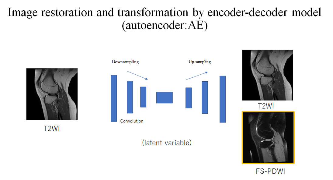
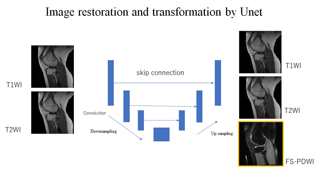
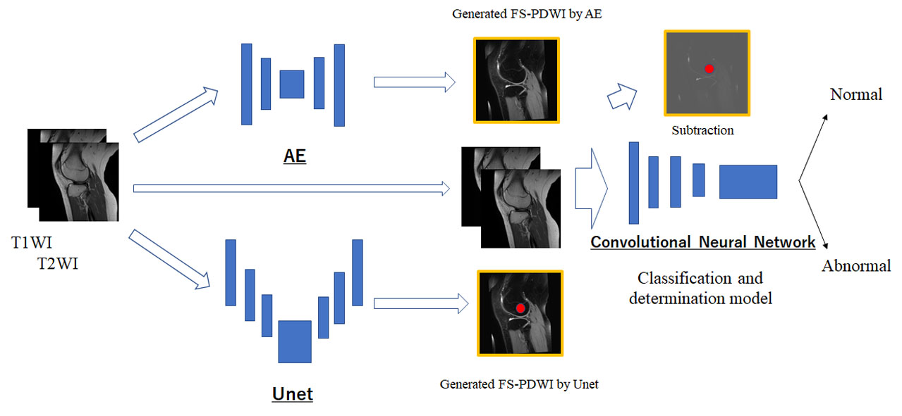
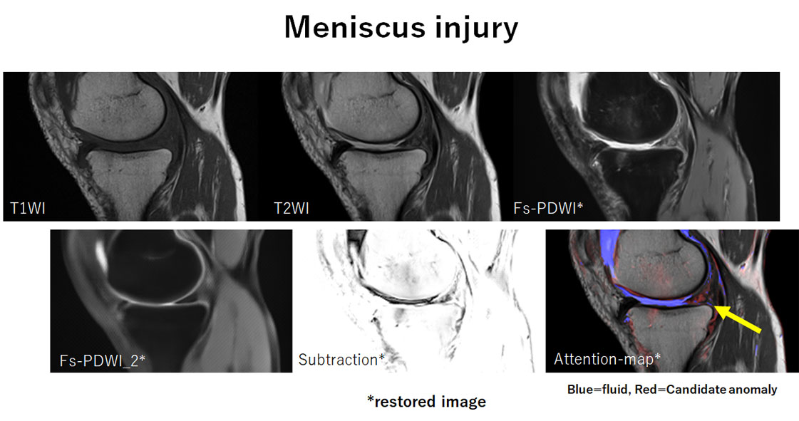
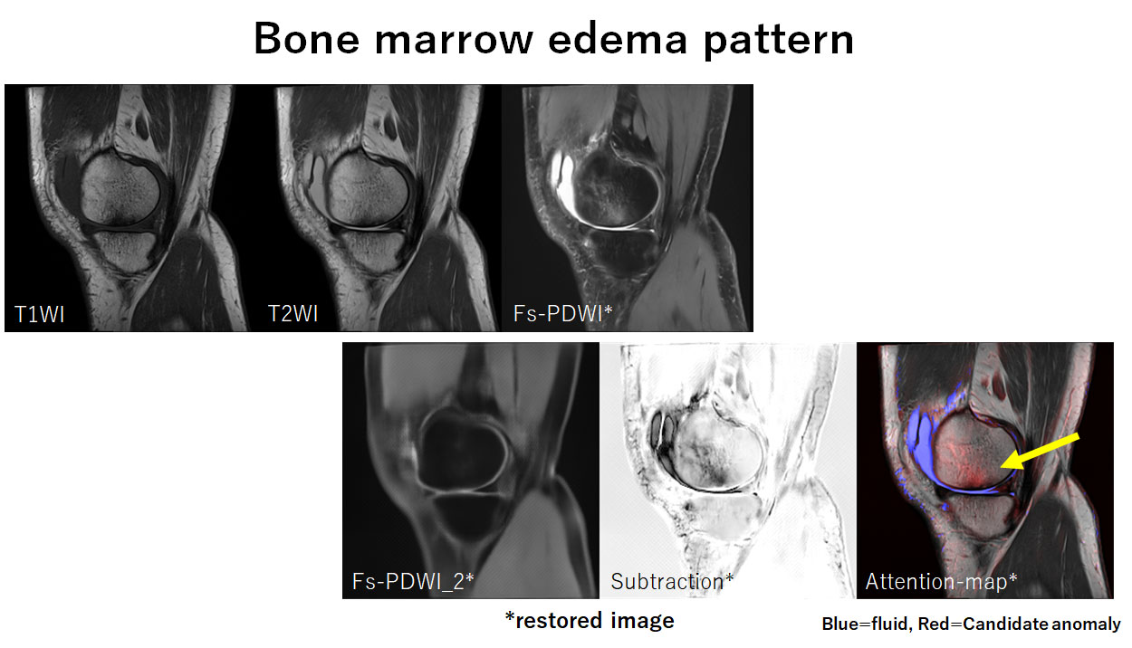
Sixty-three symptomatic knees and 12 normal knees performed in 3T MRI (Siemens, Skyra) were included. T1-weighted (T1WI), T2-weighted (T2WI), and fat-suppressed proton density-weighted (FSPDWI) sagittal images of approximately 3,000 image sets were used. Abnormal findings included bone marrow edema pattern, meniscus injury, and anterior cruciate ligament injury and those were assessed. The DML model we devised consisted of three main parts for the abnormality detection on MRI of the knee joint; (1) normal recovery model, (2) fat-suppressed contrast transformation model, and (3) abnormality classification and determination model: convolutional neural network (CNN) model. The four steps of abnormality detection we adapted were (1) normal recovery model using encoder-decoder model learned only normal MR images of the knee joint to restore the normal ones including FSPDWI (Fig.1), (2) normal and abnormal recovery model using Unet learned normal and abnormal MR images to restore normal and abnormal ones including FSPDWI (Fig.2), (3) subtraction of the restored normal fat-suppression images (FSPDWI) from the restored normal and abnormal fat-suppression images (FSPDWI), and (4) classification and determination model of the subtraction of the restored fat-suppression images (FSPDWI) (Fig.3). The accuracy of abnormality detection of the DML model was calculated by comparing with the results by one-certificated radiologist.Results
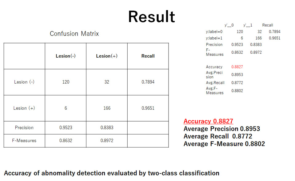
The normal recovery model using encoder-decoder model that learned only normal MR images of the knee joints could not restore unlearned abnormal findings including bone marrow edema pattern, meniscus injury, and anterior cruciate ligament injury. Therefore, the normal recovery model was able to successfully restore normal MR images including FSPDWI. The normal and abnormal recovery model using Unet was able to successfully restore normal and abnormal MR images including FSPDWI. The accuracy of abnormality detection of the DML model was 88% (Fig.4). The detections of bone marrow edema pattern and meniscus injury were better (Figs.5 and 6), but that of anterior cruciate ligament injury was poorer.Discussion
The characteristics of the DML model we devised for the abnormality detection in knee joint MRI are that it uses the model to generate fat-suppressed contrast, which is thought to be very useful for the detection of lesions in the bones and soft-tissues. This method can detect lesions showing high signal intensity on T2WI. It may be more accurate than based on T1- and T2-weighted image sets. Next, the use of the DML model trained by only normal images was significant. Therefore, subtraction of the restored normal images from the restored abnormal images with fat-suppressed contrast could be done. This time, neither learning of regions of the abnormal findings nor annotation work was done. So, only two-class labeling of normal or abnormal findings was tested. The next steps of this study are to assess the contribution of the DML model to improve the diagnostic accuracy in clinical setting, to visualize the regions that DML model would have focused on, and to modify subtraction image for the better detection of ligament injury.Conclusion
The DML model including fat-suppressed contrast generation, normal image restoration, and classification and determination models may make it possible to detect all abnormalities in knee joint MRI once.Acknowledgements
No acknowledgement found.References
1.Astuto B, Flament I, Namiri NK, Shah R, Bharadwaj U, Link TM, et al. Automatic deep learnig-assisted detection and grading of abnormalities in knee MRI studies. Radiology AI 2021; 3(3):e200165.
2. Pedoia V, Norman B, Mehany SN, Bucknor MD, Link TM, Majumdar S. 3D convolutional neural networks for detection and severity staging of meniscus and PFJ cartilage morphological degenerative changes in osteoarthritis and anterior cruciate ligament subjects. J Magn Reson Imaging 2019;49(2):400–410.
3. Liu F, Zhou Z, Samsonov A, et al. Deep learning approach for evaluating knee MR images: achieving high diagnostic performance for cartilage lesion detection. Radiology 2018;289(1):160–169.
Figures