3161
Accelerated T1 weighted PROPELLER imaging of the brain with deep-learning based parallel imaging reconstruction
Motohide Kawamura1, Daiki Tamada1, Kazuyuki Sato2, Masahiro Hamasaki2, Satoshi Funayama1, Tetsuya Wakayama3, Utaroh Motosugi4, Hiroyuki Morisaka1, and Hiroshi Onishi1
1Department of Radiology, University of Yamanashi, Chuo, Japan, 2Division of Radiology, University of Yamanashi Hospital, Chuo, Japan, 3MR Collaboration and Development, GE Healthcare, Hino, Japan, 4Department of Radiology, Kofu-Kyoritsu Hospital, Kofu, Japan
1Department of Radiology, University of Yamanashi, Chuo, Japan, 2Division of Radiology, University of Yamanashi Hospital, Chuo, Japan, 3MR Collaboration and Development, GE Healthcare, Hino, Japan, 4Department of Radiology, Kofu-Kyoritsu Hospital, Kofu, Japan
Synopsis
PROPELLER sequence is useful because of its robustness to patient motion. Longer acquisition than FSE is a major drawback limiting its wider application in clinical practice. Here, we propose an accelerated T1 weighted PROPELLER of the brain using deep learning based parallel imaging (PI) reconstruction. Our method can unfold highly undersampled aliased images (PI factor = 7), enabling 2.3 times faster acquisition than full-sampling. A preliminary reader study with prospectively undersampled data showed that the proposed method significantly outperformed a conventional SENSE reconstruction in terms of streak artifact.
Introduction
Fast spin-echo (FSE) technique is a standard pulse sequence for 2D T1 weighted (T1W) imaging of the brain. Thanks to an efficient Cartesian sampling, FSE is a fast sequence and thus adequate for routine clinical use. However, patient motion during acquisition results in artifacts including ghosts, degrading image quality of FSE especially for children, elderly or severely sick patients. To overcome this difficulty, PROPELLER sequence exploits oversampling at the center of k-space.1 It achieves signal averaging at the center of k-space, leading to robustness to patient motion. In addition, the oversampled central circle of k-space permits correction for in-plane motion and weighting for each blade. The drawback of this technique is longer acquisition time than FSE, limiting the applicability in daily clinical use. Recent advances in deep learning (DL) offer new reconstruction methods for accelerated MRI.2-4 Neural networks can learn to reconstruct fully-sampled images from undersampled k-space data. Reduction of sampling rate enables faster acquisition. However, few study investigated DL based acceleration of PROPELLER. Here, we propose a DL based reconstruction of PROPELLER with a high parallel imaging (PI) factor for blade images. PI enables shorter acquisition times by reducing the number of time-consuming phase-encoding steps in k-space. Under high PI factor, conventional reconstruction algorithms fail to unfold aliased blade images, resulting in image deterioration such as blur, noise and streak artifacts in final reconstruction results. DL based algorithms allow for successful unfolding of highly undersampled data. Therefore, it offers better quality of final images. In this study, we aim to verify the feasibility of our method for acceleration of T1W PROPELLER of the brain.Methods
This study was approved by the institutional review board. Axial images of the brain were acquired from 14 patients on a 3 Tesla MRI scanner (SIGNA Premier, GE Healthcare) using a 48-channel head coil. For 5 patients, the images were acquired without PI and used as training and validation data for DL. For the rest 9 patients, the images were acquired with PI (PI factor = 7) and used for a reader study. MR data acquisition was performed using a two-dimensional T1W PROPELLER sequence. Except for PI factor described above, we used the same parameters for both full-sampled and undersampled data; TR, 522 ms; TE, 15.3-17.1 ms; echo train length, 3. The scan times for full-sampling and undersampling were 3 min 45 s and 1 min 36 s, respectively.Input-target pairs for DL were generated by retrospective undersampling. We undersampled original full-sampled k-space data, obtaining input data with PI factor of 7. Target images were reconstructed from original data using conjugate gradient SENSE (CG-SENSE).5 The low frequency part of autocalibration signal lines was regarded as coil sensitivity map.
We adopted MOdel-based reconstruction using Deep Learned priors (MoDL) for PI reconstruction of blade images.6 Full-sampled data from 4 patients were used for training and from the remaining 1 for validation. We used a five layers convolutional neural network as DL prior. The number of iterations was set to be five for the unrolled architecture. We used the averaged mean squared errors as loss function and performed Adam optimization. After DL based PI reconstruction, the final images were reconstructed from all unfolded blade data using the nonuniform fast Fourier transform (NUFFT) with density compensation. The whole reconstruction process is summarized in Fig. 1.
For evaluation, we conducted the visual assessment with the assistance of one radiologist, who compared our proposed method to CG-SENSE. Streak artifact, blur/noise, gray-white matter contrast and overall image quality were evaluated using a 4-point scale (4, excellent; 3, good; 2, fair; 1, poor). Statistical significance was tested by Wilcoxon matched-pairs signed rank test.
Results
The average score of CG-SENSE and the proposed method was 2.0 vs. 2.78 (streak artifact, p = 0.015), 2.22 vs. 2.67 (blur/noise, p = 0.125), 2.0 vs. 2.0 (gray-white matter contrast, p = 1.000) and 2.0 vs. 2.22 (overall image quality, p = 0.500), respectively. Fig. 2 shows images reconstructed using prospectively undersampled data. Compared to the image with CG-SENSE, the proposed method successfully suppressed streak artifacts and the noises in the brain.Discussion
We proposed a DL based PI reconstruction for accelerated PROPELLER. Our approach was 2.3 times faster than full sampling. The reader study with prospective undersampling showed that the proposed method significantly outperformed CG-SENSE in terms of streak artifact. The result suggests DL enables stable unfolding of aliased images with high PI factor which allows fast PROPELLER acquisition. Blur/noise and overall image quality of the proposed method was also better than that of CG-SENSE but there was no significant difference between them. This may be attributed to the small sample size (N = 9). An extensive study with larger dataset is left for future reseach.Conclusion
DL-based PI reconstruction suggests the possibility of motion-robust PROPELLER sequence with acquisition time comparable to that of Cartesian FSE.Acknowledgements
No acknowledgement found.References
- Pipe JG. Motion correction with PROPELLER MRI: application to head motion and free-breathing cardiac imaging. Magn Reson Med. 1999;42(5):963-969.
- Hyun CM, Kim HP, Lee SM, Lee S, Seo JK. Deep learning for undersampled MRI reconstruction. Phys Med Biol. 2018 Jun 25;63(13):135007.
- Zhu B, Liu JZ, Cauley SF, Rosen BR, Rosen MS. Image reconstruction by domain-transform manifold learning. Nature. 2018 Mar 21;555(7697):487-492.
- Hammernik K, Klatzer T, Kobler E, et al. Learning a variational network for reconstruction of accelerated MRI data. Magn Reson Med 2018; 79:3055–3071.
- Pruessmann KP, Weiger M, Börnert P, Boesiger P. Advances in sensitivity encoding with arbitrary k-space trajectories. Magn Reson Med. 2001;46(4):638-651.
- Aggarwal HK, Mani MP, Jacob M. MoDL: Model-Based Deep Learning Architecture for Inverse Problems. IEEE Trans Med Imaging. 2019;38(2):394-405.
Figures
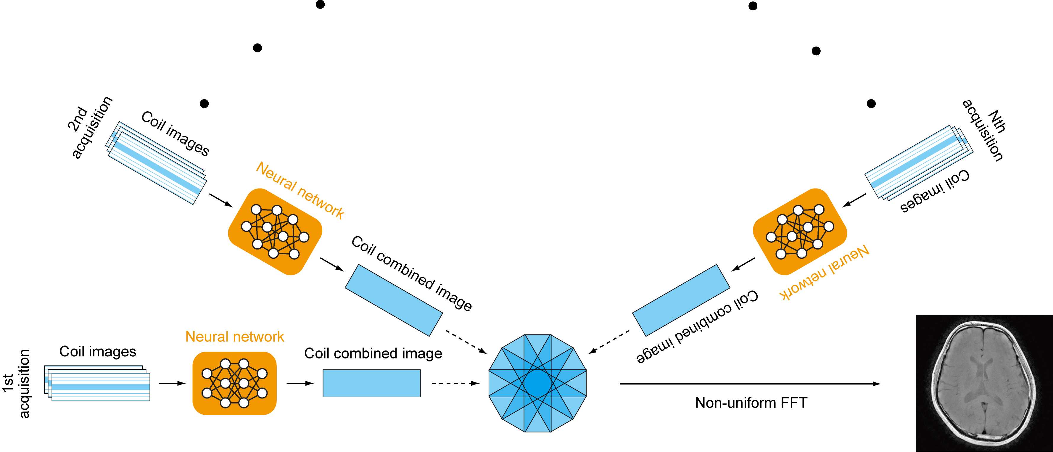
The whole reconstruction process of the proposed PROPELLER reconstruction. Deep learning based parallel imaging (PI) reconstruction can achieve a higher PI factor than the standard SENSE, allowing shorter scan times.
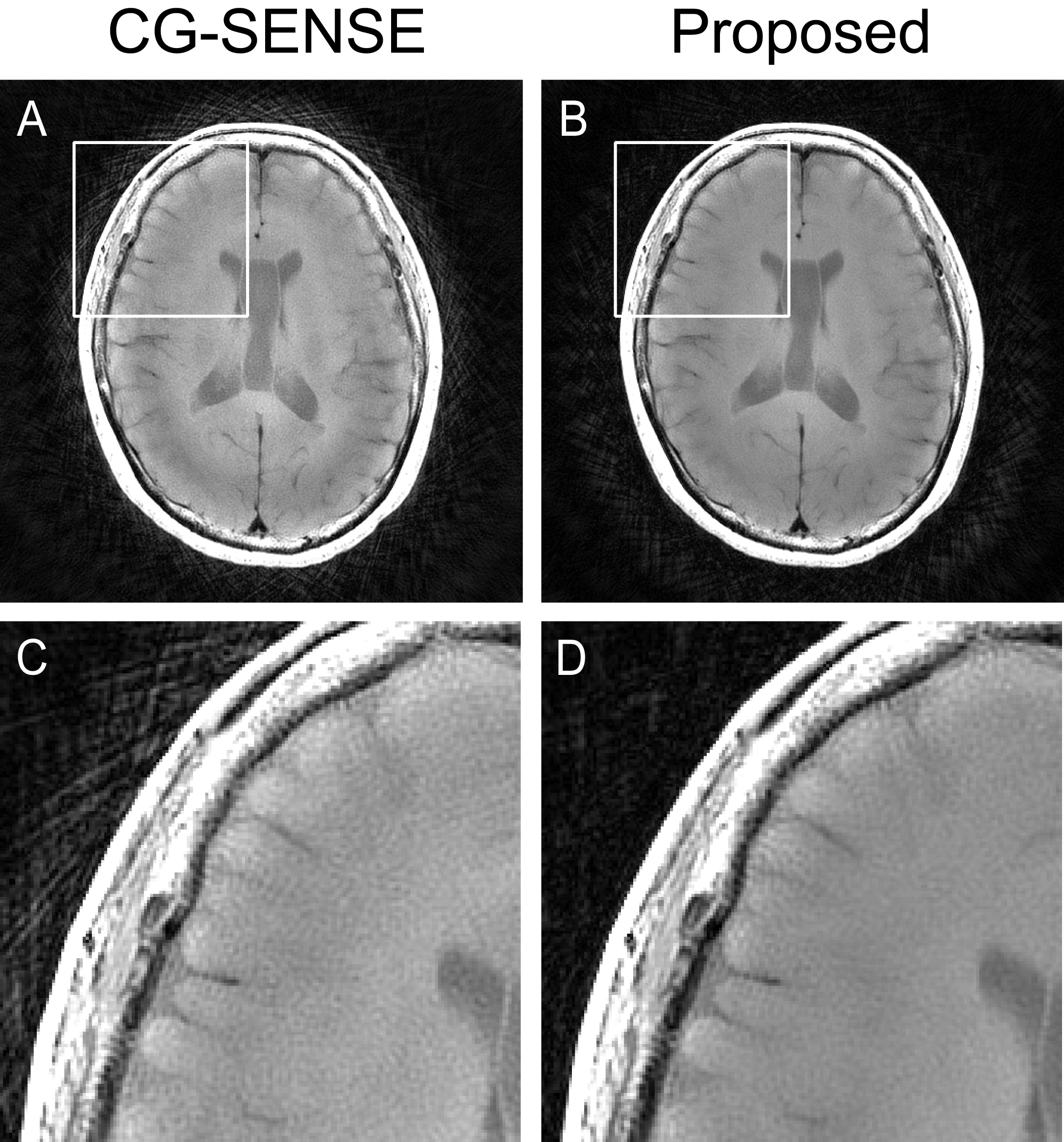
Reconstructed images from prospectively undersampled data (PI factor = 7) with CG-SENSE and the proposed method. Compared with CG-SENSE, the proposed method reduced streak artifacts and the noises in the brain. (A) CG-SENSE, (B) the proposed method. (C), (D) Magnified images of the solid boxes in (A), (B), respectively.
DOI: https://doi.org/10.58530/2022/3161