3148
Preliminary Application of Magnetization Transfer Imaging in EndometrialCarcinoma and Cervical Cancer1the First People’s Hospital of Yunnan Province, Kunming, China, 2Siemens Healthineers, Shanghai, China
Synopsis
Magnetization transfer imaging (MTI) is sensitive to reveal macromolecules in tumor tissues. This study aimed to evaluate the correlation between magnetization transfer ratio (MTR) and histological features and risk stratification in endometrial carcinoma and cervical cancer. We found that MTR value could differentiate endometrial carcinoma from cervical cancer and facilitate preoperative risk stratification of endometrial carcinoma. It could provide a preoperative basis for cancerous histologic origin and risk assessment in uterus.
Background and Objective
Magnetization transfer imaging (MTI) can indirectly reflect the content of macromolecular substances in biological tissues by quantitatively measuring magnetization transfer ratio (MTR) value1. This technique has already been well-applied in the analysis of glioma histological grade2. Endometrial carcinoma and cervical cancer are common malignant tumors of the female reproductive system, and the age of onset is gradually getting younger3. It is important to evaluate their histological features and risk stratification before operation for the individualized treatment and prognosis. The purpose of this study was to evaluate the value of magnetization transfer imaging in distinguishing histological features and preoperative risk stratification of endometrial carcinoma and cervical cancer.Methods
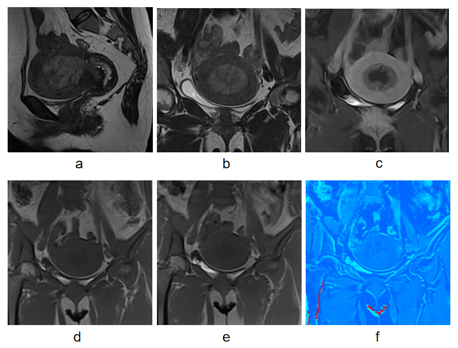
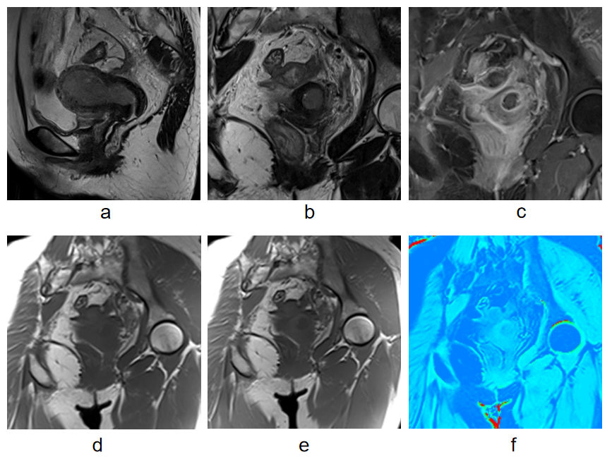
This prospective study enrolled 30 normal volunteers and 93 patients with suspicious uterine malignant tumors in our hospital. Pelvic MRI was performed on a 3.0T MRI scanner (Magnetom Prisma, Siemens Healthineers, Erlangen, Germany). A 3D fast low angle shot (FLASH) sequence was used to acquire MTI data, including two scan with (MTon) and without (MToff) MT pulse respectively. The main parameters for MTI were as follows: repetition time/echo time: 222 ms/2.35 ms; slice thickness/slice gap: 4 mm/4.8 mm; field of view: 380×380 mm2; matrix size: 256×208, and flip angle: 70°. For MT quantification, the MTR map was calculated using the following formula: MTR=(MToff-MTon)×100/MToff. MTR value was measured on the MTR map using the region of interest method. MTR values were compared among different uterine lesions.Results
A total of 90 patients (30 endometrial carcinoma, 47 cervical cancer, and 13 endometrial hyperplasia according to histopathological results) were included for the MT image quality evaluation. MTR value of endometrial carcinoma was 15.72±6.99, which was significantly lower than that of cervical cancer (22.18±10.04) (P=0.003). There were no difference among endometrial carcinoma, endometrial hyperplasia (17.81±10.13), and normal endometrium (15.62±9.52) (all P>0.05), also no difference between cervical cancer and normal cervix (P>0.05). MTR value of low-risk endometrial carcinoma (13.29±6.22) was apparently lower than that of non-low-risk endometrial carcinoma (18.49±6.98) (P=0.040). However, there were no remarkable correlation between MTR values and the different pathological grades or histological subtypes of endometrial carcinoma or cervical cancer (all P>0.05).Conclusions
The difference of macromolecular density between endometrial carcinoma and cervical cancer could be indirectly observed by measuring magnetization transfer ratio. Furthermore, MTR value could identify low-risk endometrial carcinoma.Acknowledgements
No acknowledgement found.References
1. Arlinghaus LR, Dortch RD, Whisenant JG, et al. Quantitative magnetization transfer imaging of the breast at 3.0T: reproducibility in healthy volunteers. Tomography 2016;2: 260-266.
2. Su C, Jiang J, Liu C, et al. Comparison of amide proton transfer imaging and magnetization transfer imaging in revealing glioma grades and proliferative activities: a histogram analysis. Neuroradiology 2021;63(5):685-693.
3. Siegel RL, Miller KD, Jemal A. Cancer statistics, 2019. CA Cancer J Clin 2019;69(1):7-34.
Figures