3147
Real-time MRI for assessment of patients with esophageal hiatal hernia: a feasibility research1Tianjin Medical University General Hospital, Tianjin, China, 2Siemens Healthcare, Beijing, China
Synopsis
The purpose of this study was to observe the swallowing process of hiatal hernia patients using real-time magnetic resonance imaging (MRI) and to evaluate the transport of contrast agents through the gastroesophageal junction and the induction of sliding hernia during Valsalva. Results showed that hiatal hernia was detected in all patients enrolled in the study. Therefore, real-time MRI imaging of sliding hiatal hernia has great potential for clinical application.
Introduction
Hiatal hernia is a condition in which part of the stomach enters the chest cavity through the diaphragmatic esophageal hiatus. Hiatal hernia reduces the tension of the lower esophageal sphincter, thereby losing the clamp effect that prevents acid reflux. It also acts as an acid reservoir, allowing gastric fluid to enter the esophagus, resulting in long-term exposure of the esophagus to acid, leading to gastroesophageal reflux. The disease can be divided into four types, of which Type I is also called sliding hiatal hernia, which is characterized by diaphragmatic hiatus enlargement and diaphragmatic esophageal membrane relaxation, accounting for 95% of cases 1. Gastroesophageal reflux disease (GERD) exists in most patients with sliding hiatal hernia 2. Gastroesophageal reflux symptoms include heartburn, acid reflux, belching, etc., especially sliding hiatal hernia 3. When the hernia sac compresses the heart, lungs and mediastinum, symptoms such as shortness of breath, palpitations, cough and cyanosis may occur. At present, there are three ways to diagnose hiatal hernia: CT, gastroscopy, and X-ray. However, they have limited resolution, radiation, or invasive problems, which need to be improved. MRI has potential advantages in hiatal hernia detection due to its non-invasive and high resolution. The aim of this work is to evaluate potential diagnostic value of real-time MRI for assessment of patients with esophageal hiatal hernia.Materials and methods
The study was conducted in accordance to the Declaration of Helsinki in its most recent version and received prior approval by the local ethics board. All participants gave written informed consent before each examination. All subjects were placed in supine position. All data were collected on a MAGNETOM Prisma 3T MR scanner (Siemens Healthineers, Erlangen, Germany) through an 18-element abdominal coil array. The parameters are as follows: TrueFISP: TR=741 ms, TE=2 ms, flip angle=45°, 3 slices, slice thickness=2.5 mm, distance factor=-30%, FOV=430×430mm2, measurements=10,time resolution=500ms. The TrueFISP sequence is oriented along the esophageal hiatus, and all subjects were instructed to swallow (contrast agent) during the scan. Valsalva was performed to induce sliding hiatal hernia in patients.Results
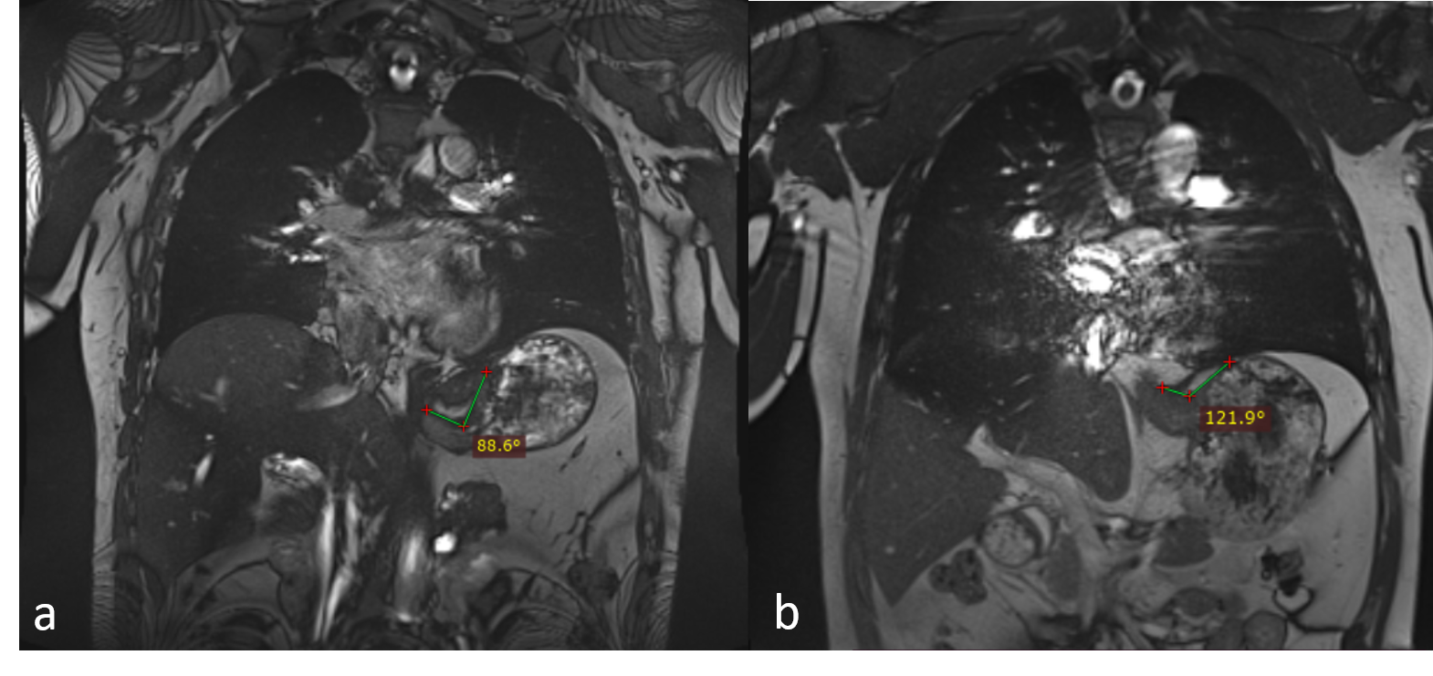
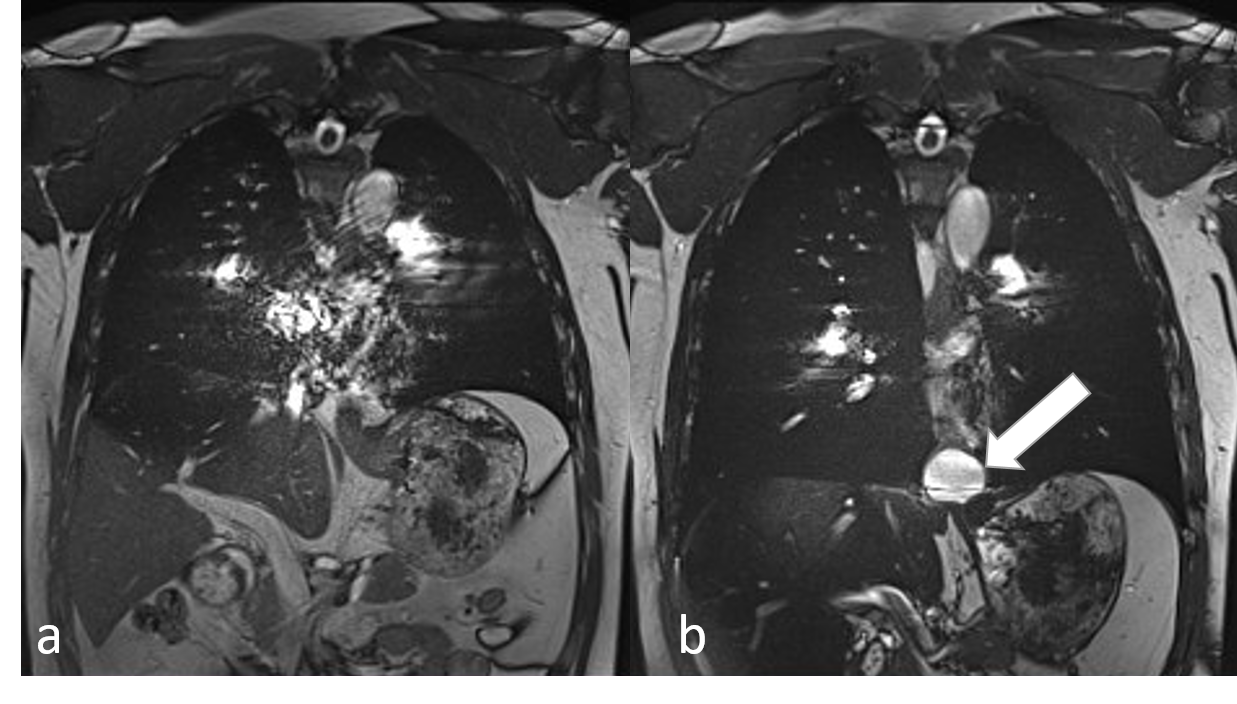
Real-time MRI techniques offered dynamic visualization of arbitrary physiologic processes during swallow and Valsalva. Gastroesophageal swallowing and hiatal hernia induction were observed in 6 volunteers and 6 patients via the TurefISP sequence. The images were evaluated by two experienced image-diagnostic physicians. Gastroesophageal reflux and hiatal hernia were detected in all patients. In a preceding application to a small series of subjects, this technique was able to identify gastroesophageal reflux by anatomical and functional visualization of the gastroesophageal junction, especially the His angle. Fig.1 showed measurements of his angle in normal volunteers (a) and patients (b) with hiatal hernia. His angle was 88.6° and 121.9° respectively. His angle was considered to be the angle between the medial border of the distal esophagus and the fundus margin bordering to the esophagus. Hernia sac was observed above the esophageal hiatus in 3 patients after Valsalva. The other 3 patients had different degrees of gastric fundus, mesangial vessels, or fatty herniation into the thorax. Fig.2 demonstrated a hernia sac in a patient with sliding hiatal hernia induced by Valsalva. TrueFISP has a high SNR and contrast ratio and no signal loss, so it is suitable for dynamic signals.Discussion and Conclusions
Real-time MRI is a new technique to evaluate the gastroesophageal junction. It can show his Angle and sliding hernia induction in patients with hiatal hernia. Therefore, real-time MRI imaging of hiatal hernia has great potential in clinical application.Acknowledgements
No acknowledgement found.References
1. Hill LD, Kozarek RA, Kraemer SJ, Aye RW, Mercer CD, Low DE, et al. The esophageal flap valve; in vitro and in vivo observations. Gastrointest Endosc. 1996, 44: 541–547.
2. Gordon C, Kang JY, Neild PJ, Maxwell JD. The role of the hiatus hernia in gastro-oesophageal reflux disease. Aliment Pharmacol Ther. 2004, 20: 719–732.
3. Shuo Zhang, Arun A. Joseph, Lisa Gross. Diagnosis of Gastroesophageal Reflux Disease Using Real-time Magnetic Resonance Imaging. Sci Rep. 2015, 15(5):12112.
Figures