3121
The value of multi-parametric magnetic resonance imaging in evaluating renal fibrosis1Diagnostic Radiology, Wuxi People's Hospital Affiliated to Nanjing Medical University, Wuxi, China, 2Diagnostic Radiology, The Affiliated Wuxi Children's Hospital of Nanjing Medical University, Wuxi, China, 3Nephrology, Wuxi People's Hospital Affiliated to Nanjing Medical University, Wuxi, China, 4Human Anatomy, Medical School, Southeast University, Nanjing, China
Synopsis
Chronic kidney disease (CKD) is chronic progressive renal parenchymal damage caused by multiple factors. Evaluation of the degree of renal interstitial fibrosis (IF) is of great importance in treatment and prognosis prediction. Herein, we use multi-parametric magnetic resonance imaging (mpMRI) to assess renal IF noninvasively with intention to identify the optimal MRI biomarkers for clinical application. Our results show that multi-parameter prediction model using cortical longitudinal relaxation time (cT1) and cortical true diffusion coefficient (cDt) can effectively assess the degree of renal IF and support clinical diagnosis, treatment strategy and risk stratification in CKD patients.
Introduction
CKD is a disease characterized by chronic progressive renal parenchymal damage and gradual loss of renal function. The histological manifestation is mainly renal interstitial fibrosis (IF). Serum creatinine (SCr) is not sensitive to early kidney damage and IF detection, although it is a common clinical parameter for assessing CKD1. Recent studies have shown that the increase in the degree of IF is more earlier than the decline in renal function 2. The early and accurate assessment of IF can guide treatment and assess the prognosis3. In recent years, the functional MRI has shown great potential in non-invasive evaluation. Studies have shown that single-parametric MRI can evaluate IF, but the sensitivity and specificity need to be further improved. While the combination of multiple MRI biomarkers can help to explain the pathophysiological effects and improve the diagnostic efficacy of renal IF4. Therefore, this study aims to use mpMRI to assess IF and find the optimal MRI biomarkers for clinical application.Methods
We prospectively recruited 28 CKD patients who were willing to accept renal biopsy and 12 healthy volunteers. All subjects underwent mpMRI examination at Siemens PRISMA 3.0T. Protocol sequence includes DWI (TR/TE: 4500ms/61ms; Voxel size: 1.6 ×1.6 × 5.0mm; matrix: 104×128; slice-thickness: 5mm; FOV: 325×400mm; b value: 50, 800), IVIM (TR/TE: 6000ms/63ms; Voxel size: 2.1 ×2.1×4.0mm; matrix: 66×120; slice-thickness: 4mm; FOV: 137×249mm; b value: 0, 10, 20, 40, 100, 150, 200, 500, 800), T1mapping (TR/TE: 5.01ms/2.3ms; Voxel size: 0.8×0.8×4.0mm; matrix: 135×224; slice-thickness: 4mm; FOV: 305×379mm; flip angle: 3 and 15 deg) and R2*mapping (TR/TE: 293ms/2.46 ms; Voxel size: 1.5×1.5×5.0mm; matrix: 154×256; slice-thickness: 5mm; FOV: 285×380mm). Two experienced radiologists used the sygno.via workstation of Siemens and measured the apparent diffusion coefficient (ADC) value, longitudinal relaxation time(T1) value and R2* value of the renal cortex and medulla. The true diffusion coefficient (Dt) value, pseudo diffusion coefficient (Dp) value and perfusion fraction (Fp) value of the IVIM sequence were measured by FireVoxel software. All patients obtained the pathological results of kidney biopsy within 2 days after MRI and were divided into IF mild group and IF moderate-severe group according to Banff 2015 renal fibrosis scoring standard. The estimated glomerular rate filtration (eGFR) value and 24 hours Urine protein value (24 h-UPRO) were recorded. One-way analysis of variance was used to compare the MRI biomarkers between different IF groups and control group. Partial correlation analysis was used to analyze the correlation between MRI parameters and eGFR. Receiver operating characteristic (ROC) curves was used to evaluate the diagnostic efficacy of MRI parameters for renal IF. Statistical analysis was performed using SPSS 17.0, and P<0.05 was considered statistically significant.Results
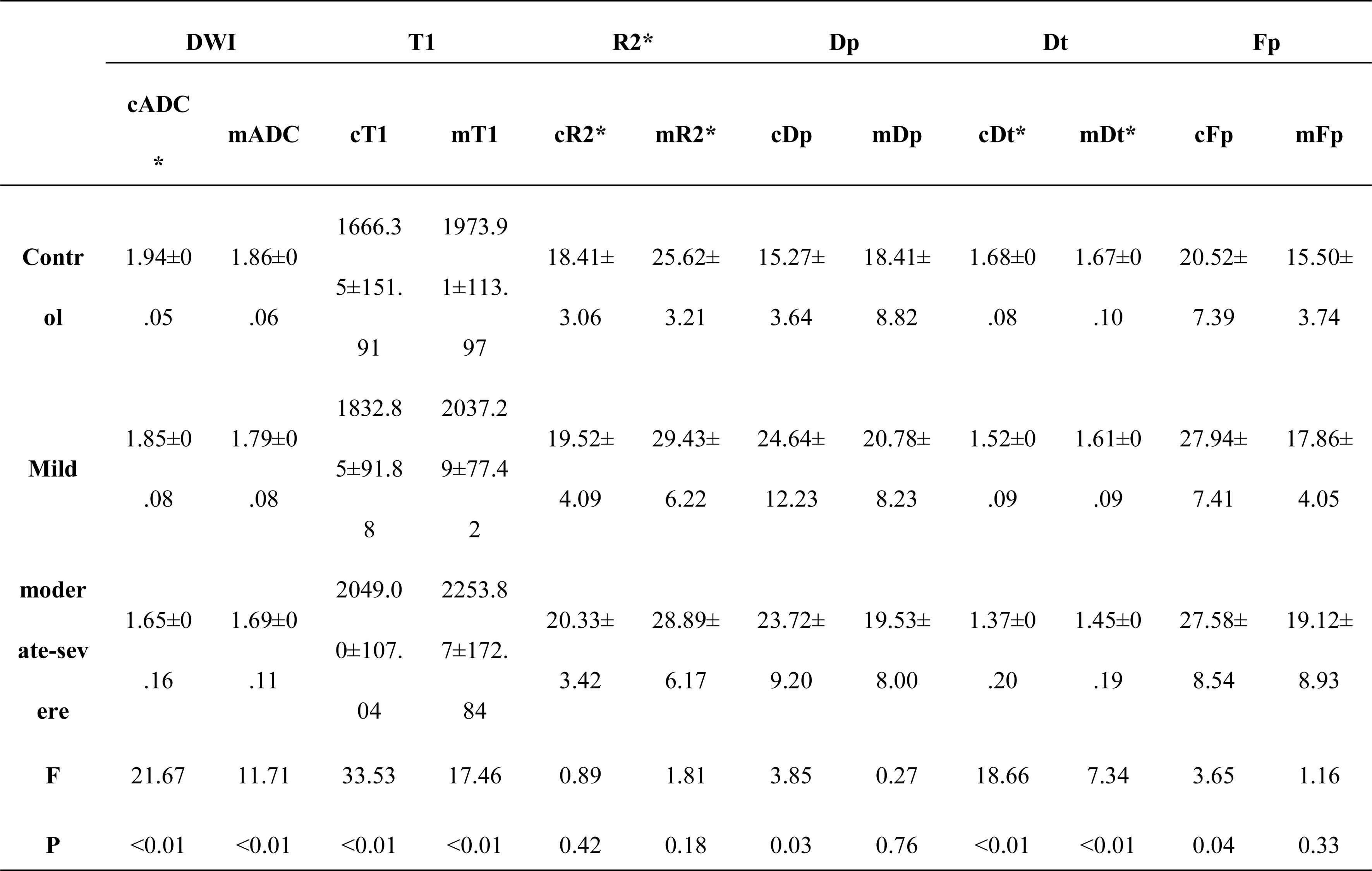
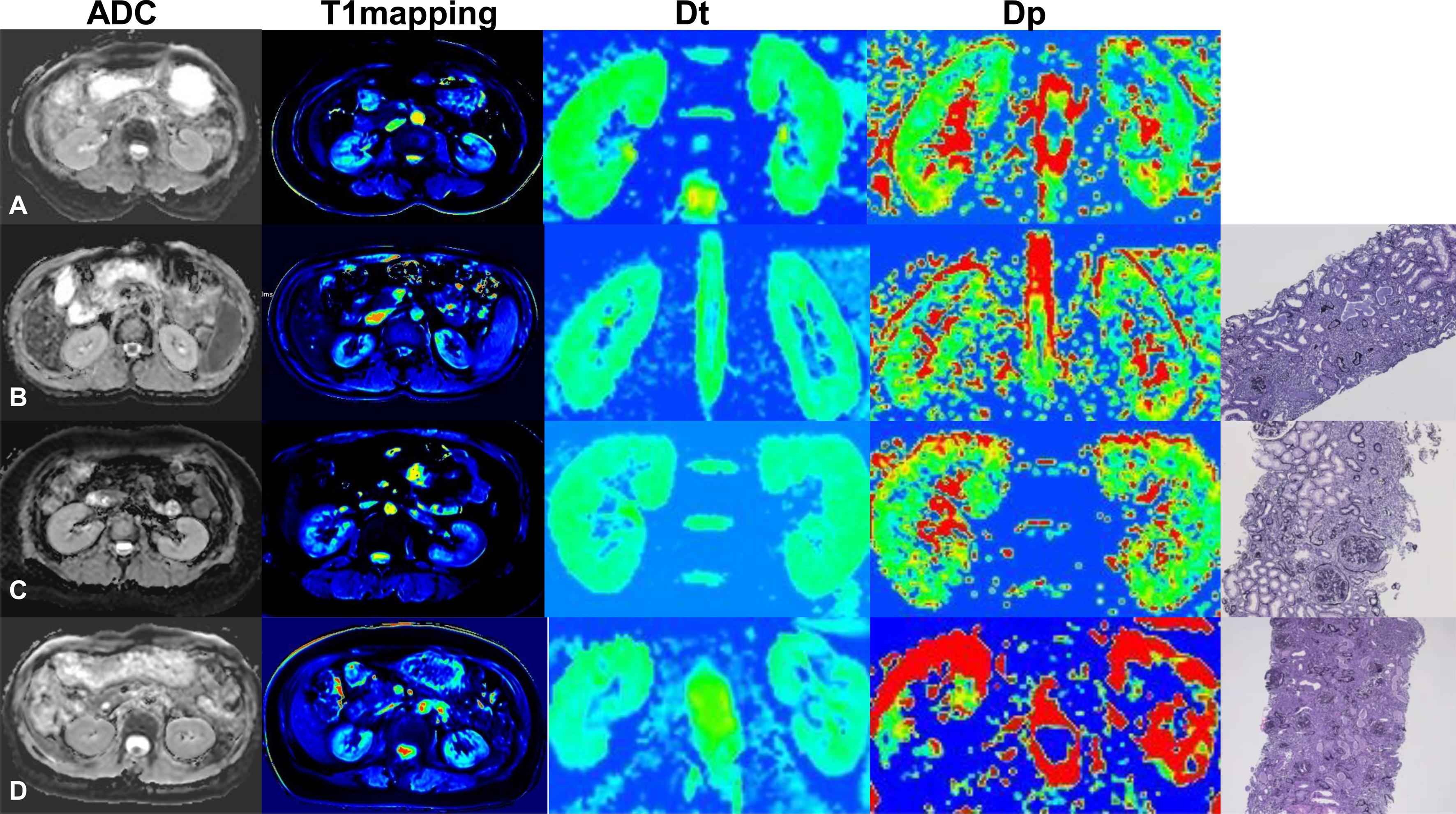
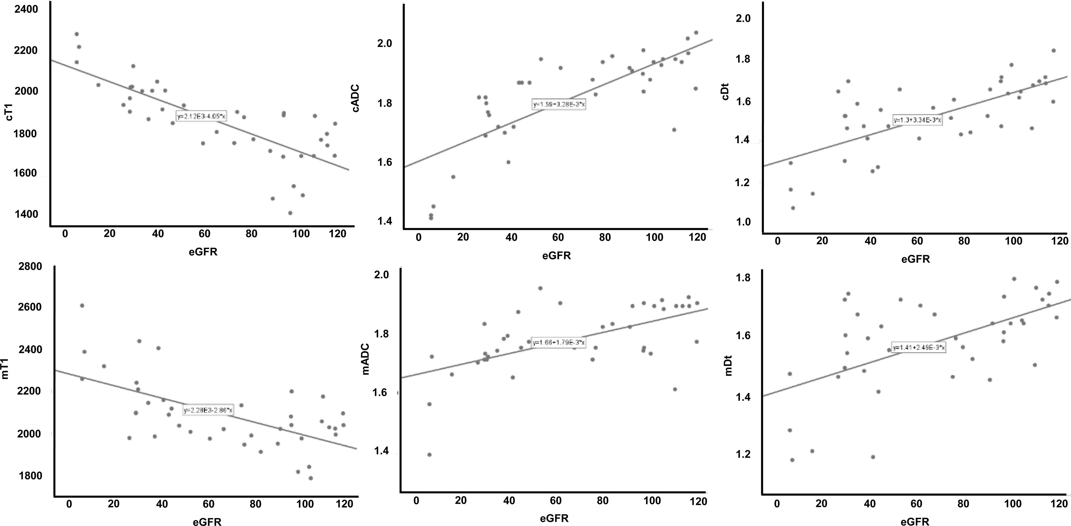
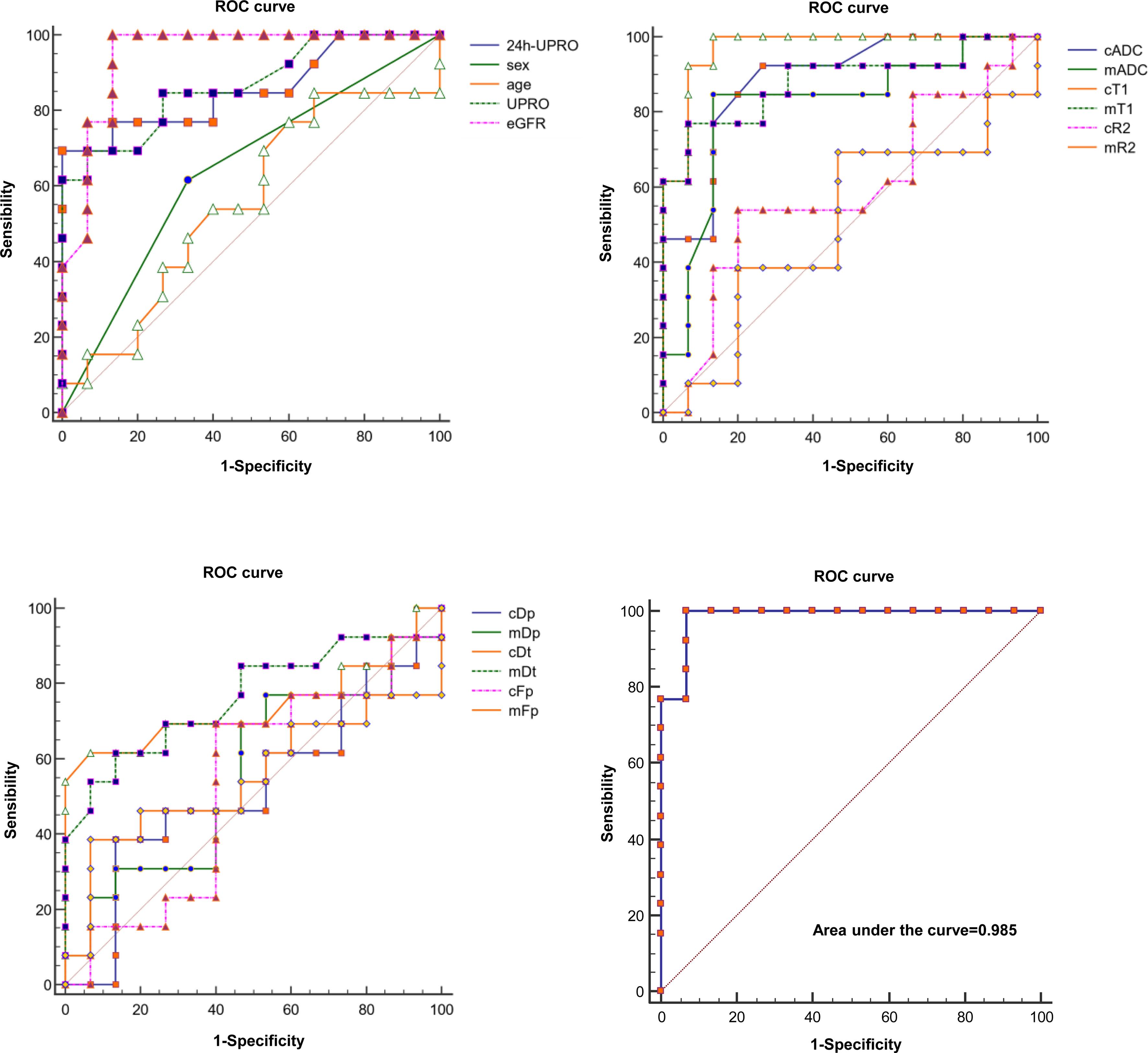
Cortical ADC (cADC), medullary ADC (mADC), cortical T1 value (cT1), medullary T1 value (mT1), cortical true diffusion coefficient (cDt) and medullary true diffusion coefficient (mDt) show significant difference among groups (P<0.01) (Fig.1-2). cADC, mADC, cDt and mDt are positively correlated with renal function eGFR. On the contrary, cT1 and mT1 are negatively correlated with renal function eGFR (Fig. 3). Furthermore, cT1 has the strong correlation with eGFR with the correlation coefficient r values of -0.81(P<0.05). The AUC values of cADC, mADC, cT1, mT1, cDt, and mDt to predict the degree of IF are 0.89, 0.82, 0.97, 0.88, 0.74, 0.76 respectively. Logistic regression analysis shows that the combination of cT1 and cDt as multi-parameter prediction model has the highest sensitivity (100%) and specificity (93.33%) for distinguishing IF with the AUC value of 0.985, which can effectively predict the degree of renal IF (Fig. 4).Discussion
This study identify that the MRI parameters cADC, mADC, cDt, mDt, cT1, and mT1 are different among groups. They are highly correlated with the renal function eGFR. The difference in MRI biomarkers reflects the progress of renal fibrosis and the pathological process of collagen fiber deposition and tissue edema5. Furthermore, our research suggest that the combination of cT1 and cDt as multi-parameter prediction model can effectively assess the degree of renal IF and support clinical diagnosis, treatment strategy and risk stratification in CKD patients.Conclusion
The combination of cT1 and cDt as biomarker has potential clinical applicability in non-invasive assessment of renal IF.Acknowledgements
This work was supported by the General Project of Shaanxi Natural Science Basic Research Plan (2021JM-558); General Cultivation Project of Xi'an Health Commission (2021ms14); Jiangsu Provincial Medical Innovation Team (CXTDB2017016).References
1. Ghazal Quinn, Amin Abedini, Hongbo Liu, et al.Renal Histologic Analysis Provides Complementary Information to Kidney Function Measurement for Patients with Early Diabetic or Hypertensive Disease. JASN August 2021, ASN.2021010044
2. Lena Berchtold, Lindsey A. Crowe, Iris Friedli, et al.Diffusion magnetic resonance imaging detects an increase in interstitial fibrosis earlier than the decline of renal function. Nephrol Dial Transplant (2020) 35: 1274–1276
3. Le Hir M, Hegyi I, Cueni-Loffing D, et al..Characterization of renal interstitial fibroblast-specific protein 1/S100A4-positive cells in healthy and inflamed rodent kidneys. Histochem Cell Biol 123: 335–346, 2005.
4. Nicholas M. Selby, Peter J. Blankestijn, Peter Boor, et al. Magnetic esonance imaging biomarkers for chronic kidney disease: a position paper from the European Cooperation in Science and Technology Action PARENCHIMA. Nephrol Dial Transplant (2018) 33: ii4–ii14
5. Octavia Bane, Stefanie Hectors, Sonja Gordic, et al.Multiparametric magnetic resonance imaging shows promising results to assess renal transplant dysfunction with fibrosis. Kidney Int. 2020 February ; 97(2): 414–420.
Figures