3101
Preliminary assessment of 3D APT-weighted combined with diffusion weighted imaging in characterization of bone and soft tissue tumors1The First Affiliated Hospital of Zhengzhou University, Zhengzhou, China, 2Philips Healthcare, Beijing, 100102, China, Beijing, China
Synopsis
Amide proton transfer weighted imaging (APTWI) is a noninvasive emerging molecular MRI technique based on chemical exchange saturation transfer (CEST) between amide protons of proteins and polypeptides and free water protons. This work investigated and evaluated the ability of APT parameter in distinguishing benign from malignant bone and soft tissue tumors. Results indicated that APTWI combined with diffusion weighted imaging showed a significantly improved differentiation between benign and malignant tumors.
Introduction
Although bone and soft-tissue tumors are rare, early diagnosis and prompt referral to a specialized unit offers the best chance of a successful outcome both in terms of survival and surgical resection. Previous studies have shown that the amide proton transfer weighted imaging (APTWI) provides a new method to evaluate grading gliomas and distinguishing benign from malignant tumors [1]. However, APTWI is still limited evaluated in clinical practice and few analyses have been performed in distinguishing benign from malignant bone and soft tissue tumors. The present study aims to evaluate and compared the diagnostic performance of APTWI and the conventional diffusion weighted imaging (DWI) in differentiation between benign and malignant tumors.Methods
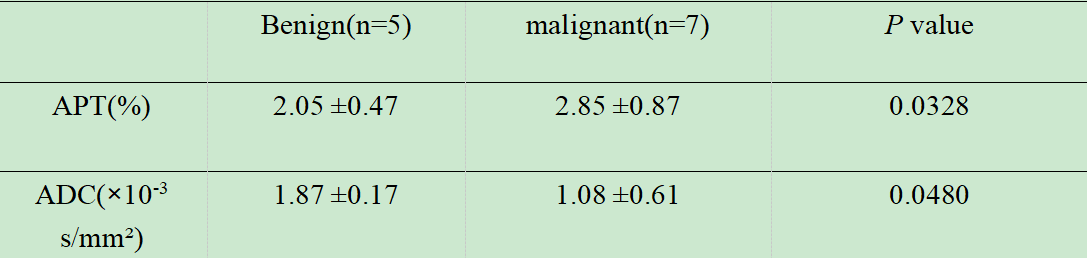
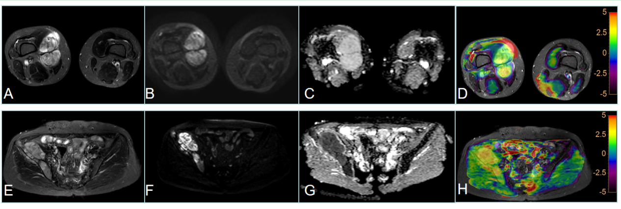
Twelve patients (8 males and 4 females, age ranged from 13 to 70) with soft tissue or bone tumors (all have been diagnosed according to pathological biopsy) underwent MR scans with a 3T MR scanner (Ingenia CX, Philips Healthcare, Best, the Netherlands) in this work. According to the WHO classification of tumors of soft tissue and bone (2020) criteria, 12 patients were divided into two groups:5 for benign tumors and 7 for malignancies. All patients were scanned by MR APTWI and DWI sequence. APT imaging was acquired using the 3D turbo spin echo sequence, and performed using four interleaved saturation pulses with a total duration of 2.0 s and a saturation power (B1) level of 2.0 μT. The other parameters were: TR/TE = 6540/8.3 ms; FOV = 230×181 mm; scan matrix = 116×90; voxel size = 2×2×5 mm3; 9 slices; TSE factor = 174; acquisition time = 6 min 0 sec. The axial DWI sequence (TR/TE=3700/85ms and NSA=4) was taken with b values of 0 and 800 s/mm², and the corresponding apparent diffusion coefficient (apparent diffusion coefficient, the ADC) map was calculated. ADC and APT values were measured by drawing ROIs (Region of Interest) within the periphery and center of the lesions, and get the average respectively. And the measured parameters in benign and malignant lesions were compared by using Mann-Whitney U test with SPSS software (version 20.0). A P value of less than 0.05 was considered statistically significant. Furtherly, the receiver operating characteristic (ROC) curve was used to access the diagnostic performance of APT and ADC values in differentiation between benign and malignant tumors.Results
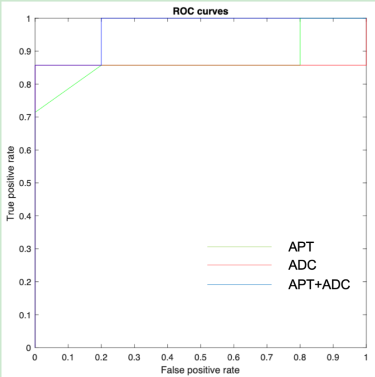
Both the APT and ADC values show significant differences between benign and malignant lesions (Table 1). APT values were significantly higher, whereas ADC values were lower in malignant tumors. The ROC analyses show that both APT and ADC values performed well in the differentiation between malignant and benign tumors, with the area under the ROC curves (AUCs) of 0.871 and 0.857, respectively. The combination of APT and ADC values could further improve the diagnostic performance with an AUC of 0.971, but without significant difference to those by APT or ADC alone.Discussion
In our study, we first observed that APT signal intensities were significantly higher for malignant tumors than benign tumors. APT imaging is a novel molecular MRI method and provides information predominantly based on the amide protons in cellular proteins and peptides in the intracellular space and extracellular space [1]. Higher peptide or protein concentration may result from higher cell density in malignant tumors. Previous studies have demonstrated that APT signal intensity was positively correlated with the tumor grade, such as gliomas [2]. Similarly, our results showed higher APT signal intensity in malignant tumors than benign tumors, indicating different mobile protein and peptide concentrations in the tumor according to the grade malignancy. On the other hand, we also observed that lower ADC intensities in malignant tumors compared with benign tumors. The literature contains many studies that have evaluated the role of DWI in musculoskeletal diseases. It was consistent with pervious published paper that ADC was correlated with tumor grade and reflect tumor cellularity and water content in interstitial space [3]. The combination of APT and ADC values may give a more comprehensive characterization of tumor tissues, thus showing an improved diagnostic performance in this study.Conclusions
APTWI combined with DWI is shown to be valuable in differentiation between malignant and benign bone and soft tissue tumors, which can provide useful information on changes of diffusion properties and cellular proteins and peptides in tissues.Acknowledgements
No acknowledgement found.References
1.Togao O, Hiwatashi A, Yamashita K, Kikuchi K, Keupp J, Yoshimoto K, et al.Grading diffuse gliomas without intense contrast enhancement by amide proton transfer MR imaging: comparisons with diffusion- and perfusionweighted imaging. Eur Radiol. 2017;27(2):578–88.
2.Togao O, Yoshiura T, Keupp J, Hiwatashi A, Yamashita K, Kikuchi K, et al. Amide proton transfer imaging of adult diffuse gliomas: correlation with histopathological grades. Neuro Oncol. 2014;16(3):441–8.
3. Dallaudière B, Lecouvet F, Vande Berg B, Omoumi P, Perlepe V, Cerny M, et al. Diffusion-weighted MR imaging in musculoskeletal diseases: current concepts. Diagn Interv Imaging. 2015;96(4):327-40.
Figures