3095
Shoulder PROPELLER MRI with Deep learning-based reconstruction: image quality and agreement between standard and accelerated sequence1Radiology, Haeundae Paik Hospital, Inje University College of Medicine, Busan, Korea, Republic of, 2GE Healthcare, Seoul, Korea, Republic of, 3GE Healthcare, Houston, TX, United States, 4GE Healthcare, New York, NY, United States
Synopsis
Although the shorter scan time enabled by DLRecon has potential to reduce motion related image degradation, it cannot remove or mitigate such artifacts. The periodically rotated overlapping parallel lines with enhanced reconstruction (PROPELLER) or similar techniques can reduce motion artifacts in shoulder MRI, however usually takes a longer time to acquire. Recently, DLRecon for PROPELLER has been developed. Accelerated PROPELLER sequence with DLRecon showed improved image quality and comparable interobserver agreement for shoulder pathology compared to standard PROPELLER acquisition with conventional reconstruction, despite the shorter scan time (45% reduction).
Introduction
Shoulder MRI is susceptible to image quality degradation from patient’s motion, including respiration. Cartesian acquisition-based accelerated shoulder MRI sequences with deep learning-based reconstruction (DLRecon) reported comparable diagnostic performance without sacrificing image quality while decreasing scan time 1. Although the shorter scan time enabled by DLRecon has the potential to reduce motion-related image degradation, it cannot remove or mitigate such artifacts 2. The periodically rotated overlapping parallel lines with enhanced reconstruction (PROPELLER) or similar techniques can reduce motion artifacts in shoulder MRI, however usually takes a longer time to acquire 3-5. Recently, DLRecon for PROPELLER has been developed. In this study, we aim to investigate the image quality and agreement for anatomic evaluation of shoulder among standard and accelerated PROPELLER sequence reconstructed with conventional and DLRecon methods 6-8.Methods
Between March 2021 and May 2021, thirty examinations of twenty-nine patients who underwent shoulder MRI. Scans were acquired using a 3T MRI scanner (SIGNATM Architect, GE Healthcare, Waukesha, USA) with 2 schemes of axial intermediate weighted (IW) fat suppressed (FS), coronal and sagittal T2 weighted (T2-W) FS images, conventional and accelerated, were included in this study. The typical image parameters are specified in Table 1.Two sets of images for each scheme were generated: one with conventional reconstruction and the other with a 2D DLRecon algorithm, with denoising and sharpening properties 2, compatible with PROPELLER. The network offered tunable noise reduction levels (25, 50 and 75%; higher levels indicated higher levels of denoising). A factor of 75% was chosen for this study.
Two musculoskeletal radiologists with 6 and 7 years of subspecialty experience independently assessed the pathology of the subscapularis tendon (SSC), supraspinatus-infraspinatus tendon (SIT), and glenoid labrum (GL) in shoulders without a history of a previous operation (n=20). Inter-observer and inter-image set agreement for the evaluated pathologies was assessed using weighted kappa statistics.
For quantitative analysis, the representative slices were chosen from axial (Figure 1A), coronal (Figure 1B), and sagittal (Figure 1C) images, respectively. Signal to noise levels was calculated by dividing the mean signal of the slice by the estimated standard deviation of the noise. The noise levels were estimated by a hybrid Discrete Wavelet Transform (DWT) and edge information removal based algorithm which is described in detail, elsewhere 9.
Results
Table 2 shows the mean signal-to-noise ratio (SNR) for four image sets in all three planes. The mean SNR was worst in accelerated sequence with conventional reconstruction (mean SNR of axial, 22.447±2.929; mean SNR of coronal, 11.644±1.379; mean SNR of sagittal, 15.495±3.904) and best in standard sequence with DLRecon (mean SNR of axial, 64.948±10.004; mean SNR of coronal, 66.094±8.591; mean SNR of sagittal, 61.087±10.042). There was a statistically significant difference among all four image sets (p<.001). The SNR of accelerated sequence with DLRecon (mean SNR of axial, 52.754±6.832; mean SNR of coronal, 36.656±3.872; mean SNR of sagittal, 39.725±7.887) was even better than standard sequence with conventional reconstruction (p<.001 for the axial and coronal plane, p=0.008 for the sagittal plane) with post-hoc analysis.The inter-observer agreement was almost perfect for SIT (k=0.894) and substantial for SSC (k=0.785) and GL (k=0.726) in all four image sets.
Discussion
Accelerated sequences had 45% lower scan time than standard sequences and showed worse SNR compared to standard sequences. The use of DLRecon with both standard and accelerated sequences result in improved image quality compared to those with conventional reconstruction pipeline. Also, the image quality of the accelerated sequence with DLRecon was comparable to the standard sequence.With the use of accelerated PROPELLER sequence, the chance of shoulder MRI suffering from motion artifacts would be much decreased without trading-off with image quality.
Conclusion
DLRecon can improve the image quality of PROPELLER sequence for the shoulder. Accelerated PROPELLER sequence with DLRecon showed better image quality and comparable interobserver agreement for shoulder pathologies compared to standard PROPELLER acquisition with conventional reconstruction, despite the shorter scan time.Acknowledgements
No acknowledgement found.References
1. Hahn, Seok, et al. "Image Quality and Diagnostic Performance of Accelerated Shoulder MRI With Deep Learning-Based Reconstruction." American Journal of Roentgenology (2021)
2. Lebel RM. Performance characterization of a novel deep learning-based MR image reconstruction pipeline. arXiv preprint arXiv:200806559 2020)
3. Dietrich, Tobias J., et al. "PROPELLER technique to improve image quality of MRI of the shoulder." American Journal of Roentgenology 197.6 (2011): W1093-W1100.
4. Nagatomo, Kazuya, et al. "Efficacy of periodically rotated overlapping parallel lines with enhanced reconstruction (PROPELLER) for shoulder magnetic resonance (MR) imaging." European journal of radiology 85.10 (2016): 1735-1743.
5. Lavdas, E., et al. "Elimination of motion and pulsation artifacts using BLADE sequences in shoulder MR imaging." Skeletal radiology 44.11 (2015): 1619-1626.
6. Xinzeng Wang, Daniel Litwiller, Marc Lebel, Ali Ersoz, Lloyd Estkowski, Jason Stafford, and Ersin Bayram. High Resolution T2W imaging using Deep Learning Reconstruction and Reduced Field-of-View PROPELLERProc. Intl. Soc. Mag. Reson. Med. 28 (2020) 0820
7. Mohammed Saleh, Sanaz Javadi, Manoj Mathew, Jong Bum Son, Jia Sun, Ersin Bayram, Xinzeng Wang, Jingfei Ma, Janio Szklaruk, and Priya Bhosale. Proc. Intl. Soc. Mag. Reson. Med. 29 (2021) 3000
8. Ali Pirasteh, Lloyd Estkowski, Daniel Litwiller, Ersin Bayram, and Xinzeng Wang. Motion Robust High-Resolution Pelvic Imaging using PROPELLER and Deep Learning ReconstructionProc. Intl. Soc. Mag. Reson. Med. 29 (2021) 2998
9. Pimpalkhute, VA, Page R, Kothari A, Bhurchandi KM, Kamble VM. Digital Image Noise Estimation Using DWT Coefficients. IEEE Transactions on Image Processing 2021;30:1962-1972.
Figures
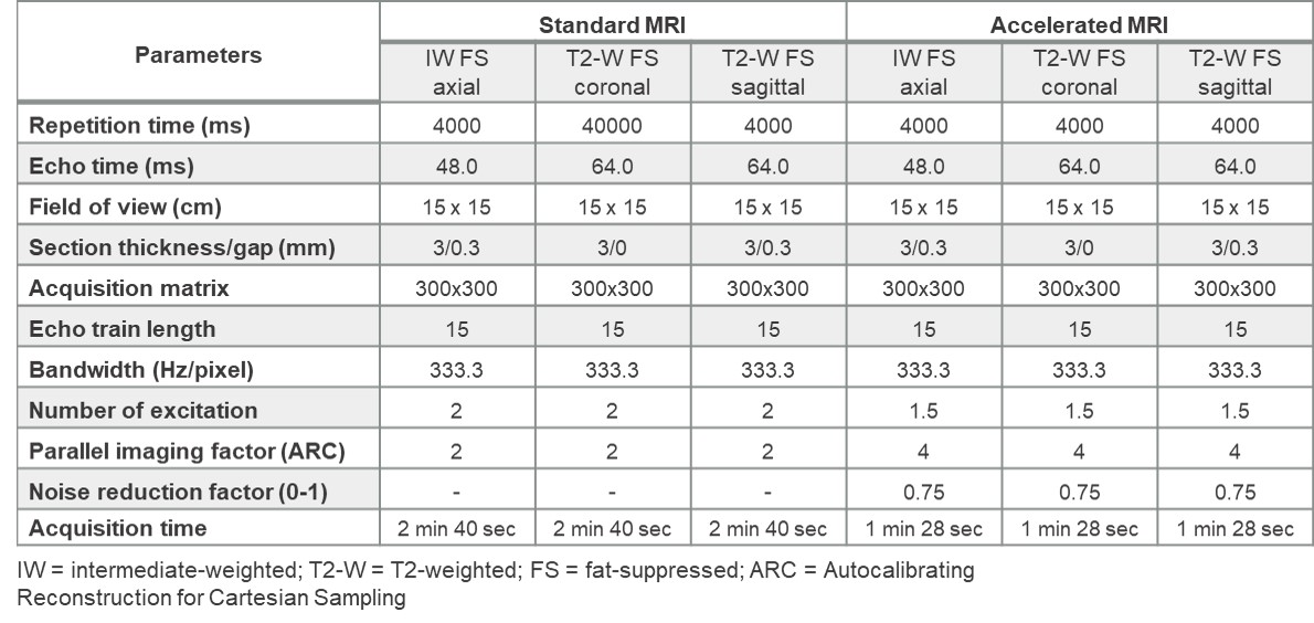
Table 1. MRI parameters. Total acquisition time was 8 minutes for standard sequences and 4 minutes and 24 seconds for the accelerated sequences.
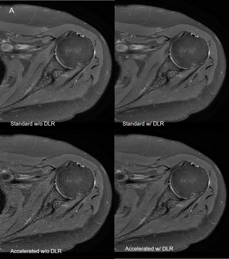
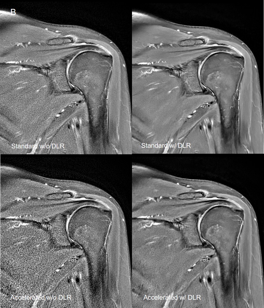
Figure 1. Representative slices for quantitative analysis of signal to noise ratio of four image sets. (B) The coronal image was selected in the midplane which shows a superior glenoid labrum.
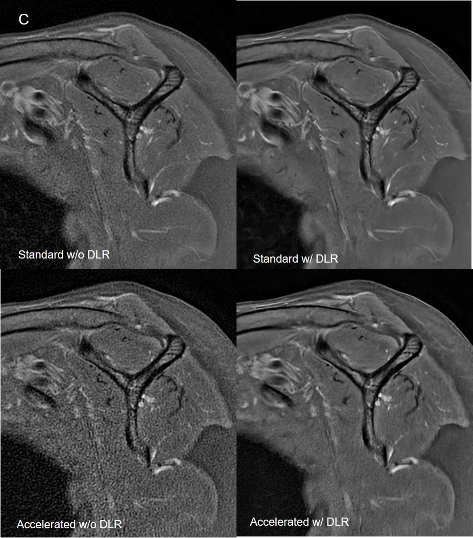
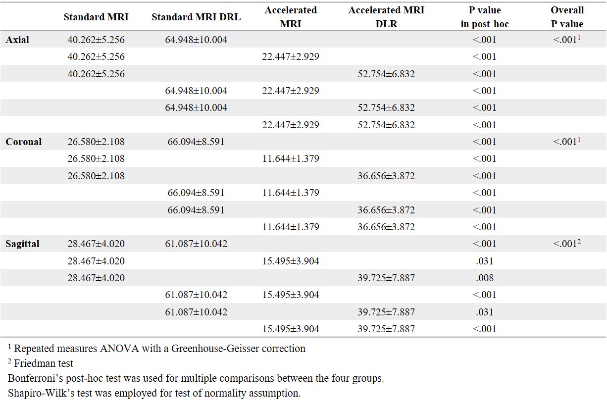
Table 2. Comparison of the mean signal to noise ratio of the four image sets