3093
Feasibility of ZTE-MRI on wrist in comparison with routine MR imaging and radiography1Biomedical Engineering College, Hubei University of Medicine, Shiyan, Hubei, China, 2GE Healthcare, Beijing, China, 3Taihe Hospital, Hubei University of Medicine, Shiyan Hubei, China
Synopsis
ZTE-MRI have high feasibility of angle measurements such as femoral neck-shaft angle as well as bone erosions on temporomandibular joint in reflection of altered bone morphometry and function. In our study, X-ray taken as reference, good consistency of ulnar deviation angle and the length of ulnar styloid process was shown between each two modalities, but better consistency between ZTE-MRI processed with average intensity projection and X-ray. Significantly different number of bone erosions between ZTE-MRI and T1 weighted images were found. To sum up, ZTE-MRI might have potential in elevating diagnosis efficiency of detecting altered bone morphometry and function.
Introduction
Manifestations in wrist articular diseases such as bone fracture, dislocation and bony structure destruction can be clearly seen on X-ray with the advent of superior displaying cortical bone[5]. In recent years, a novel zero echo time magnetic resonance imaging (ZTE-MRI) is known to overcome conventional MRI limitations and have well demonstrated tissues with short transverse relaxation time such as cortical bone with the feature of nominal zero echo time, acquisition over-sampling and 3D radial center-out k-space filling[6]. ZTE-MRI have high feasibility of angle measurement such as acetabular version and femoral neck-shaft angle as well as bone erosion counts on temporomandibular Joint and sacroiliac joint bone . Since rheumatoid arthritis (RA) has a typical manifestation of bone erosion, progresses to the late stage in accompanies with edema and serious articular destruction, and leads to carpal instability[7], feasibility of ZTE-MRI in diagnosis and long-term RA follow-ups might help orthopedists in diagnosis and timely treatment-planning. Therefore, our study explored feasibility of ZTE-MRI in evaluation of carpal morphometry and detection rate of bone erosion in comparison of routine MR imaging and X-rayMaterials and methods
This study was approved by our institutional review board approval and written informed consent was given and signed by each participant. 21 patients who conform to the American rheumatism association in 1987 (ACR) classification of rheumatoid arthritis were recruited in this study. All patients underwent MRI and X-ray examination on the same day. The MRI examination includes routine T1 weighted imaging and ZTE-MRI. The angle measurements including ulnar deviation, ulnar wrist and carpal, and length measurements including length of radial styloid process and length of ulna styloid process were measured and measured on radiographs, ZTE-MRI and post-processed average intensity projection (AIP) of ZTE-MRI by two radiologists (Illustrated in Figure 1). Wrist morphometry such as bone erosion counts were assessed on ZTE-MRI, routine T1-weighted imaging and X-ray. Bone erosion is defined as focal signal loss area with clear edge and low signal at the center on T1WI and ZTE-MRI[8]. Inter-observer and inter-modality agreement of measurements was tested using intraclass correlation coefficient (ICC). Paired T test or nonparametric Wilcoxon signed rank test depending on normality distribution were performed on the bone erosion counts between different sequences.Results
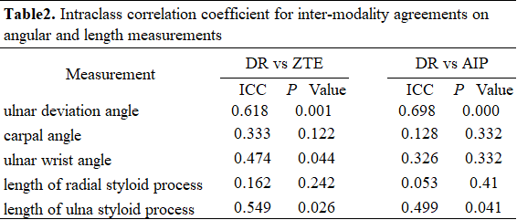
All angle and length measurements showed good to excellent inter-observer consistency on X-ray, ZTE and AIP ZTE images (ICC range =[0.855–0.990], [0.733-0.928], [0.892-0.980], p = 0.000)(Table 1). The measurements of ulnar deviation angle and the length of ulnar styloid process showed good consistency between each two modalities (Table 1). Other angle and length measurements showed low to moderate consistency between ZTE-MRI and X-ray (Table 2). There were significantly different number of bone erosions between ZTE-MRI and T1W images (p<0.001) shown in Figure 2Discussion
In our study, the measurements of ulnar deviation angle and ulnar styloid process on ZTE-MRI, AIP ZTE-MRI and digital X-ray showed good to excellent consistency under the condition of very careful palm positioning during both MR and X-ray examinations. The reason for relatively higher consistency of angle and length measurements between AIP ZTE-MRI and X-ray than between ZTE-MRI and X-ray, especially ulnar deviation angle and length of ulnar styloid process, might be that AIP ZTE-MRI is a kind of projection image as X-ray. In addition, only ulnar deviation angle with good inter-modality consistency might be caused by different wrist joint placement between the palms lying on the scan bed and putting down. As carpal bone marrow edema and synovial thickness accumulated in wrist, counting bone erosion became more difficult to carried out especially on routine-used T1WI respectively as clinical diagnosis reference for erosion detection and edema (Figure 3). The major reason for the finding of wrist deformity including bone erosion clearly on ZTE-MRI and slightly clearly on T1WI images but poorly on X-ray films was the feature of nominal zero echo time, indicating ZTE-MRI was less sensitive to signals with longer T2 relaxation time such as edema and thus elevated erosion detection. Consistent with lower sensitivity of T1WI in detection of bone erosions in previous study[4], we also discovered ZTE-MRI has superior performance on bone erosions with existence of edema Invisible and blurred edges of bone surface due to lack of signals from any short-T2* tissues on conventional MRI were compensated by ZTE-MRI now.Conclusions
ZTE-MRI might have potential in elevating diagnosis efficiency of altered bone morphometry and function via measuring angle and length and counting the number of bone erosion.Acknowledgements
No acknowledgement found.References
[1] W.Y. Liu, H.T.Wu, C.E. Lin, H.L. Lee, W.Y. Guo Evaluation of the Condyle Position and Volume in the Temporomandibular Joint Using Zero Echo Time MRI. In: Annual Meeting of the International Society for Magnetic Resonance in Medicine (ISMRM) May 2018.
[2] Breighner RE, Bogner EA, Lee SC, Koff MF, Potter HG. Evaluation of Osseous Morphology of the Hip Using Zero Echo Time Magnetic Resonance Imaging. Am J Sports Med 2019; 47(14):3460-3468.
[3] Breighner RE, Endo Y, Konin GP, Gulotta LV, Koff MF, Potter HG. Technical Developments: Zero Echo Time Imaging of the Shoulder: Enhanced Osseous Detail by Using MR Imaging. Radiology 2018; 286(3):960-966.
[4] Y. Li SH, W.V. Liu, X. Li Diagnostic performance of zero echo time imaging and T1-weighted fast spin echo on sacroiliac joint bone erosions using CT as the gold standard. . Annual Meeting of the International Society for Magnetic Resonance in Medicine (ISMRM) May 2020.
[5] Bruno F, Arrigoni F, Palumbo P, Natella R, Maggialetti N, Reginelli A, et al. The Acutely Injured Wrist. Radiol Clin North Am 2019; 57(5):943-955.
[6] Weiger M, Pruessmann KP, Hennel F. MRI with zero echo time: hard versus sweep pulse excitation. Magn Reson Med 2011; 66(2):379-389.
[7] Muramatsu K, Ihara K, Tanaka H, Kawai S. Carpal instability in rheumatoid wrists. Rheumatol Int 2004; 24(1):34-36.
[8] Perry D, Stewart N, Benton N, Robinson E, Yeoman S, Crabbe J, et al. Detection of erosions in the rheumatoid hand; a comparative study of multidetector computerized tomography versus magnetic resonance scanning. J Rheumatol 2005; 32(2):256-267.
Figures
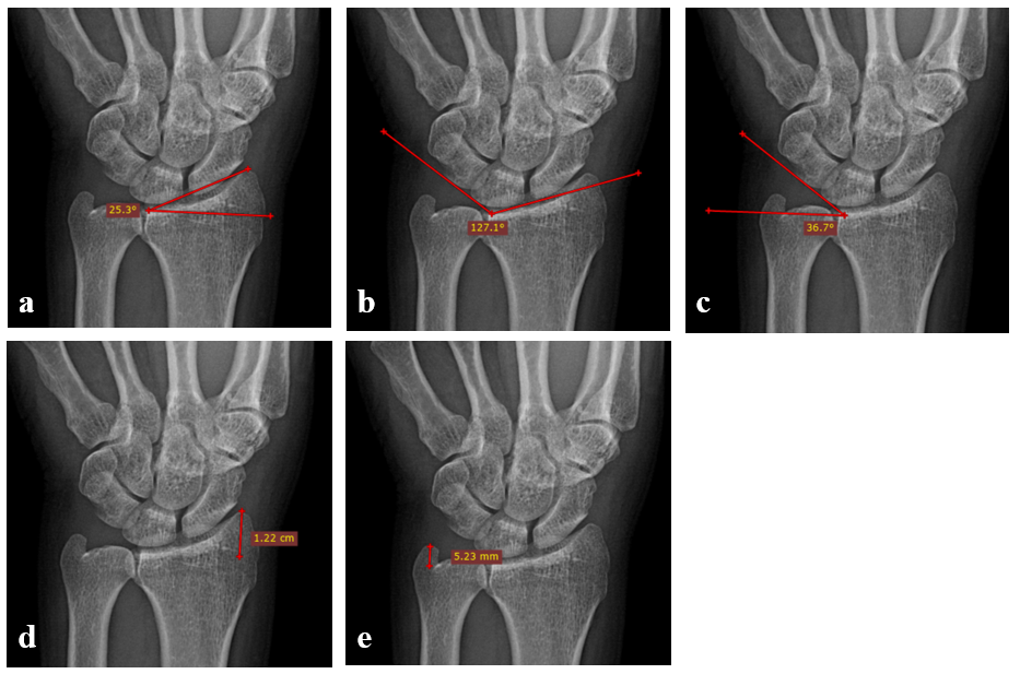
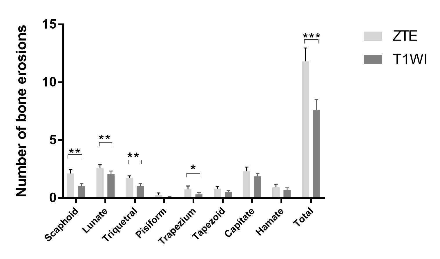
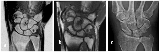
Figure 3. Bone erosions on a) CT-like ZTE, b) T1WI, c) X-ray.
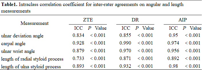
Table1. Intraclass correlation coefficient for inter-rater agreements on angular and length measurements