3091
The Trajectory of Knee Structures Before The Onset of Rheumatoid Arthritis: A 4-Year Longitudinal Magnetic Resonance Imaging Study1Department of Medical Imaging, The Third Affiliated Hospital of Southern Medical University, Guangzhou, China, 2Philips Healthcare, Guangzhou, China
Synopsis
The knee joint is one of the most commonly affected large joint in rheumatoid arthritis (RA). Its progression often causes joint destruction and deformity. The cartilage injury, bone marrow lesions and synovitis scores of the knees at baseline, P-1 (1 year prior to P0), and P0 (time of onset of RA) were evaluated using the MOAKS score. The generalized estimation equation (GEE) was used to compare the MR scores of knee joints at three time points to observe the longitudinal changes. The changes of bone marrow lesions and synovitis scores in knee joints were significant before the occurrence of RA.
Introduction
Rheumatoid arthritis (RA) is a chronic, synovitis-based autoimmune disease that mainly affects small joints such as wrist, finger and toe joints, and can also damage the structure of large joints as the disease progresses [1]. The pain and deformity of the large joints affect the patients' daily activities more than small joints. What’s more, the radiographic recovery of large joints is worse than that of small joints, especially the knee, even in patients treated with bDMARD. Long-term radiological damage studies on RA indicate that the knee joint is one of the most commonly affected large joints. The progression of the RA structural injury of the knee joint usually causes joint destruction and joint deformity [2]. However, MRI is mainly used to observe the inflammation of small joint tissues such as synovitis, bone marrow lesions (BMLs), and tenosynovitis, etc. after the occurrence of RA to longitudinally study the occurrence and development of RA [3, 4]. There is still a lack of assessment of the joint structural changes in RA patients before the occurrence of the disease. Therefore, the purpose of this study is to longitudinally study the MRI structural characteristics of the knee joint before the occurrence of RA.Methods
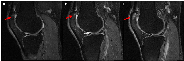
Thirty-six knees with RA were included from the Osteoarthritis Initiative (OAI) database. These cases were diagnosed during follow-up from 12m to 48m, and all the knee joint KL grades were 0 or 1 before the diagnosis of RA. All MR images of the knees at baseline, P-1 (1 year prior to P0), and P0 (time of onset of RA) were evaluated by two radiologists using the MOAKS score. Cartilage injury, BMLs and synovitis scores were evaluated, and the generalized estimation equation (GEE) was used to compare the MR scores of knee joints at three time points to observe the longitudinal changes.Results
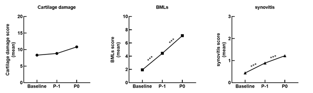
Before the occurrence of RA, the scores of the three MR characteristics of the knee joints showed an increasing trend at three time points. Compared with baseline, there is no significant difference in the total score of cartilage damage in RA patients at P-1 (P = 0.473), while the total score of BMLs and synovitis were significantly higher (baseline vs P-1: 1.94 vs 4.44, P < 0.001; 0.44 vs 0.89, P < 0.001). From P-1 to P0, the total score of cartilage injury increased from 8.83 to 10.81 (P = 0.065), the total score of BMLs increased from 4.44 to 7.11 (P < 0.001), and the total score of synovitis increased from 0.89 to 1.22 (P < 0.001), Figure 1. The GEE model was constructed. After adjusting for gender, age and BMI at the time of inclusion, time of RA diagnosis and total score of each feature at baseline, the results showed that only BMLs and synovitis scores were different between the three time points.Conclusion
The changes of BMLs and synovitis scores in knee joints were significant before the occurrence of RA. This study suggests that we should pay attention to the changes of BMLs and synovitis of the knee joint even if there are no clinical symptoms before the occurrence of RA.Acknowledgements
This study was supported by National Natural Science Foundation of China (grant No. 81801653), and Guangdong Science and Technology Department (grant No. 2017B090912006).References
[1] Schett G, Firestein GS. Mr Outside and Mr Inside: classic and alternative views on the pathogenesis of rheumatoid arthritis. Ann Rheum Dis. 2010 May;69(5):787-9. doi: 10.1136/ard.2009.121657. Epub 2010 Mar 18.
[2] Nieuwenhuis WP, van Steenbergen HW, Stomp W, Stijnen T, Huizinga TW, Bloem JL, van der Heijde D, Reijnierse M, van der Helm-van Mil AH. The Course of Bone Marrow Edema in Early Undifferentiated Arthritis and Rheumatoid Arthritis: A Longitudinal Magnetic Resonance Imaging Study at Bone Level. Arthritis Rheumatol. 2016 May;68(5):1080-8. doi: 10.1002/art.39550.
[3] Sidhu N, Wouters F, Niemantsverdriet E, van der Helm-van Mil AHM. MRI detected synovitis of the small joints predicts rheumatoid arthritis development in large joint undifferentiated inflammatory arthritis. Rheumatology (Oxford). 2021 Jun 23:keab515. doi: 10.1093/rheumatology/keab515. Epub ahead of print.
[4] Ten Brinck RM, van Steenbergen HW, van der Helm-van Mil AHM. Sequence of joint tissue inflammation during rheumatoid arthritis development. Arthritis Res Ther. 2018 Nov 21;20(1):260. doi: 10.1186/s13075-018-1756-z.
Figures
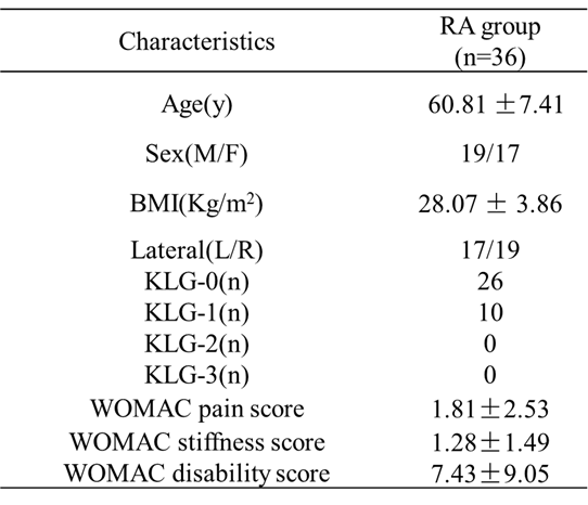
Table 1. Participant characteristics at Baseline
BMI: body mass index; KLG: Kellgren Lawrence grades