3076
Anomalous functional connectivity of amygdala subregional networks in adolescents with major depressive disorder1Huaxi MR Research Center (HMRRC), Functional and molecular imaging Key Laboratory of Sichuan Province, Department of Radiology, West China Hospital, Sichuan University, Chengdu, China
Synopsis
Using seed based FC analysis, we investigated alterations in functional connectivity of amygdala subregional networks in a relatively large sample of adolescent MDD. Our findings identified adolescent MDD patients, compared to HC, have anomalous amygdala subregional functional network connections to several brain regions that play a pivotal role in emotional and cognitive processing, which may contribute to revealing the pathophysiology underlying MDD. These findings were not detected when using the whole amygdala as seeds, further verify the significance of analyses at the subregion level.
Synopsis
Using seed based FC analysis, we investigated alterations in functional connectivity of amygdala subregional networks in a relatively large sample of adolescent MDD. Our findings identified adolescent MDD patients, relative to HC, have anomalous amygdala subregional functional network connections to several brain regions that play a pivotal role in emotional and cognitive processing, which may contribute to revealing the pathophysiology underlying MDD. These findings were not detected when using the whole amygdala as seeds, further verify the significance of analyses at the subregion level.Introduction
The amygdala is the key structure of brain emotional circuit and amygdala-centered network dysfunction has been highlighted in the neurobiological mechanisms of major depressive disorder (MDD). The amygdala is composed of four main subregions, each of which contribute to distinct affective functions via their unique connectivity patterns. A previous study has found amygdala subregional‐network dysfunction in adult MDD [1], however, this level of investigation has been lacked in adolescent MDD, who is at such a critical period for brain development. We address this gap by examining the differences in the resting-state functional connectivity of amygdala subregions in adolescents with MDD versus healthy controls (HC) to characterize the neurobiological mechanisms and to better understand the developmental effect of this disorder.Methods
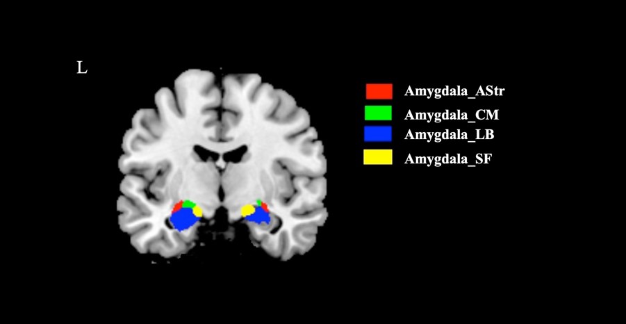
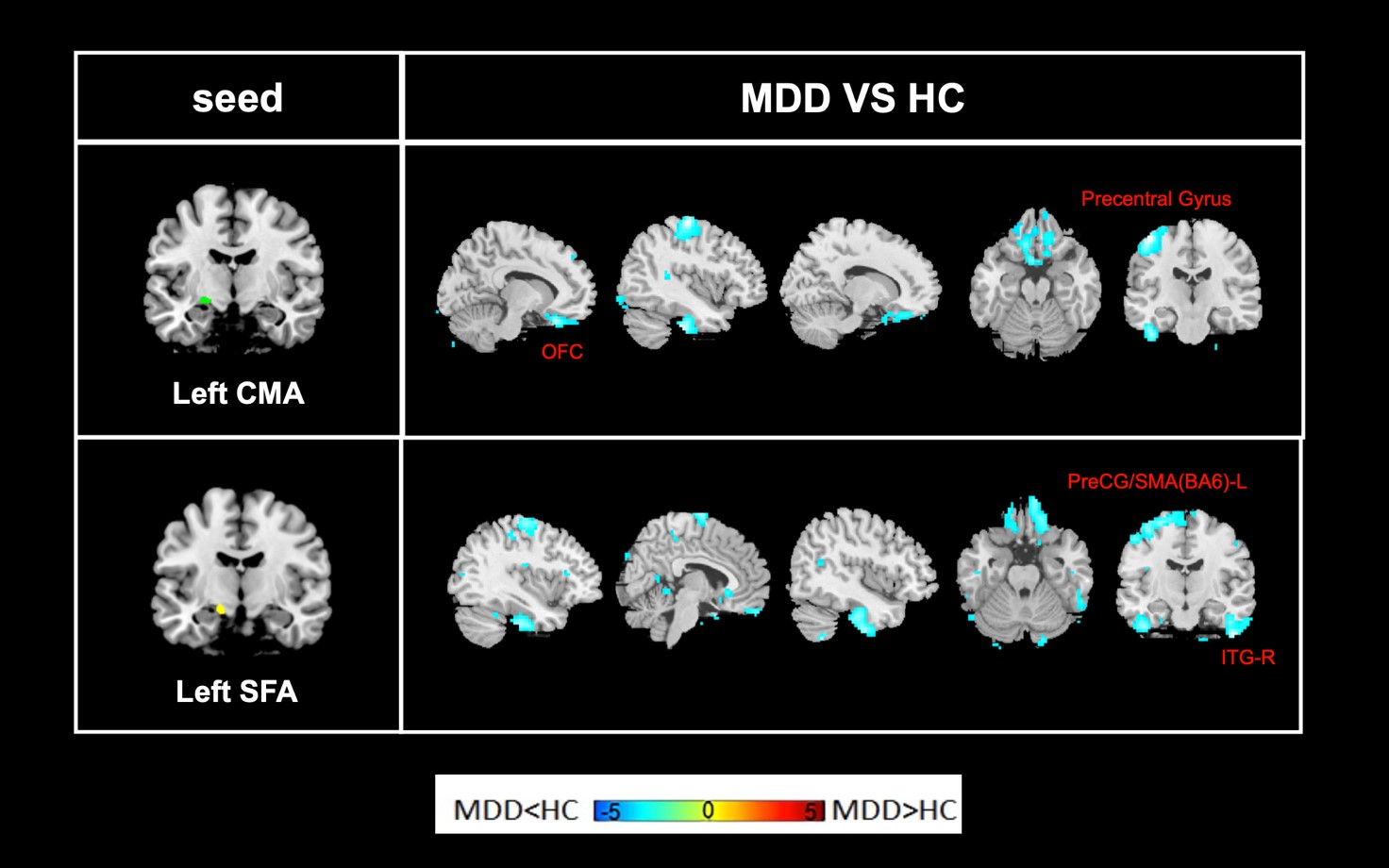
A total of 68 medication-naive adolescents with MDD and 43 age- and sex-matched healthy controls (HC) were recruited in this study after giving written informed consent. Imaging data were acquired using 3.0-Telsa Siemens magnetic resonance imaging system with a 20-channel phased-array head coil. The preprocessing of resting-state functional images was performed using DPABI software including excluding the first 10 time points, slice timing, realignment, regressing out nuisance signals, spatial normalization, smooth, detrend and filtering. The masks of amygdala four subregions, including the basolateral (BL), centromedial (CM), superficial (SF) amygdala and amygdalostriatal transition area (AStr), were created using cytoarchitectonically-defined probabilistic maps (Figure 1) and the whole amygdala created using the AAL map. Functional connectivity analyses with the amygdala and its four subregions as seeds were performed using REST software. For each subject, averaged time course of each seed were extracted from the preprocessing data and then correlated (Pearson correlation) with the rest voxels of the whole brain. The correlation coefficients were standardized using Fisher’s r-to-z transformation to improve normality and z-value FC maps per subject were obtained for further statistical analyses.Voxel‐based comparison of z‐value maps were conducted between the two groups using a random‐effects two‐sample t test in SPM12, which identifies brain regions showing significant connectivity differences with each seed in MDD patients relative to HC. The significance threshold was set to P<0.005 at the voxel level, and FWE correction was set to p<0.05 at the cluster level.Results
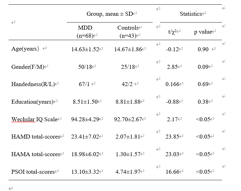
The demographic information and clinical characteristics of the participants are shown in Table 1. Relative to HC, patients with MDD showed decreased functional connectivity between the left CMA and the orbitofrontal cortex (OFC) and the precentral gyrus (PreCG). We also found decreased functional connectivity between the left SFA and the Left PreCG, supplementary motor area (SMA) and right inferior temporal gyrus (ITG) in MDD patients compared with HC (Figure 2). No significant group difference was identified when using the whole amygdala as seed.Discussion and Conclusion
We found adolescent MDD patients, relative to HC, showed anomalous amygdala subregional functional network connections to several regions including OFC, PreCG, SMA and ITG, which involved in emotional and cognitive processing. Decreased amygdala subregional FC with OFC and PreCG may account for the emotional disturbances typical of MDD. Previous meta-analyses revealed decreased amygdala FC with ITG in adolescent MDD [2], while our results specify the association to SFA subregion which may play a vital role in amygdala-ITG connectivity dysfunction in this disorder. Decreased amygdala subregional FC with SMA may reflect dysregulated cognitive control and converge with a theoretical model in which adolescents with depression are inclined to become trapped in cognitive dysregulation. Besides, these findings were not detected when using the whole amygdala as seeds, which verify the importance of analyses at the subregional level.Acknowledgements
This study is supported by grants from 1.3.5 Project for Disciplines of Excellence, West China Hospital, Sichuan University (ZYJC21041) and Clinical and Translational Research Fund of Chinese Academy of Medical Sciences (2021-I2M-C&T-B-097).References
[1] Tang S, Li H, Lu L, et al. Anomalous functional connectivity of amygdala subregional networks in major depressive disorder. Depress Anxiety. 2019;36(8):712-722. doi:10.1002/da.22901
[2] Tang S, Lu L, Zhang L, et al. Abnormal amygdala resting-state functional connectivity in adults and adolescents with major depressive disorder: A comparative meta-analysis. EBioMedicine. 2018;36:436-445. doi:10.1016/j.ebiom.2018.09.010
Figures