3075
Disrupted white matter network of brain structural connectomes in bipolar disorder patients revealed by q-ball imaging1Department of Medical Imaging and Radiological Sciences, Graduate Institute of Artificial Intelligence, Chang Gung University, Taoyuan, Taiwan, 2Department of Psychiatry, National Cheng Kung University Hospital, College of Medicine, National Cheng Kung University, Tainan, Taiwan, 3Institute of Behavioral Medicine, College of Medicine, National Cheng Kung University, Tainan, Taiwan, 4Department of Psychiatry, Chang Gung Memorial Hospital, Chiayi, Taiwan, 5Medical Imaging Research Center, Institute for Radiological Research, Chang Gung University and Chang Gung Memorial Hospital at Linkou, Taoyuan, Taiwan
Synopsis
Bipolar disorder (BD) is a major psychiatric disorder associated with structural and functional brain alterations and cognitive deficits. This study used q-ball imaging and graph theoretical analysis to investigate both neurological structural change and network alterations between BD patients and healthy controls. The results showed the alterations in several brain regions including the corpus callosum and cingulate gyrus, which are associated with executive, cognitive, emotional function, and memory. We found the BD group demonstrated higher global integrity than the HC group, but they remained small-world properties. It indicated that white matter integrity and network alterations were associated with bipolar disorder.
Introduction
Bipolar disorder (BD) is a major psychiatric illness that affects 1% of the population and has its onset during adolescence or early adulthood 1. In recent years, many diffusion tensor imaging (DTI) investigations have indicated that BD is commonly associated with white matter (WM) abnormalities. Studies showed that these alterations may even be progressive in some individuals. However, DTI cannot resolve neural fibers crossing within an individual voxel due to the constraints of the tensor model 2. Hence, we use an advanced method called q-ball imaging (QBI), which can resolve multiple intravoxel fiber orientations 2. Two QBI indices, including generalized fractional anisotropy (GFA) and normalized quantitative anisotropy (NQA), were reconstructed for the assessment of WM connectivity. For network measurement, graph theoretical analysis (GTA) from QBI tractography data was performed and three topological parameters were acquired.Methods
We recruited 25 BD patients and 43 healthy controls (HCs) from the community. All participants were acquired diffusion MRI images using 3T MRI scanner (MR750, GE Medical Systems) with an 8-channel head coil. Echo planar imaging (EPI) pulse sequence was performed with parameters: TR / TE = 8000 / 115 ms, voxel size = 223 mm3, and 96different diffusion-sensitive gradient directions with b = 2500 s/mm2. In voxel-based statistical analysis (VBA), all diffusion images of participants have processed eddy current correction by FSL (FMRIB Software Library), which could decrease eddy current artifacts caused by EPI sequence. We then normalized all imaging data by using Statistical Parametric Mapping 8 (SPM8; Wellcome Department of Cognitive Neurology, London, UK). Next, we used QBI methods to reconstruct two indices, including GFA and NQA, by applying DSI studio. Finally, a two-sample t-test was performed to evaluate the values of QBI indices within different regions of the brain between the BD and HC groups. Gender, age, years of education, and BMI were used as the covariates. For network measurement, the GTA was performed with QBI tractography data. We divided the brain into 90 regions and reconstructed neural fiber pathways using fiber assignment by DSI studio. Three topological parameters were then calculated from the structural connectivity matrix, including small-worldness, global efficiency, and characteristic path length indices. The participants’ gender, age, years of education, and BMI were regarded as the covariates of no interest.Results
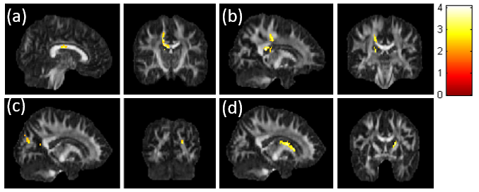
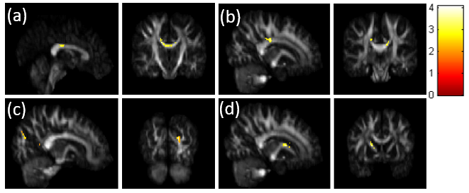
In results of VBA, the BD group had significantly lower GFA and NQA in the brain regions of corpus callosum, cingulate gyrus, cuneus, and caudate than that of the HC group (Fig. 1 and 2). As for the results of GTA, we found the small-worldness index was more than 1 in both BD and HC groups (Fig. 3a). Furthermore, the BD group demonstrated higher global efficiency and lower characteristic path length index compared with the HC group (Fig. 3b and 3c).Discussion
The GFA and NQA indices can represent the integrity and the compactness of white matter, respectively. Our study found the smaller GFA and NQA of the corpus callosum in BD compared with HC. The corpus callosum is the largest interhemispheric commissure for higher cognitive functions associated with emotion, which involves sustaining attention, approach-related motivation and behavior, inhibition and excitation of the contralateral hemisphere, affective prosody, and memory 3. We also found that the GFA and NQA of the BD group were lower than the HC group in the cingulate gyrus and cuneus. From previous studies, the cingulate gyrus and cuneus are playing important roles in the default mode network (DMN) which extends from the prefrontal medial cortex (mPFC) to the precuneus, posterior cingulate cortex (PCC), and inferior parietal cortex 4. The global DMN is involved in multiple cognitive and affective functions such as emotional processing, self-referential mental activity, mind wandering, and recollection of experiences 4. Additionally, we found significant differences between BD and HC in caudate, which is a part of basal ganglia. Basal ganglia structures are particularly closely networked with both cortical and subcortical regions involved in mood expression and regulation, including the amygdala and ventral prefrontal cortex (VPFC), and appear to be integral to effective regulation 5,6. Alterations in the corpus callosum, cingulate gyrus, cuneus, and caudate may cause cognitive and affective changes by BD. Our results of GTA showed the small-worldness index of the BD and HC group was higher than 1, which indicated that the small-world properties remained in both groups. Larger global efficiency indices and shorter characteristic path lengths were found in the BD group compared with the HC group, which indicated white matter integrity and network alterations were associated with bipolar disorder.Conclusion
Our study investigated the neurological structural change and network alterations between BD patients and the HC group. We found both white matter integrity and network alterations which are associated with BD. These abnormalities may have the potential to be used as the imaging marker in diagnosing BD.Acknowledgements
This study was supported by the research program, MOST110-2221-E-182-027, which was sponsored by the Ministry of Science and Technology, Taipei, Taiwan.References
1. Bellani M., et al. DTI and Myelin Plasticity in Bipolar Disorder: Integrating Neuroimaging and Neuropathological Findings. Front Psychiatry. 2016 Mar 1; 7: 21.
2. Tuch DS. Q-ball imaging. Magn Reson Med. 2004 Dec; 52(6): 1358-72.
3. Zhang R., et al. White matter abnormalities of corpus callosum in patients with bipolar disorder and suicidal ideation. Ann Gen Psychiatry. 2019 Sep 10;1 8: 20.
4. Zovetti N., et al. Default mode network activity in bipolar disorder. Epidemiol Psychiatr Sci. 2020 Sep 8; 29: e166.
5. Shahana N., et al. Neurochemical alteration in the caudate: implications for the pathophysiology of bipolar disorder. Psychiatry Res. 2011 Aug 30; 193(2): 107-12.
6. Watson CG., et al. Graph theory analysis of DTI tractography in children with traumatic injury. Neuroimage Clin. 2019; 21: 101673.
Figures