3074
Myelin abnormalities in patients with bipolar disorder: an inhomogeneous magnetization transfer study1Shenzhen Mental Health Center/Shenzhen Kangning Hospital, Shenzhen, China, 2MR Research, GE Healthcare, Beijing, China
Synopsis
Previous DTI studies have revealed widespread increased radial diffusivity, which tend to be associated with myelin alteration, in patients with bipolar disorder (BD). This study utilizes a surrogate measure of myelin content termed inhomogeneous magnetization transfer (ihMT) to investigate myelin abnormalities in patients with BD. Our results demonstrate that regions with myelin deficits are mainly concentrated in the premiddle corpus callosum and left anterior white matter tracts, suggesting that interhemispheric communication and information transmission of left anterior brain areas may be most affected in patients with BD. Overall, this study provides new insight into the neuropathological mechanisms underlying BD.
Introduction
There is an increasing number of diffusion imaging studies that reported the abnormalities of white matter in patients with bipolar disorder (BD)[1]. Particularly, widespread increases in radial diffusivity (RD), which tend to associate with myelin alteration, were reported in BD[2; 3]. Here, we use a surrogate measure of myelin content, namely inhomogeneous magnetization transfer (ihMT) technique[4], to investigate the myelin abnormalities in patients with BD.Method
The ihMT data from 31 patients with BD (8 male and 23 female) and 42 age, sex, and years of education-matched health controls (HC, 12 male and 30 female) were acquired. Then four indices, including ihMT ratio (IHMTR), quantitative ihMT (qIHMT), magnetization transfer ratio (MTR), and quantitative magnetization transfer (qMT) were estimated. Subsequently, the mean values of 50 white matter bundles were extracted based on the JHU-ICBM white matter template[5] for each subject. The differences of ihMT indices between BD and HC groups were compared using a general linear model with age, sex, and years of education as nuisance covariates. Partial correlation analyses were conducted after removing the effects of age, sex, and years of education to investigate associations between the significantly altered ihMT indices and clinical measures, including the Hamilton Depression Rating Scale (HAMD), Hamilton Anxiety Scale (HAMA), and duration of illness, in BD group.Results
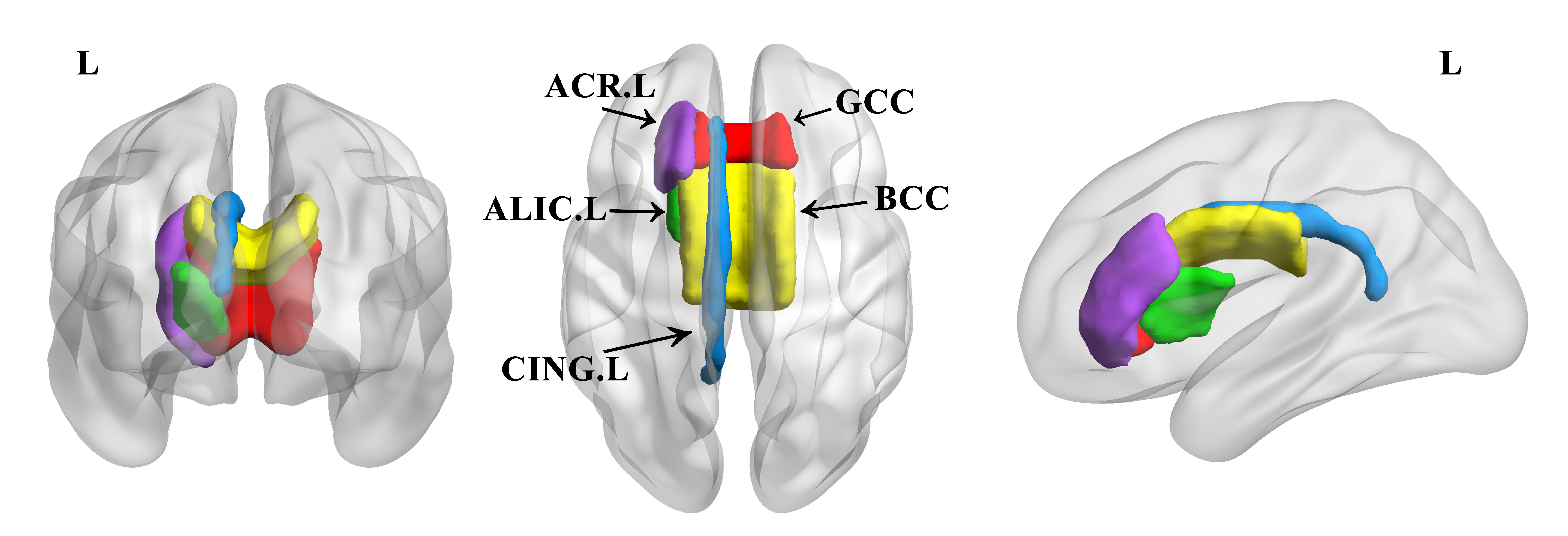
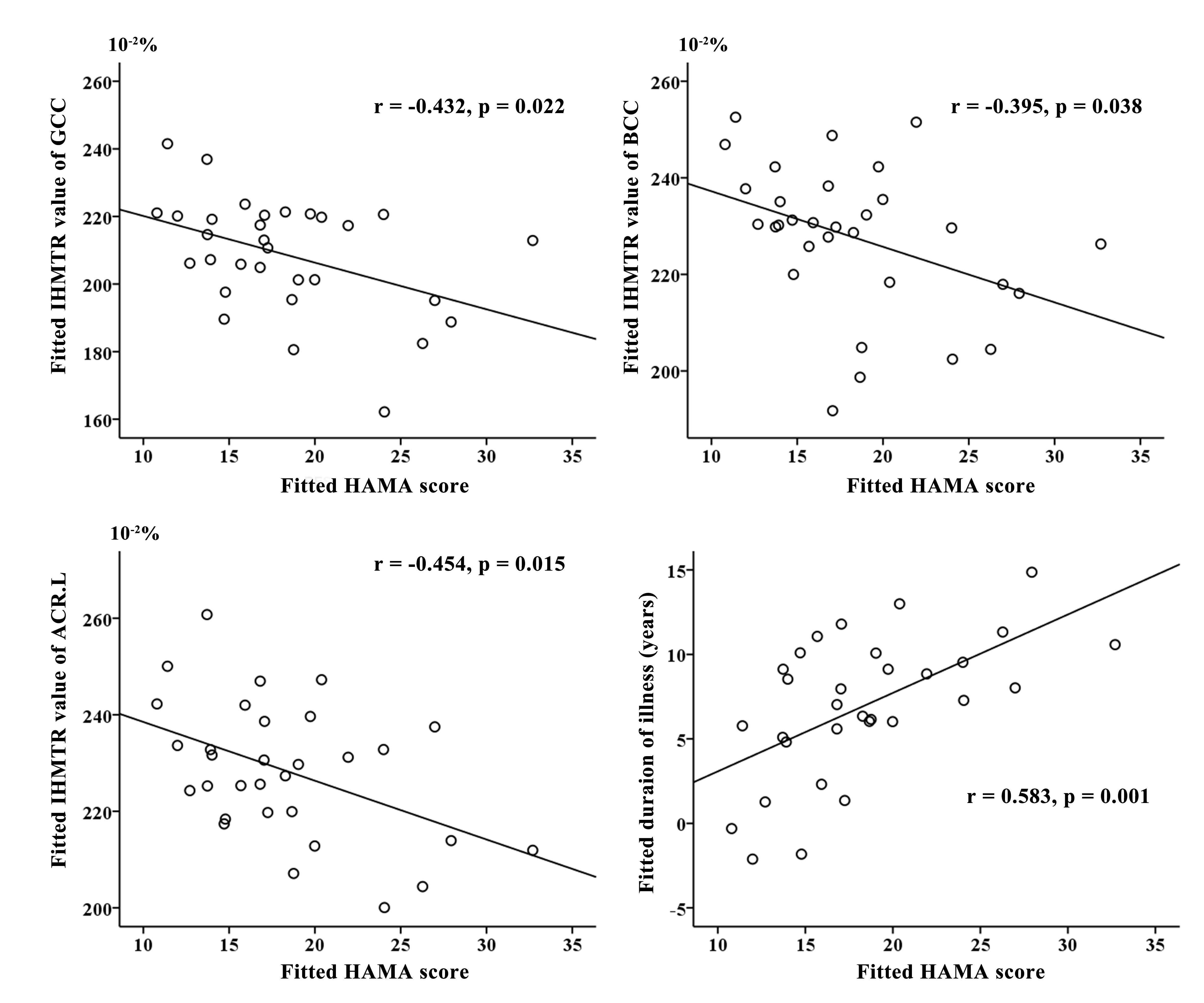
Compared to HC group, the BD group revealed significantly reduced IHMTR values in the genu and body of corpus callosum, left anterior limb of internal capsule, left anterior corona radiate, and left cingulate gyrus, reduced qIHMT values in the genu and body of corpus callosum, and left cingulate gyrus, and reduced qMT in the left cingulum (FDR corrected, p<0.05, Figure 1). Partial correlation analyses showed that the IHMTR values of genu and body of corpus callosum, and left anterior corona radiate were negatively correlated with HAMA scores, and the duration of illness were positively correlated with HAMA scores (Figure 2).Discussion
The ihMT technique is a modification on the basis of magnetization transfer pulses to alter the image contrast according to the large macromolecules that constitute large part of myelin[4; 6]. Previous studies reveal that ihMT is a more specific measure pointed to myelin content than diffusion tensor imaging (DTI), magnetization transfer imaging, and myelin water imaging[7; 8]. In the present study, we found reduced ihMT indices in the genu and body of corpus callosum, left anterior limb of internal capsule, left anterior corona radiate, and left cingulate gyrus in BD group compared with HC group. These findings are consistent with previous DTI studies which reveal increased RD in patients with BD. Our study provides more direct imaging evidence for myelin deficits in patients with BD. We found the regions with abnormal myelin content are mainly concentrated in the premiddle corpus callosum and the left anterior white matter tracts, suggesting that interhemispheric communication and information transmission of left anterior brain areas may be most affected in patients with BD. Moreover, myelin abnormalities in genu and body of corpus callosum, and left anterior corona radiate are negatively associated with anxiety state in patients with BD, meaning that the more anxious the patient was, the more myelin in the corresponding brain region was damaged. In addition, the duration of illness was positively correlated with HAMA scores in patients with BD, suggesting that the longer the course of the disease, the more likely it is to cause more serious anxiety.Conclusion
In summary, this study provides new insight into the neuropathological mechanisms underlying BD.Acknowledgements
No acknowledgement found.References
[1] Yang C, Li L, Hu X, et al. Psychoradiologic abnormalities of white matter in patients with bipolar disorder: Diffusion tensor imaging studies using tract-based spatial statistics[J]. J Psychiatry Neurosci, 2019, 44(1): 32-44.
[2] Linke J O, Adleman N E, Sarlls J, et al. White matter microstructure in pediatric bipolar disorder and disruptive mood dysregulation disorder[J]. J Am Acad Child Adolesc Psychiatry, 2020, 59(10): 1135-1145.
[3] Brown J A, Jackson B S, Burton C R, et al. Reduced white matter microstructure in bipolar disorder with and without psychosis[J]. Bipolar Disord, 2021.
[4] Varma G, Duhamel G, de Bazelaire C, et al. Magnetization transfer from inhomogeneously broadened lines: A potential marker for myelin[J]. Magn Reson Med, 2015, 73(2): 614-622.
[5] Mori S, Oishi K, Jiang H, et al. Stereotaxic white matter atlas based on diffusion tensor imaging in an icbm template[J]. Neuroimage, 2008, 40(2): 570-582.
[6] Varma G, Girard O M, Prevost V H, et al. Interpretation of magnetization transfer from inhomogeneously broadened lines (ihmt) in tissues as a dipolar order effect within motion restricted molecules[J]. J Magn Reson, 2015, 260: 67-76.
[7] Ercan E, Varma G, Mädler B, et al. Microstructural correlates of 3d steady-state inhomogeneous magnetization transfer (ihmt) in the human brain white matter assessed by myelin water imaging and diffusion tensor imaging[J]. Magn Reson Med, 2018, 80(6): 2402-2414.
[8] Geeraert B L, Lebel R M, Mah A C, et al. A comparison of inhomogeneous magnetization transfer, myelin volume fraction, and diffusion tensor imaging measures in healthy children[J]. Neuroimage, 2018, 182: 343-350.
Figures