3049
Assessment of relationship between chemotherapy-induced brain structural alteration and neuropsychological scale revealed by GQI1Department of Medical Imaging and Radiological Sciences, Graduate Institute of Artificial Intelligence, Chang Gung University, Taoyuan, Taiwan, 2School of Medicine, Chang Gung University, Taoyuan, Taiwan, 3Department of Psychiatry, Chang Gung Memorial Hospital, Chiayi, Taiwan, 4Department of Diagnostic Radiology, Chang Gung Memorial Hospital, Chiayi, Taiwan, 5Medical Imaging Research Center, Institute for Radiological Research, Chang Gung University and Chang Gung Memorial Hospital at Linkou, Taoyuan, Taiwan
Synopsis
Breast cancer is the most prevalent cancer among women, and most of them suffered from chemotherapy-related cognitive impairment (CRCI). In this study, we aimed to use generalized q-sampling imaging (GQI) to detect the relationship between GQI indices and mood symptoms caused by chemotherapy, post-traumatic stress disorder (PTSD), or post-traumatic growth (PTG). Using statistical multiple regression, we found the relationship change between GQI indices and depression, and a consistent relationship was obtained between GQI indices and anxiety with or without chemotherapy. The results indicated that neurotoxicity or traumatic stressors may affect brain structures.
Introduction
Breast cancer (BC) is the most prevalent and has the highest incident rate worldwide in 2020 1. One well-known side effect of BC survivors is chemotherapy-related cognitive impairment (CRCI). The cause of CRCI remains ambiguous, and some review articles ascribe these symptoms to neurotoxicity; another suggests that BC may be a traumatic stressor that results in post-traumatic stress disorder (PTSD) and/or post-traumatic growth (PTG) which lead to CRCI 2,3,4. A previous study showed that cerebral white matter integrity decreased after receiving chemotherapy using diffusion tensor imaging (DTI) 5. In this study, we tried to use generalized q-sampling imaging (GQI), regarded as a technique that can deal with the problem of complex fibers that DTI cannot do, to explore the association in participants with or without chemotherapy between cerebral white matter changes and mood symptoms.Methods
This study included 65 women with a history of BC but did not receive chemotherapy (BB), 35 of BB came back to have follow-up assessment after completing chemotherapy less than 12 months (BBF), and 60 women who had completed their chemotherapy less than 12 months (BA). All subjects were scanned on 3T MRI (Verio, Siemens). There were total 193 different diffusion-sensitive gradient directions with b-value = 0, 1000, 1500 and 2000 s/mm2. The FSL (FMRIB Software) and SPM (Statistical Parametric Mapping) were used for eddy current correction and normalization. Afterward, we calculated GQI indices, generalized fractional anisotropy (GFA) and normalized quantitative anisotropy (NQA), by DSI studio with a model-free reconstruction method. Participants have also measured mood symptoms including depression and anxiety. We used the depression module of Patient Health Questionnaire-9 (PHQ9) and the anxiety subscale of the Hospital Anxiety and Depression Scale (HADS-A) to evaluate the extent of depression and anxiety, respectively. Statistical multiple regression was used to evaluate the correlation between GQI indices and mood symptoms.Results
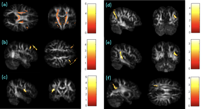
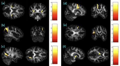
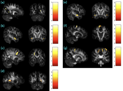
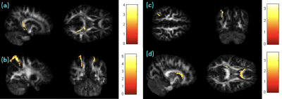
Statistically significant correlations were shown in Fig. 1. Both GFA and NQA showed a positive correlation with depression in corona radiata, internal capsule, and frontal gyrus of BB; superior longitudinal fasciculus (SLF) of group BBF and BA; angular gyrus of BBF; and cuneus of BA. Figure 2 demonstrated the negative correlation with depression for both GQI indices in SLF, angular gyrus and cuneus of BB; frontal gyrus of BBF; and internal capsule of BA group. Besides, corona radiata in BBF showed a negative correlation with depression only for GFA. In Fig. 3, the positive correlations were found in precuneus between NQA and anxiety; temporal and frontal gyrus between GFA and anxiety of BB; precuneus, temporal and frontal gyrus of BBF; and frontal gyrus of BA between both GQI indices and anxiety. In Fig. 4, the negative correlations between NQA and anxiety were shown in corpus callousum (CC) of group BB and BA, and parietal gyrus of BBF. Both GFA and NQA correlated negativity in the frontal gyrus of BB.Discussion
GQI was used to assess changes in white matter integrity, and higher values represent greater integrity. PHQ9 and HADS-A were used to evaluate the extent of depression and anxiety, respectively, and higher values represent the severity of depression and anxiety, respectively. In the association between GQI indices and depression, we observed that correlation changed from a positive to a negative in corona radiata, internal capsule, and frontal gyrus. These regions play an important role in nerve conduction and emotion. Our results suggested that chemotherapy might affect the corticospinal tract and frontal gyrus 6. On the contrary, in SLF, angular gyrus, and cuneus, they changed from a negative to a positive correlation. Previous research showed that most of the participants experienced self-improving and start a new life, these differences might due to PTG 7. In the association between GQI indices and anxiety, positive correlations were found in the precuneus, temporal and frontal gyrus, and negative correlations were obtained in CC and parietal gyrus. It indicated participants before chemotherapy were consistent with participants after chemotherapy. It might probably be because the chemotherapy-induced changes in white matter and anxiety were not huge enough to be detected.Conclusion
We found the correlation altered in some regions associated with emotion, memory, and motor function. The results might indicate potential effects of chemotherapy, PTSD, and PTG on cerebral white matter and depression.Acknowledgements
This study was supported by the research grant MOST107-2221-E-182-054-MY3 from the Ministry of Science and Technology, Taipei, Taiwan.References
1. International Agency for Research on Cancer. Global Cancer Observatory. https://globocan.iarc.fr/factsheets/cancers/breast.
2. Parikh, D., et al. Post-traumatic stress disorder and post-traumatic growth in breast cancer patients--a systematic review. Asian Pac J Cancer Prev. 2015; 16(2): 641-646.
3. Pendergrass, J. C., et al. Cognitive Impairment Associated with Cancer: A Brief Review. Innov Clin Neurosci. 2018; 15(1-2): 36-44.
4. Tong, T., et al. Chemotherapy-related cognitive impairment in patients with breast cancer based on MRS and DTI analysis. Breast Cancer. 2020; 27(5): 893-902.
5. Deprez, S., et al. Longitudinal assessment of chemotherapy-induced structural changes in cerebral white matter and its correlation with impaired cognitive functioning. J Clin Oncol. 2012; 30(3): 274-281.
6. Simo, M., et al. Chemobrain: a systematic review of structural and functional neuroimaging studies. Neurosci Biobehav Rev. 2013; 37(8): 1311-1321.
7. Niebauer, E., et al. Patient perspectives on the causes of breast cancer: a qualitative study on the relationship between stress, trauma, and breast cancer development. Int J Qual Stud Health Well-being. 2021; 16(1): 1983949.
Figures


