3048
An evaluation of the caput nuclei caudati in children with autism spectrum disorder and language deficits with DKI and Synthetic MR1the Third Affiliated Hospital of Zhengzhou University, Zhengzhou, China, 2GE Healthcare, Beijing, China
Synopsis
In this study, DKI combined with synthetic MRI of magnetic resonance imaging compilation (MAGiC) was used to assess the developmental integrity of the caput nuclei caudati of children with ASD and language deficits, and the relationship between DKI parameters and language test scores were explored.
Synopsis
The problem of language deficits in children with autism spectrum disorder (ASD) has been explored. The caput nuclei caudati (CNC) is involved in the language processing process and is closely related with ASD. Effective language-related magnetic resonance markers could help evaluate the language ability of children with ASD, which can benefit for identifying and intervening earlier. In this study, DKI combined with synthetic MRI of magnetic resonance imaging compilation (MAGiC) was used to assess the developmental integrity of the CNC of children with ASD and language deficits, and the relationship between DKI parameters and language test scores were explored. We found there were significant differences of gray matter volume, white matter volume and Mk ratio in bilateral CNC (R0-Mk) between ASD and typical developmental(TD)group and they can be used as sensitive biomarkers to reflect the microstructural changes in the CNC in children with language defects in ASD.Introduction and Purpose
ASD is a neurodevelopmental disease(1).Children with ASD usually have the first symptoms of language degradation as early as 2 years old(2),which may leading poor language effects(3).Studies have shown that normal children’s language development is affected by the lateralization of the left side of the caudate nucleus(4).The CNC,as a part of the close connection with the ventrolateral prefrontal cortex,is involved in all aspects of speech production and language understanding(5).However,the CNC changes in children with ASD and language deficiency are still unknown.In this study,the brain volume changes of children in the ASD were evaluated using the MAGiC,while the microstructural changes using DKI in the CNC of ASD children.The relationship between the DKI parameters of the CNC of ASD children and language assessment was also explored.Materials and Methods
A total of 30 typical developmental group(F23/M7)aged 26-52 months(34.80±8.90 months)and 34 ASD patients(F26/M8)with language defects,aged 24-56 months(34.97±9.72 months)were collected.PEP-3(6)was used to assess language ability.The communication score is recognized by the deputy test,cognitive verbal/preverbal(CVP),receptive language(RL),and expressive language(EL) scores are combined.The routine T1 and T2 weighted MRI,MAGiC and DKI sequence were performed for all subjects.DKI:TR/TE=8200ms/minimum,FOV=200×200mm,matrix=256×256,layerthickness/spacing=4mm/0mm.MAGiC:TR/TE=4266ms/19.4ms,FOV=200×200mm,Pixel Size=0.8×0.9 mm, layer thickness/spacing=4mm/1.0mm.The FA,MD,DA,Dr,Mk,Ka and Kr maps were obtained with DKI in iQuant Worktation(GE Healthcare,Beijing,China),and the ROI in the units of 6 voxels in bilateral CNC were manually delineated by using ITK-SNAP 3.8.0.(Figure1).Then the right-left CNC ratio R0 (R0=right CNC /left CNC) were calculated, noted with R0 for each parameter.The MAGiC images were processed to generate the volume of cerebrospinal fluid,myelination,gray matter,white matter,non-white matter/gray matter/myelination/cerebrospinal fluid tissue volume with SyMRI. All data were statistically analyzed with SPSS version 25.The data were compared using t test or the Mann-Whitney U test after the normal distribution and homogeneity of variances were checked.The enumeration were compared with the chi-square test.Covariance analysis with the volume of gray and white matter as the covariants was used to compare the DKI parameters of the CNC in the two groups,spearman was used for correlation analysis.P<0.05 indicates that the difference is statistically significant.Results
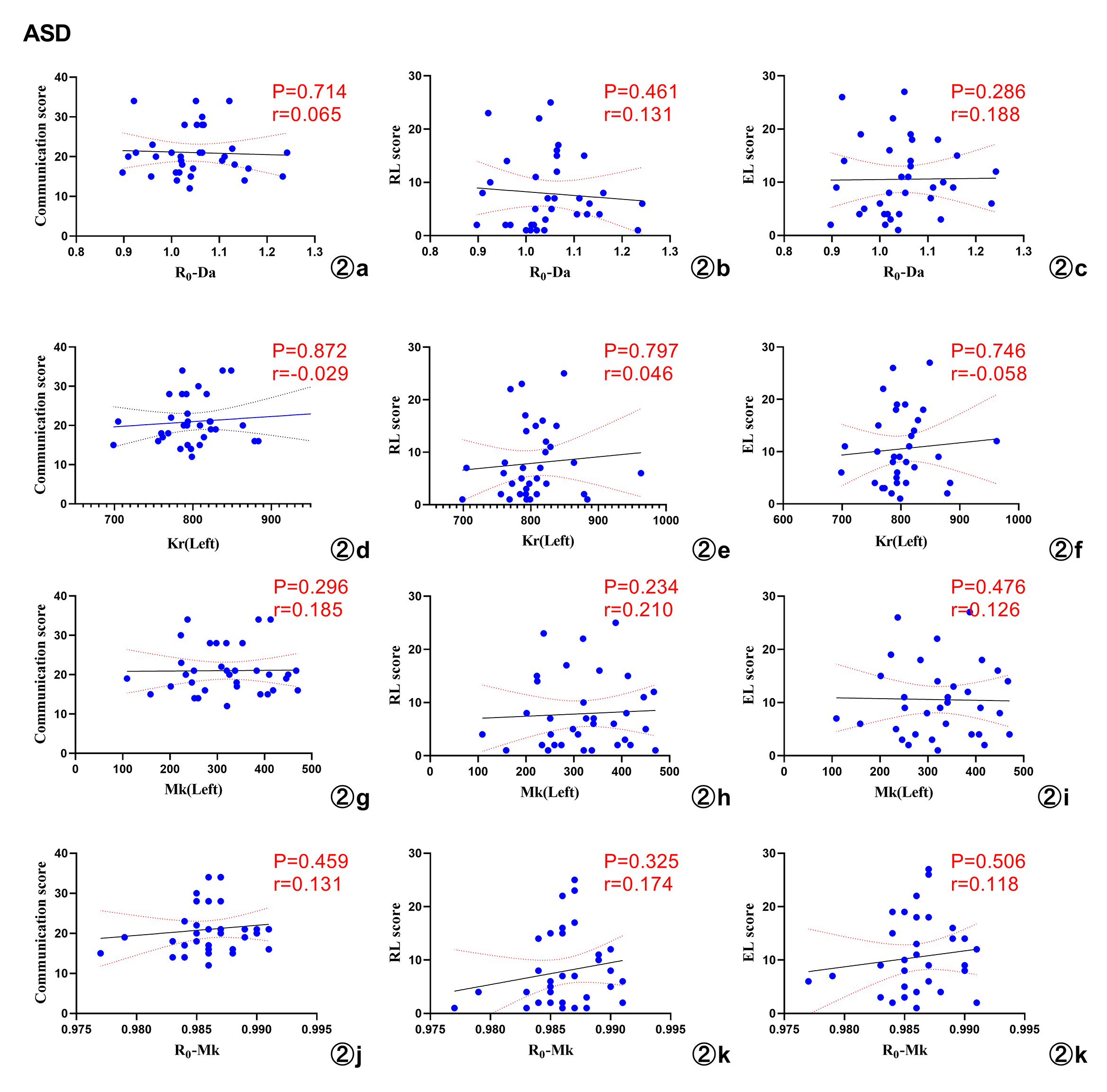
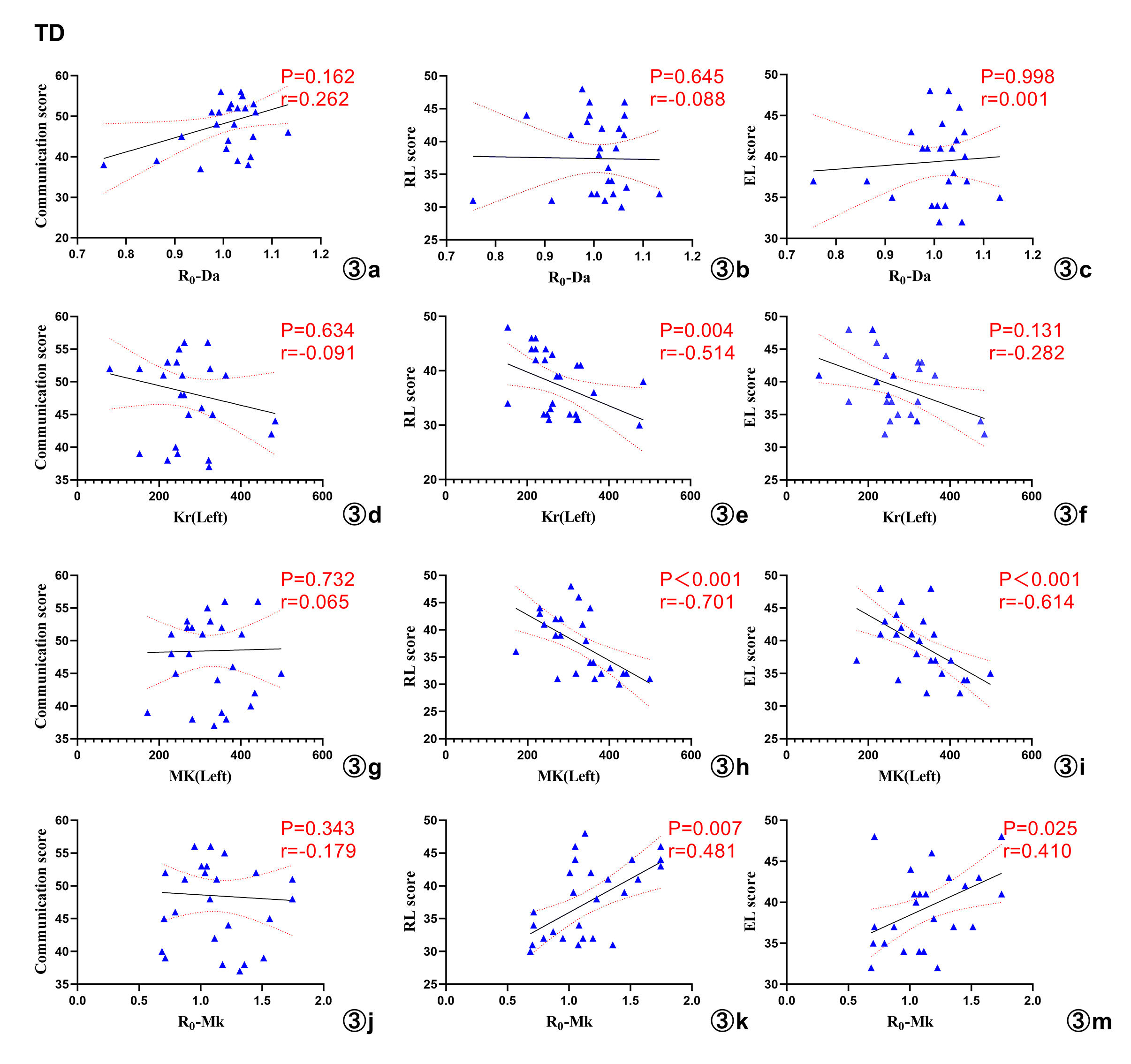
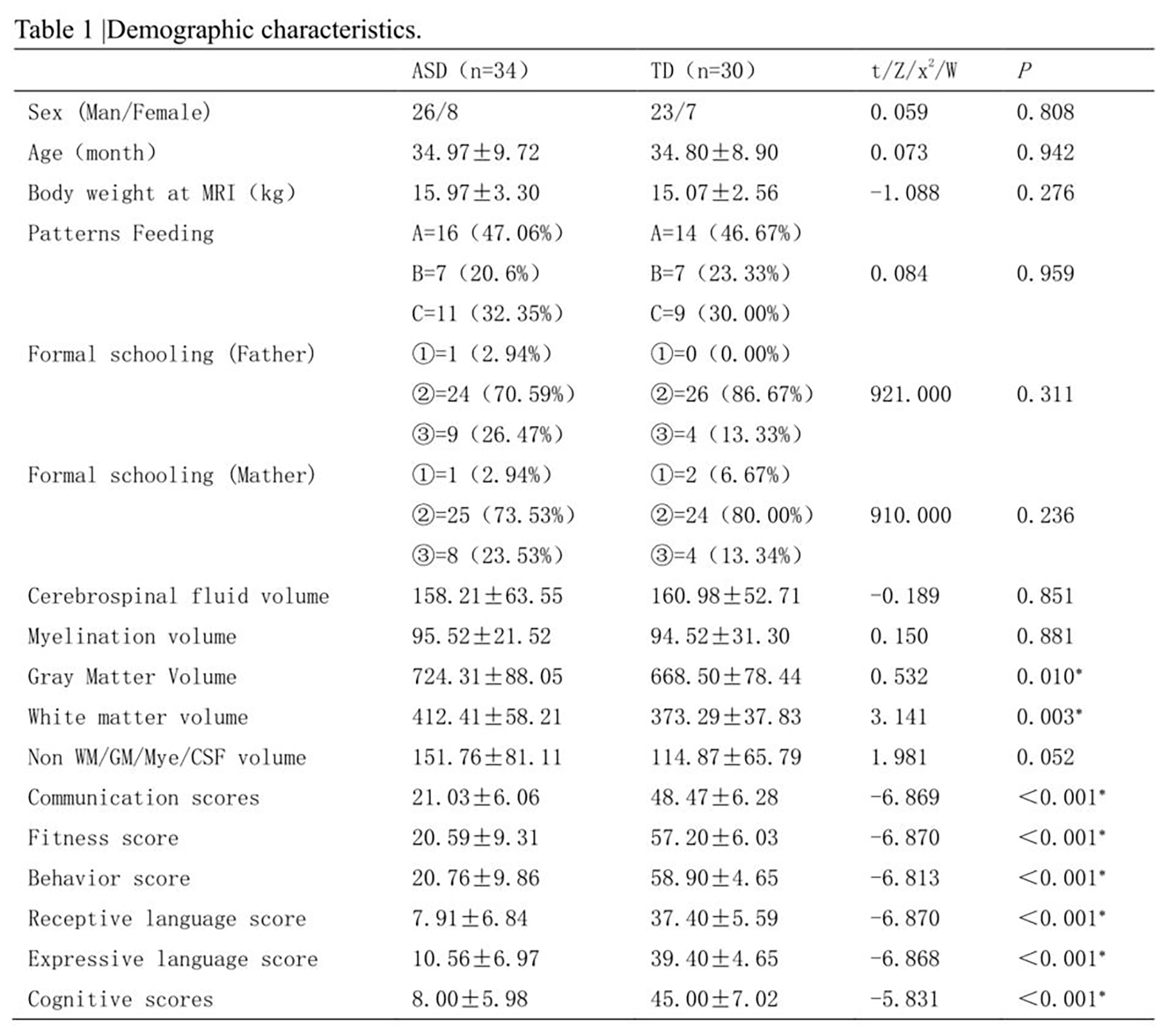
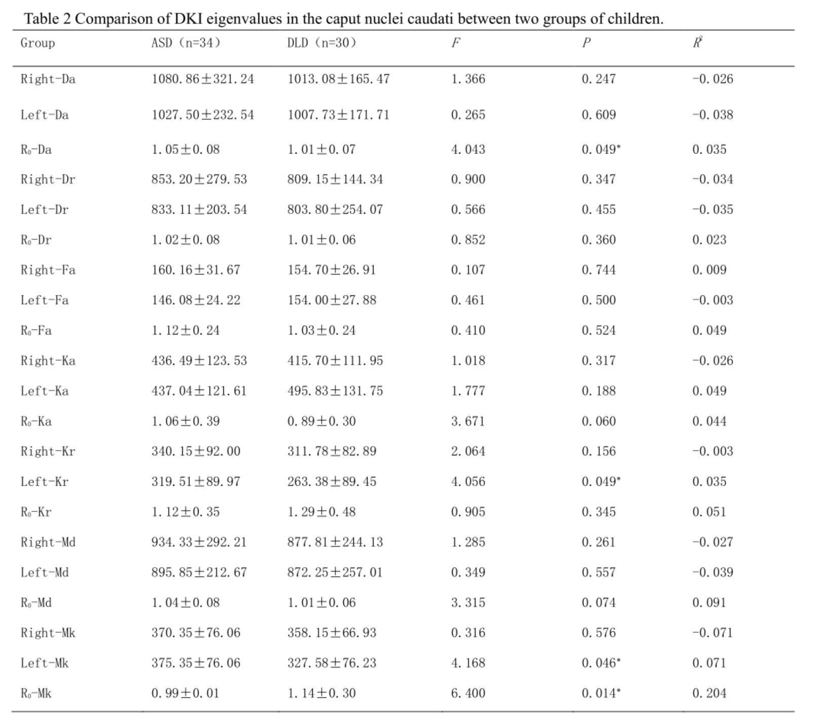
Compared with the TD group,the white matter volume and gray matter volume were found significantly increased while the communication score,physical fitness score,behavior score,language comprehension,language expression and cognitive scores of children in the ASD group were significantly lower (P<0.05),as shown in Table.After controlling the volume of gray matter and white matter, increased R0-Da value of the ASD group(P=0.049),increased Kr value (P=0.049) and Mk value (P=0.046) of the left CNC (LCNC) (P=0.049),and decreased R0-MK value (P=0.014) were found (Table 2). The correlation analysis showed that Kr in the LCNC and RL scores of children in the TD group were negatively correlated (r=-0.514,P=0.004), the Mk in the LCNC was negatively correlated with RL ( r=-0.701, P<0.001) and EL scores (r=-0.614,P<0.001), and the R0-Mk values were positively correlated with RL (r=0.481, P=0.007) and EL scores (r=0.410,P=0.025), which were not found in ASD with language deficits (P>0.05), as shown in Figure 2 and Figure 3.Discussion
It reported that the intracranial volume,as well as the white matter and gray matter volumes of ASD patients increases(7,8),which has also been found in this study.The increased volume may be associated with pathological proliferation of fiber tracts leading to abnormal brain connectivity, and information transmission(9).Increased Mk,Kr and Da reflected possible increased complexity and heterogeneity of microstructure(10,11).As shown in our results,the increased Kr of LCNC,Mk of LCNC and R0-Da, the decreased R0-Mk in ASD group could suggest that the integrity of the CNC axon may be impaired(12,13), leading to more diffusion along the radial direction and larger bias from Gaussian distribution.R0-Mk is a more sensitive parameter to tell the differences. Larger differences on the LCNC could be considered as the lateralization of the LCNC in the language development investigated in study(14).The existed correlation between DKI parameters and language proficiency assessment in TD group, but not in the ASD group may indicate that structural changes in the CNC affect brain information processing and thus affect normal language function(15).Conclusion
The volume of gray matter and white matter,and Mk ratio in bilateral CNC (R0-Mk) can be used as sensitive biomarkers to reflect the microstructural changes in the CNC in children with language defects in ASD and the R0-Mk can be potentially helpful in quantitatively evaluating the children’s language function.Acknowledgements
No acknowledgement found.References
1、Joyce-Beaulieu D., Sulkowski M.L. The Diagnostic and Statistical Manual of Mental Disorders: Fifth Edition (DSM-5) Model of Impairment. In: Goldstein S., Naglieri J. (eds) Assessing Impairment 2016. Springer, Boston, MA. DOI:10.1007/978-1-4899-7996-4-8.
2、Barger BD, Campbell JM, McDonough JD. Prevalence and onset of regression within autism spectrum disorders: a meta-analytic review. J Autism Dev Disord. 2013 Apr;43(4):817-28. doi: 10.1007/s10803-012-1621-x.
3、D'Mello AM, Moore DM, Crocetti D, Mostofsky SH, Stoodley CJ. Cerebellar gray matter differentiates children with early language delay in autism. Autism Res. 2016 Nov;9(11):1191-1204. doi: 10.1002/aur.1622.
4、Tan AP, Ngoh ZM, Yeo SSP, Koh DXP, Gluckman P, Chong YS, Daniel LM, Rifkin-Graboi A, Fortier MV, Qiu A, Meaney M. Left lateralization of neonatal caudate microstructure affects emerging language development at 24 months. Eur J Neurosci. 2021 Jul;54(2):4621-4637. doi: 10.1111/ejn.15347.
5、Leh SE, Ptito A, Chakravarty MM, Strafella AP. Fronto-striatal connections in the human brain: a probabilistic diffusion tractography study. Neurosci Lett. 2007 May 29;419(2):113-8. doi: 10.1016/j.neulet.2007.04.049.
Figures