3045
What happens to the brain over a single season of playing high school rugby: structural and white matter fibre tract changes related to impact1Auckland Bioengineering Institute, University of Auckland, Auckland, New Zealand, 2Mātai Medical Research Institute, Gisborne, New Zealand, 3Liggins Institute, University of Auckland, Auckland, New Zealand, 4Department of Ophthalmology, University of Auckland, Auckland, New Zealand, 5School of Computer Science, University of Auckland, Auckland, New Zealand, 6General Electric Healthcare, Victoria, Australia, 7Faculty of Medical and Health Sciences & Centre for Brain Research, University of Auckland, Auckland, New Zealand
Synopsis
We conducted a multimodal MRI study on high school rugby players with bespoke kinematic mouthguard sensors to investigate the correlation between the cumulative effects of subconcussive head impact exposure and any change in the structure of the brain. Despite being underpowered, the study found that the volume of the corpus callosum changed the most over a season of rugby (PCA); Axial Diffusivity of the UF and ILF tracts negatively correlated with the cumulative impact sustained by the athletes. Further investigation of the cumulative effect of playing high-contact sports over an extended period is required, especially for children with developing brains.
Introduction
Contact sports athletes experience repeated head impacts with various ranges of magnitude and acceleration1 which might lead to either concussive or subconcussive head injury2,3 . Rugby has one of the highest rates of concussion of all full-contact sports4. Wearing a mouthguard is the only mandatory protective equipment in this sport which has not shown to have much difference in the rates of concussion 5,6,7. To improve the sensitivity to subtle structural changes expected from subconcussive impacts, a method is needed which is independent to the spatial co-localization of the impact 8. Emerging evidence has shown a correlation between the repeated exposure to impact and structural changes in the volume or integrity of fibre tracts over a year of playing contact-sport9. Therefore, we conducted a multimodal MRI study on high school rugby players with bespoke kinematic mouthguard sensors to investigate the correlation between the cumulative effects of subconcussive head exposure and any change in the structure of the brain.Method
37 male high school rugby players (15 - 18 years old) were recruited for this study. Using a 3T MRI scanner (GE SIGNA Premier; General Electric, MI, USA), T1-MPRAGE (isotropic voxel size 0.5 mm3) and multi-shell diffusion MRI scans (b-values (gradient directions) = 0 (4), 1000 (15), 2000 (15), 3000 (20); isotropic voxel size 2 mm3) were acquired on the brains of all players to investigate the structural changes on the volume and white matter integrity. Scans were acquired at three time points over the season; pre-season, mid-season and post-season. To measure the extent of repetitive head impacts, a bespoke kinematic sensor mouthguard HitIQ® was moulded to fit the oral cavity of each player to record the linear and angular acceleration of the impacts. All the T1-weighted images were processed using FSL10 and AccuBrain®11 to segment and measure the volume of the brain and subcortical structures. MRtrix12 and TractSeg13 were used to segment eight bundles of fibre tracts and measure the diffusion indices (FA, MD, AD, RD). Statistical tests were conducted, including PCA, correlation, and linear regression.Results
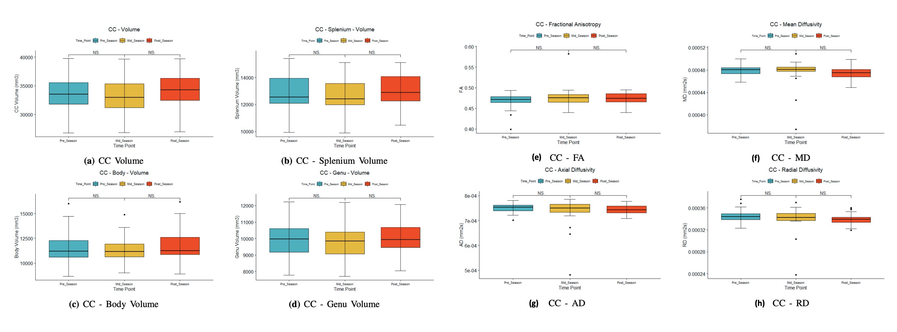
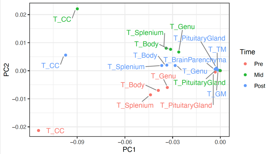
The distribution of the impact data were visually checked for the periods between the MRI scans. The impact distributions were collated for 1) mid-pre season for the first half of the season, 2) post-mid season for the second half of the season, 3) pre-post season showing the overall seasonal distribution of the impact data (Figure 1). Most of the impacts recorded were below the threshold value and were filtered out, leaving only impacts over the threshold to be summated (i.e., cumulative impact received by each athlete during practice and games for the season). The MRI volumetric data and tractography measurements for the CC are shown in Figure 2. The t-test of both volumetric and tractography measurements did not show a significant difference over the time points. In addition, Voxel-wise analysis failed to show any significant difference in FA value across the season after correcting for family-wise error.The PCA of the volumetric MRI measurements showed the regions that contributed the most to the variability of the MRI measurements over time were the CC and its constitutive parts (Figure 3). The correlation between the MRI measurements (volumetric and tractography) and impact measurements over the season (pre-post) showed 1 region and 3 tracts is presented in Figure 4. The linear regression predicting the relationship between the identified region/tracts and cumulative impacts showed uncorrected p < 0.01. As the impact distribution for the first and second half were similar, only the overall seasonal correlations are presented.Discussion and Conclusion
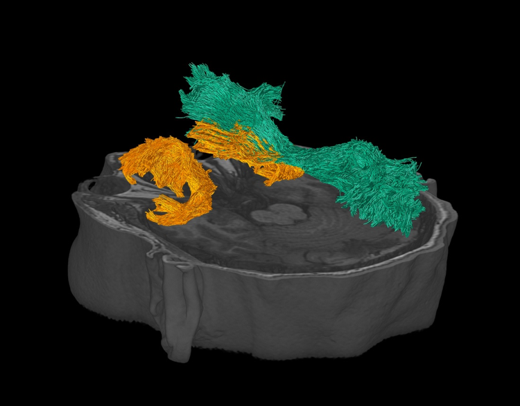
The t-test did not show significant differences between the timepoints. However, the PCA, correlation, and linear regression analyses show trends that indicate the possible per-season brain changes that accumulate over a lifetime of contact sports. Specifically, volumetric PCA showed that changes in the CC volume at different time points had the largest variability, confirming that CC is the structure of interest in analysing the influence of head impacts. However, the changes to the brain structure from cumulative subconcussive impacts over a single season have effects too small to be conclusively detected in this cohort with low sample numbers. Thus, studies with larger cohorts are required to confirm the findings presented here.Despite being underpowered, the findings here agreed with previous studies that found that the CC is the region that changed the most over a season of rugby, and the tracts UF and ILF have been negatively correlated with the average AD of the tracts. The correlation between cumulative impacts and AD in the right IFOF suggests potential axonal injury in young athletes, even without a clinically diagnosed concussion15. Particularly, the IFOF is a direct path that feeds visual information to the prefrontal cortex for higher cognitive function, leading to verbal and memory neurocognitive deficits. This is consistent with previous studies showing IFOF and UF are vulnerable to impacts16,17.
Therefore, our results show that individuals that play contact sports frequently may be exposed to a higher risk of cumulative brain damage without a defining impact to qualify as a 'traumatic brain injury'. Further investigation of the cumulative effect of playing high-contact sports over an extended period is required, especially for children with developing brains.
Acknowledgements
Supported by Kānoa - Regional Economic Development & Investment Unit, New Zealand; and the Catalyst Strategic Fund from Government Funding administered by the New Zealand Ministry of Business Innovation and Employment. We would like to acknowledge the support of GE Healthcare for assistance with the MRI protocol.References
1. Wilson, L., Stewart, W., Dams-O’Connor, K., et al. The chronic and evolving neurological consequences of traumatic brain injury. Lancet Neurol. 2017. 16, 813–825.
2. Committee on Sports-Related Concussions in Youth; Board on Children, Youth, and Families; Institute of Medicine; National Research Council; Graham R, Rivara FP, Ford MA, et al., editors. Sports-Related Concussions in Youth: Improving the Science, Changing the Culture. Washington (DC): National Academies Press (US); 2014 Feb 4. 5, Consequences of Repetitive Head Impacts and Multiple Concussions.
3. M, Haque M, Johal L, et al. Maturation of the adolescent brain. Neuropsychiatr Dis Treat. 2013;9:449-461.
4. Gardner AJ, Iverson GL, Williams WH, Baker S, Stanwell P. A systematic review and meta-analysis of concussion in rugby union. Sports Med. 2014;44(12):1717-31.
5. Barbic D, Pater J, Brison RJ. Comparison of mouth guard designs and concussion prevention in contact sports: a multicenter randomized controlled trial. Clin J Sport Med. 2005;15(5):294-8.
6. Blignaut JB, Carstens IL, Lombard CJ. Injuries sustained in rugby by wearers and non-wearers of mouthguards. Br J Sports Med. 1987;21(2):5-7.
7. Ratka J, Mansell J, Russ A. Use of Mouthguards and Association With Concussion Rates in Rugby: A Critically Appraised Topic. International Journal of Athletic Therapy & Training. 2018;23(6):226-229.
8. Davenport N.D., Lim K.O., Armstrong M.T., and Sponheim S.R. Diffuse and spatially variable white matter disruptions are associated with blast-related mild traumatic brain injury. Neuroimage . 2012;59, 2017–2024
9. Davenport M. E, Whitlow C. T, Urban J. E, et al. Abnormal White Matter Integrity Related to Head Impact Exposure in a Season of High School Varsity Football. Journal of Neurotrauma. 2014 (31):19, 1617-1624
10. M. Jenkinson, C.F. Beckmann, T.E. Behrens, M.W. et al. FSL. NeuroImage, 2012; 62:782-90
11. Abrigo J, Shi L, Luo Y, et al. Standardization of hippocampus volumetry using automated brain structure volumetry tool for an initial Alzheimer’s disease imaging biomarker. Acta Radiol. 2019;60(6):769–76
12. J.-D. Tournier, R. E. Smith, D. Raffelt, R. et al. MRtrix3: A fast, flexible and open software framework for medical image processing and visualisation. NeuroImage, 2019; 202. 116–37.
13. Wasserthal J, Neher P, Klaus H. Maier-Hein K. H, TractSeg - Fast and accurate white matter tract segmentation. NeuroImage. 2018; 183, 239-253
14. Mykol Larvie, Bruce Fischl, Chapter 3 - Volumetric and fiber-tracing MRI methods for gray and white matter, Editor(s): Joseph C. Masdeu, R. Gilberto González, Handbook of Clinical Neurology, Elsevier, Volume 135, 2016, Pages 39-60
15. Santos J. P. M, Anthony P. K, Sarrah M, et al. White Matter Abnormalities Associated With Prolonged Recovery in Adolescents Following Concussion. Frontiers in Neurology. 2021;12, 1024
16. Bahrami N, Sharma D, Rosenthal S, et al. Subconcussive Head Impact Exposure and White Matter Tract Changes over a Single Season of Youth Football. Radiology. 2016 Dec;281(3):919-926.
Figures