3036
Altered spontaneous brain activity in patients with comitant exotropia before and after surgery: a resting-state fMRI study1Department of Radiology, The First Affiliated Hospital of Nanjing Medical University, Nanjing, China
Synopsis
We investigate the brain functional alterations in patients with comitant exotropia (CE) before and after surgery, using resting-state functional magnetic resonance imaging with amplitude of low-frequency fluctuation. Our study suggested that CE may lead to decreased brain functional activities in visual-associated areas and the brain function could partially restore along with recovery of exodeviation after strabismus surgery. Besides, our results provided an explanation to the postoperative remnant of stereopsis impairment, which would be helpful to improve the clinical interventions for patients with CE.
INTRODUCTION
Comitant strabismus has been identified as the most common type of strabismus, among which comitant exotropia (CE) is twice as common as esotropia subgroup1.2. Patients with CE would usually develop compromised binocular vision and impaired stereoscopic depth perception, which could result in a profound decrease of quality of life3. Currently, corrective surgery is the main treatment approach for patients with CE. Although the deviated optic axis could be corrected surgically, the impaired stereovision may remain remnant in individuals with early-onset and long-standing strabismus4. Hence, the underlying organic basis of the remnant stereopsis disturbance need to be elucidated.Previous neuroimaging studies demonstrated that patients with CE possessed both structural and functional brain alterations5.6. However, previous studies only focused on the preoperative brain alterations of patients with CE. The cerebral functional alterations along with corrective surgery remain largely unknown.
As a reliable and reproducible functional magnetic resonance imaging approach, amplitude of low-frequency fluctuation (ALFF) can be used to reveals the neural activity of specific regions and physiological state of the brain7. Therefore, the present study was conducted to (1) investigate the differences of ALFF values between patients with CE and healthy controls (HCs), (2) explore the spontaneous brain activities alterations along with surgery in patients with CE.
METHODS
Thirty-two patients with CE were recruited to undergo a preoperative fMRI scan, as well as 20 HCs. Twenty-four among the patients were available for rescanned fMRI one month after surgery. The ALFF method was used to evaluate the group differences of spontaneous brain activity. The correlations between ALFF values and clinical variables were analyzed in the patient group.RESULTS
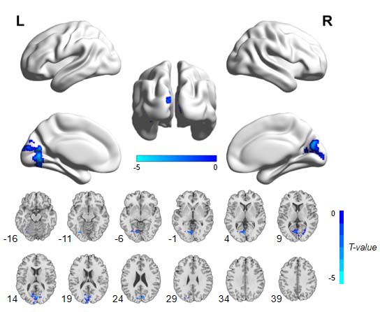
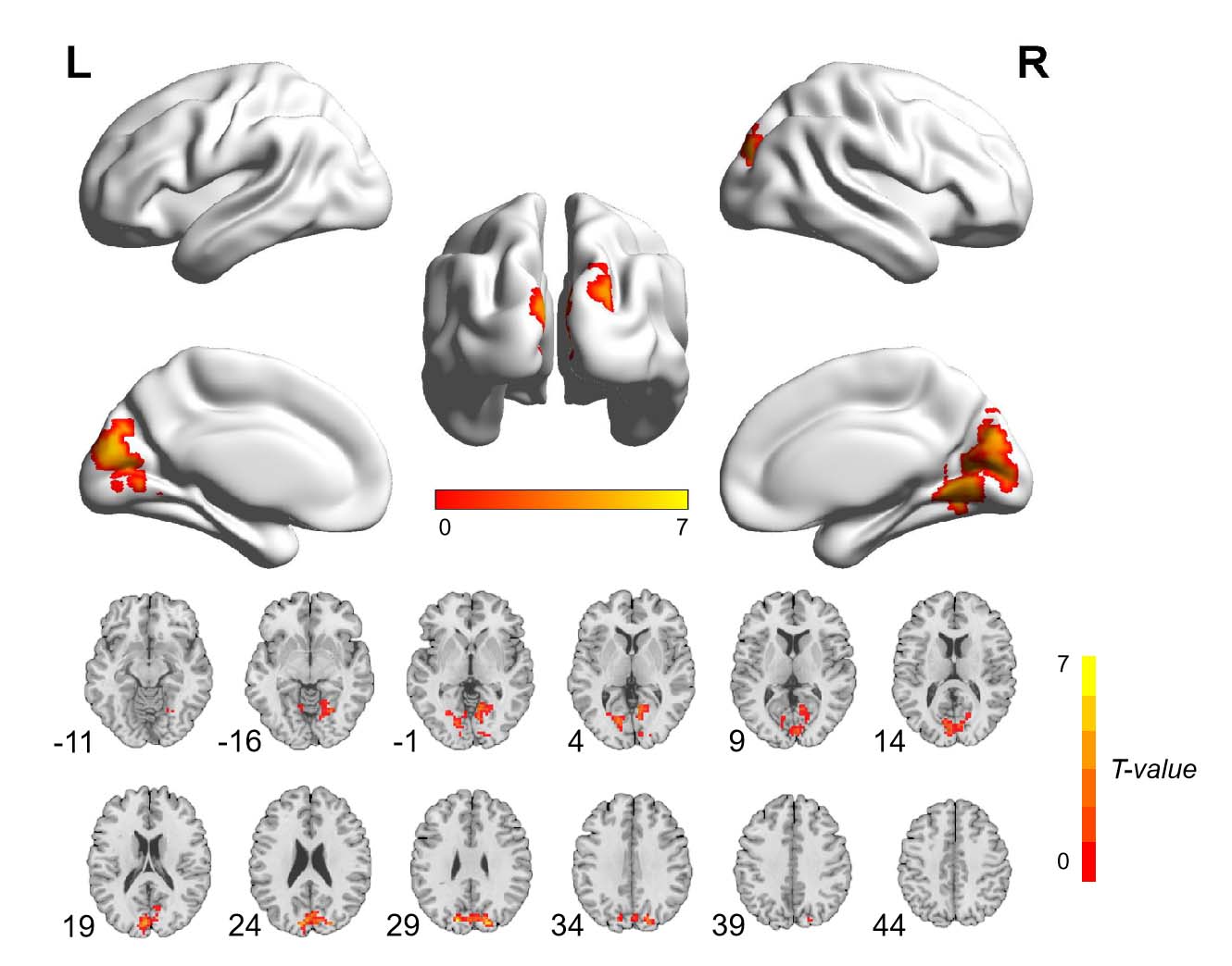
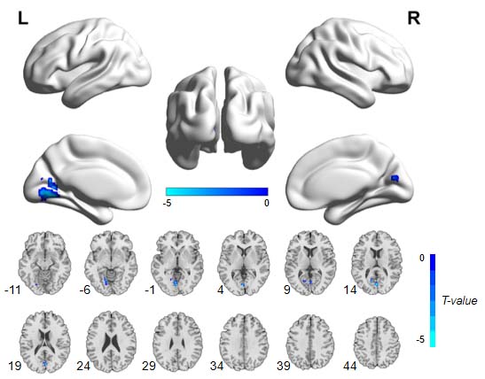
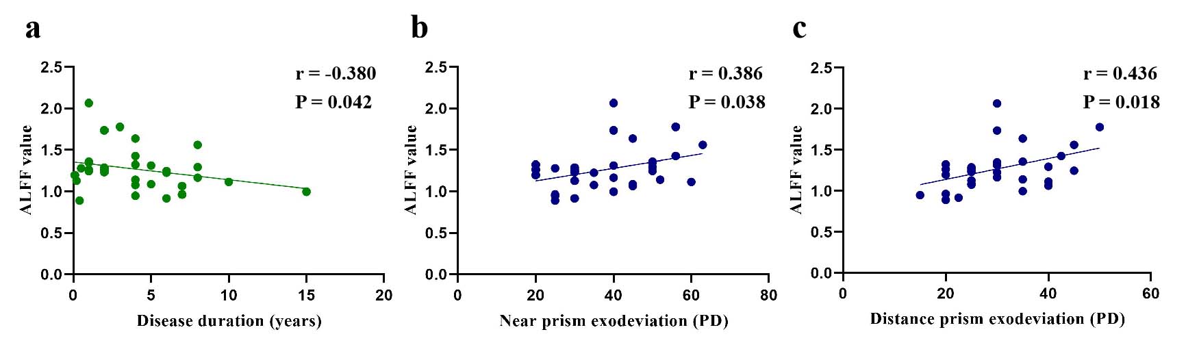
The preoperative patient group showed significantly higher scores of stereoacuity than HCs (P < 0.001). Compared with corresponding preoperative information, the 24 patients with follow-up showed significantly lower scores of stereoacuity (P = 0.027) as well as lower near (P < 0.001) and distance (P < 0.001) prism exodeviation. Besides, the postoperative patients showed significantly higher scores of stereoacuity than HCs (P < 0.001).Preoperatively, compared with HCs, 32 patients with CE showed significantly decreased ALFF values in one cluster involving bilateral cuneus, bilateral calcarine sulcus and left lingual gyrus. The ALFF values in the above cluster were negatively correlated with disease duration (r = -0.380, P = 0.042), while positively correlated with near and distance prism exodeviation (r = 0.386, 0.436; P = 0.038, 0.018, respectively). One month after surgery, 24 patients with available rescanned fMRI demonstrated increased ALFF values in one cluster involving bilateral cuneus, calcarine sulcus and lingual gyrus relative to preoperative collection, while still reduced ALFF values in the cluster involving left lingual gyrus and calcarine sulcus compared with HCs.
DISCUSSION
The cuneus, calcarine and lingual gyrus are major components of secondary visual cortex (V2), which plays a critical role in visual memory and various higher order functions of vision, including processing of spatial visual information, perception of color and identification of facial expressions of emotions8.9. The decreased ALFF values in bilateral cuneus, calcarine sulcus and left lingual gyrus in preoperative patient group observed in our study may be an expression of the disturbed processing of stereo visual information, which was in line with the impaired stereopsis of patient group. The postoperative augmented ALFF values in bilateral V2, accompanied by postoperatively improved stereopsis in our study cohort, may indicate the functional restoration of visual cortex. Additionally, the scores of stereoacuity were still higher in the postoperative patients than in HCs, which was in accordance with the decreased brain activities. Therefore, we supposed that the decreased ALFF values in left lingual gyrus and calcarine sulcus postoperatively could be associated with the residuary stereopsis disturbance in the patients with CE after the exodeviation was corrected sugically.CONCLUSION
Our study suggested that CE may lead to decreased brain functional activities in visual-associated areas and the brain function could partially restore along with recovery of exodeviation after strabismus surgery. Besides, our results provided an explanation to the postoperative remnant of stereopsis impairment. Understanding the neuropsysiological alterations of CE after surgery would be helpful to improve the clinical interventions for patients with CE. Moreover, the treatment response could be displayed objectively using fMRI examinations, which may contribute to evaluate the effect of the treatment.Acknowledgements
No acknowledgement found.References
1. Matsuo T, Matsuo C: The prevalence of strabismus and amblyopia in Japanese elementary school children. Ophthalmic Epidemiol 2005; 12(1):31-36.
2. Yu CB, Fan DS, Wong VW, et al. Changing patterns of strabismus: a decade of experience in Hong Kong. Br J Ophthalmol 2002; 86(8):854-856.
3. Adams DL, Economides JR, Horton JC. Contrasting effects of strabismic amblyopia on metabolic activity in superficial and deep layers of striate cortex. J NEUROPHYSIOL 2015; 113(9):3337-3344.
4. Zhou J, Wang Y, Feng L, et al. Straightening the Eyes Doesn't Rebalance the Brain. FRONT HUM NEUROSCI 2017; 11:453.
5. Yan X, Lin X, Wang Q, et al. Dorsal visual pathway changes in patients with comitant extropia. PLOS ONE 2010; 5(6):e10931.
6. Huang X, Zhou S, Su T, et al. Resting cerebral blood flow alterations specific to the comitant exophoria patients revealed by arterial spin labeling perfusion magnetic resonance imaging. MICROVASC RES 2018; 120:67-73.
7. Wang P, Yang J, Yin Z, et al. Amplitude of low-frequency fluctuation (ALFF) may be associated with cognitive impairment in schizophrenia: a correlation study. BMC PSYCHIATRY 2019; 19(1):30.
8. Roland PE, Gulyas B. Visual memory, visual imagery, and visual recognition of large field patterns by the human brain: functional anatomy by positron emission tomography. CEREB CORTEX 1995; 5(1):79-93.
9. Kitada R, Johnsrude IS, Kochiyama T, et al. Brain networks involved in haptic and visual identification of facial expressions of emotion: an fMRI study. NEUROIMAGE 2010; 49(2):1677-1689.
Figures