3030
Association Between the Recall Verbal Memory and Side of Asymptomatic Carotid Artery Stenosis1Department of Biomedical Imaging and Radiological Sciences, National Yang Ming Chiao Tung University, Taipei, Taiwan, 2School of Medicine, National Yang-Ming University, Taipei, Taiwan, 3Neurological Institute, Taipei Veterans General Hospital, Taipei, Taiwan, 4Institute of Brain Science, National Yang Ming Chiao Tung University, Taipei, Taiwan
Synopsis
Previous studies reported that the patients with asymptomatic internal carotid stenosis (aICS) had a higher risk of stroke and recall verbal memory impairment. However, whether the side of aICS could cause different verbal memory outcomes was less explored. In this study, significant difference of functional connectivity (FC) was found between the left and right aICS groups. The correlation analysis estimating the association between FC and recall verbal memory also presented different profiles between two aICS groups.
Background and Purpose
Patients with asymptomatic internal carotid artery stenosis (aICS) have a higher risk of ischemic stroke. With the progression of plaque, the interruption of blood flow might even make patients suffer from dizziness and cognitive impairment (such as the verbal memory function). However, comparisons of functional connectivity (FC) and corresponding symptoms between left and right aICS groups were less explored. In our study, we aimed to investigate the altered FCs and the extent of cognitive impairment associated with the laterality of aICS.Materials and Methods
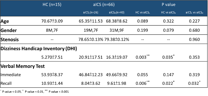
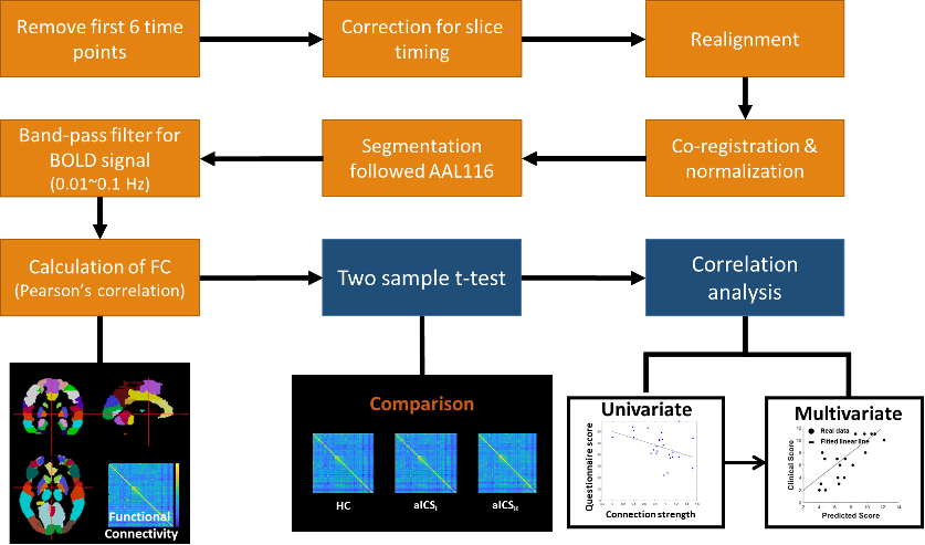
We recruited 15 healthy controls (HCs), 26 patients with left aICS (aICSL), and 40 patients with right aICS (aICSR). Inclusion criteria of patients were the degree of stenosis greater than 60% and no neurodegenerative diseases. Dizziness handicap inventory (DHI) and verbal memory test were used to evaluate the severity of dizziness and level of working (immediate) and short-term (delayed recall) verbal memory, respectively (Table 1).[1] MRI data, including 3D BRAVO T1-weighted images (TR/TE: 12.2/5.2 ms; voxel size: 1.0×1.0×1.0 mm3) and EPI BOLD fMRI (TR/TE: 3000/30ms; voxel size: 1.73×1.73×3mm3) during resting state (124 volumes) were acquired on a 3T MR scanner (GE, Discovery 750).The fMRI data went for standard FC extraction with Data Processing Assistant for Resting-State fMRI (DPARSF) toolbox.[2] The extraction included correction for slice timing, realignment, co-registration between 3D-T1WI and fMRI, regression to confounding effects of head motion and signals from white matter and cerebrospinal fluid, spatial normalization, and spatial smoothing with a 6-mm full-width half-maximum Gaussian kernel. The Automatic Anatomical Labeling atlas (AAL116) was used as template to parcel out the cortical and subcortical region into 116 regions.[3] FC between each pair regions was estimated with Pearson’s correlation coefficient on regional BOLD signals (with a bandpass filtering between 0.01 and 0.10 Hz) followed by the Fisher’s r-to-z transform.
The altered FC induced by the interrupted perfusion of left or right aICS would be identified using a two-sample t-test with the comparison to the healthy control group (p<0.05). Pearson’s correlation analysis was applied to identify the potential FCs associated with recall verbal memory function (p<0.05). By further considering the brain as an integrated network, we employed multivariate stepwise regression model to investigate the relation between multiple FCs (identified by the Pearson’s correlation analysis) and the recall verbal memory function. The processing workflow is shown in Figure 1.
Results and Discussion
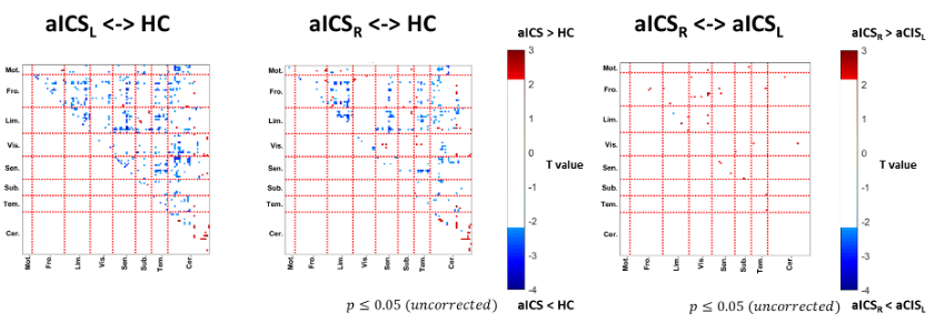
The patients with aICS had significantly more severe dizziness and deficit on recall verbal memory test compared to the HCs (Table 1). For the comparison between aICSL and aICSR groups, the performance of recall verbal memory was significantly poorer in the aICSL group compared to that in the aICSR group (Table 1).Figure 2 shows the comparisons of FCs between the HC, aICSL, and aICSR groups. Similar patterns were found when comparing the FCs of HC group with two aICS groups. Frontal and limbic related FCs showed lower coupling strength in aICS groups than that in the HC group. Higher FCs within the cerebellum regions representing a potential neuro-plastic compensation after stenosis were also observed in aICS groups. Compared to aICSR, the significantly reduced FCs of limbic regions with visual and sensory regions were found in the aICSL group.
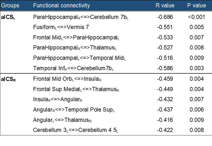
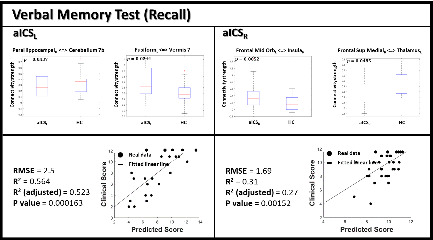
To identify the potential FCs related with recall verbal memory function, we listed the significant correlations in the aICSL and aICSR groups (Table 2). In general, the aICSL group exhibited stronger correlations (higher absolute correlation coefficients) between the FC and recall verbal memory function compared to the aICSR group. In the aICSL group, the FC between right parahippocampal gyrus and left cerebellum 7b showed a negative correlation (R=-0.686) with recall verbal memory function. The connection between left fusiform and vermis 7 also showed a significantly negative correlation (R=-0.551). In the aICSR group, the significant correlation with recall verbal memory function was observed on FC between left orbital part of middle frontal gyrus and right insula (R=-0.459). The FC between left medial superior frontal gyrus and right thalamus also presented a significant correlation (R=-0.449). Accordingly, the alteration of the highlighted FCs might be associated with verbal memory loss after aICS.
The relations between integrated brain network and recall verbal memory were revealed based on the stepwise linear regression. [4]
$$aICSLScorePredict=13.356-7.307×FCParaHippocampalL<->Cerebellum 7bR-5.390×FCFusiformR<->Vermis7$$
$$aICSRScorePredict=11.278-2.8879×FCFrontal Sup MedialR<->ThalamusL-2.2385×FCFrontal Mid OrbL<->InsulaR$$
Above equations showed the relations between selected FCs and the recall verbal memory score in the aICSL and aICSR groups, respectively. The predicted score showed a higher consistence with clinical score in the aICSL group (R2=0.564) compared to the aICSR group (R2=0.310) (Figure 3).
Conclusions
In this study, patients with aICSL showed poorer performance of recall verbal memory, significant reduction of FCs, and stronger correlations between FCs and recall verbal memory compared to the patients with aICSR. We also reported higher consistence between the observed scores and predicted scores using a regression model in the aICSL group. Based on our results, the laterality of aICS can be a critical factor to link with the decline of recall verbal memory function and guide the treatment for patients with aICS.Acknowledgements
This work was supported by the Ministry of Science and Technology, Taiwan (MOST 109-2314-B-010-022-MY3) and the National Yang-Ming University (VGHUST110-G7-2-2).References
1. Jacobson, G.P. and C.W. Newman, The development of the Dizziness Handicap Inventory. Arch Otolaryngol Head Neck Surg, 1990. 116(4): p. 424-7.
2. Chao-Gan, Y. and Z. Yu-Feng, DPARSF: A MATLAB Toolbox for "Pipeline" Data Analysis of Resting-State fMRI. Front Syst Neurosci, 2010. 4: p. 13.
3. Tzourio-Mazoyer, N., et al., Automated anatomical labeling of activations in SPM using a macroscopic anatomical parcellation of the MNI MRI single-subject brain. Neuroimage, 2002. 15(1): p. 273-89.
4. Liebscher, E., A universal selection method in linear regression models. Open Journal of Statistics, 2012. 2(02): p. 153.
Figures