3029
fMRI response to individualized cue-reactivity paradigm of males with gaming behavior1School of Physics and Engineering, ITMO University, Saint-Petersburg, Russian Federation, 2Department of Radiology, Federal Almazov North‐West Medical Research Center, Saint-Petersburg, Russian Federation
Synopsis
Task-related fMRI studies are providing increasing amount of information on the neurobiological aspects of the Gaming Disorder. This study aims to Explore both the activation patterns and the functional connectivities present in the gaming disorder-like subject brains via task-based fMRI study using individualized visual stimuli. 24 male test participants and 25 male control participants took part in the fMRI scanning with gaming-related and neutral visual stimuli. Data analysis showed altered nucleus accumbens connectivity and amygdala resembling that in cases of substance addiction1,2.
Introduction
Gaming Disorder (as defined in the ICD-11) or Internet Gaming Disorder (as defined in the DSM-5) has attracted a lot of attention in recent years. With the ongoing debate on the origins and personal and social implications of compulsive gaming behavior3–6 studies have been undertaken to explore the neurobiological aspects of the disorder. A number of works explore the brain functional networks via functional MRI (fMRI), particularly with task-related fMRI8,9 and functional connectivity estimation using generalized Psychophysiological Interactions (gPPI) analysis. The findings in these include altered functional connectivity and task-related activation patterns similar to the ones observed in substance addiction, particularly related to the mechanisms of craving and reward5. These studies have demonstrated the efficiency of cue-response task-related fMRI experiments in detecting the altered activation and connectivity patterns. This study aims to further explore the functional connectivities present in the subjects with gaming (or internet gaming) disorder-like behavior via task-based fMRI study using visual associative stimuli.Methods
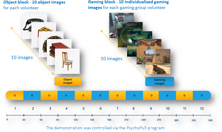
The participants for the study were recruited via social media by announcing a volunteer survey in the local academic and gaming social groups. Among the 49 volunteers participating in the study, 24 males were initially labelled as gamers (mean age 24.9 ± 3.1 years) based on the self-reported average gaming time (>20 hour per week) and 25 male volunteers formed the control (<10 gaming hours per week) group (mean age 23.6 ± 3.9 years). The declared game time was verified via an analysis of gaming platform accounts provided in the survey. All surveyed gamers displayed a preference for of different game genres and titles. The 49 male participants were selected from the larger (234) volunteer group by vetting out non-responding participants, participants with borderline gaming time, and female participants. The latter were excluded for sample homogenization due to a critically low response rate in gaming group (only 2 female participants with gaming time >20 hours per week). All the participants were provided a written informational guide in accordance with local ethics committee. All the selected participants have filled out the IGD-20 test9, which resulted in the 28.9 ± 7.8 score for the control group and 46 ± 13.3 score for the gaming group, with the difference between the two groups confirmed by the paired t-test with p=0.000004.The fMRI experiment used a paradigm of 12 consecutive alternating blocks: "Object images" and "Gaming images". The " Object images l" block consisted of 10 random non-game-related images. The "Gaming images" block consisted of 10 random individualized game images. Individual stimuli were selected for each gamer in accordance to the preferred games listed in the survey. The control group stimuli in the “Gaming images” block were randomly selected from the gamer group image pool. The “Object images” stimuli were the same for all participants and consisted of 40 images unrelated to video games. The duration of one image presentation was 3 seconds, the duration of the block was 30 seconds, the duration of the entire paradigm was 360 seconds (Fig. 1). The demonstration was controlled via the PsychoPy3 program. The images were projected onto a screen visible to the subject via a mirror system. MRI data were obtained on a 1.5T scanner using a 12-channel head coil.
The scan protocol comprised structural MPRAGE and functional EPI-FID sequences. The structural 3D scans were obtained in the sagittal plane with the following parameters: matrix 192×192×160, 240×240×192 mm FoV, TE/TR = 3.7/2400 ms. Functional (BOLD contrast) scans were obtained in the axial plane during the presentation of the visual stimuli. The protocol parameters for functional scans were: matrix 64×64, 230×230 mm FoV, TE/TR = 50/3000 ms, slice thickness = 5 mm, slice gap = 1.3 mm. Functional connectivity analysis was performed using the CONN 18b toolbox (MATLAB R2020b), activation patterns were detected with SPM12 toolbox (MATLAB R2020b). In all cases the first two scans were discarded to avoid the effects of triggering a scan. All scans were normalized to MNI-space and converted to an isotropic 2 mm voxel space, local averaging with Gaussian 8 mm FWHM window was applied during functional connectivity analysis. We performed ROI-to-ROI analysis with task modulation effects (gPPI) as well as activation pattern detection in the GLM with p-FDR<0.05.
Results
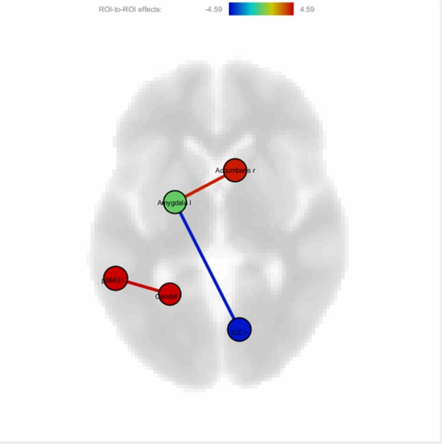
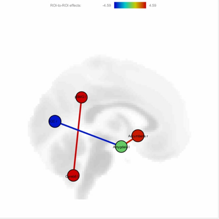
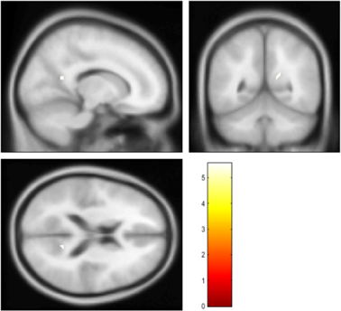
Gaming group participants, compared to controls group participants, exhibited significantly increased functional connectivity (Fig 2., Fig. 3.) between: amygdala (left hemisphere) and nucleus accumbens (right hemisphere), 8 area in left cerebellar hemisphere and posterior supramarginal gyrus (left hemisphere). On the other side, functional connectivity amygdala (left hemisphere) and Intracalcarine cortex was decreased.The results of the activation pattern analysis via the GLM model with p-FWE<0.05 are presented on Fig.4.Discussion and conclusion
While the survey results do not directly indicate the presence of IGD in any of the subjects as the IGD is suggested with test scores over 71 points, the detection of addiction-like patterns in functional connectivity analysis suggests the presence of additiction-like state in gaming volunteers.Acknowledgements
The experiment was founded by the Russian Science Foundation (Grant No. 18-79-10167);
The processing results is funded by RPMA grant of School of Physics and Engineering of ITMO University.
References
1. Chen C.-Y., Yen J.-Y., Wang P.-W., Liu G.-C., Yen C.-F., · Ko C.-H. Altered Functional Connectivity of the Insula and Nucleus Accumbens in Internet Gaming Disorder: A Resting State fMRI Study. Eur Addict Res. 2016; 22:192-200
2. Cheng, H., Liu, J. Alterations in Amygdala Connectivity in Internet Addiction Disorder. Sci Rep. 2010; 10, 2370.
3. Hull JG, Brunelle TJ, Prescott AT, Sargent JD. A longitudinal study of risk-glorifying video games and behavioral deviance. Journal of Personality and Social Psychology. 2014;107(2):300-325.
4. Jeong EJ, Ferguson CJ, Lee SJ. Pathological Gaming in Young Adolescents: A Longitudinal Study Focused on Academic Stress and Self-Control in South Korea. J Youth Adolescence. 2019;48(12):2333-2342.
5. Weinstein AM. An Update Overview on Brain Imaging Studies of Internet Gaming Disorder. Front Psychiatry. 2017;8.
6. Latham AJ, Patston LLM, Tippett LJ. The virtual brain: 30 years of video-game play and cognitive abilities. Front Psychol. 2013;4.
7. Ko C-H, Liu G-C, Hsiao S, et al. Brain activities associated with gaming urge of online gaming addiction. Journal of Psychiatric Research. 2009;43(7):739-747.
8. Ma S-S, Worhunsky PD, Xu J, et al. Alterations in functional networks during cue-reactivity in Internet gaming disorder. Journal of Behavioral Addictions. 2019;8(2):277-287.
9. Halley M. Pontes, Orsolya Király, Zsolt Demetrovics, Mark D. Griffiths. The Conceptualisation and Measurement of DSM-5 Internet Gaming Disorder: The Development of the IGD-20 Test. 2014; PLoS ONE 9(10): e110137.
Figures