3026
Effects of Dobutamine on Neural Activity in Normal Subjects: A Exploratory Resting-State Functional MRI Study.1School of Biological Science and Medical Engineering, Beihang University, Beijing, China, 2Department of Radiology, Beijing Friendship Hospital, Capital Medical University, Beijing, China, 3National Space Science Center, Chinese Academy of Sciences, Beijing, China, 4Philips Heathcare, Beijing, China, 5National Cancer Center/Cancer Hospital and Shenzhen Hospital, Shenzhen, China
Synopsis
To explore the alternations in spontaneous brain activities in normal subjects after dobutamine infusion, the regional homogeneity (ReHo) method was used to analyze the interregional synchronized activity of all participants. The results showed that compared with normal resting state, significantly decreased ReHo was found after dobutamine infusion in bilateral frontal lobe, while increased in left temporal lobe and right parietal lobe, suggesting that dobutamine may have an effect on functional brain activity.
Introduction
Dobutamine is used clinically to increase cardiac output and increase blood pressure during shock and heart failure. Several experimental and clinical studies on different species have shown that dobutamine can mobilize neurovascular protection mechanisms or affect cerebral perfusion, thereby improving systemic and regional hemodynamics 1–4. However, dobutamine has not been well studied in normal subjects, and in particular, there is a lack of research on the effects of dobutamine on neural activity in the brain. The purpose of this study was to explore the alternations in spontaneous brain activities reflected by regional homogeneity (ReHo) in normal subjects with dobutamine infusion using blood oxygen level dependent functional magnetic resonance imaging (BOLD-fMRI).Methods
Twenty-seven male healthy volunteers were recruited in our study. Participants who found plaque or premature beats through carotid ultrasound and routine electrocardiogram (ECG) monitoring were excluded. In total, 22 subjects were included in this study (27 ±2.96 years). All of them received resting-state functional magnetic resonance imaging (rs-fmri) scanning twice on a 3.0T Philips Ingenia scanner with a head-neck coil: normal resting states (Scan1) and dobutamine infusion states (Scan2). Two scans underwent the same MRI acquisition protocol: TR = 2016.2 ms, TE = 35 ms, FOV = 240 mm × 240 mm, Matrix = 64 × 64, slice thickness = 4 mm. In total, 28 slices were obtained for each subject. For dobutamine infusion, the appropriate dose of dopamine was configured before the experiment according to the height and weight of different subjects. In the experiment, the following physiological parameters were monitored and recorded electronically before and after dobutamine infusion: (1) heart rate (HR), (2) SpO2, (3) blood pressure (BP) from BP cuff, and (4) ECG. All physiological data were sampled automatically. Image data preprocessing was performed using data processing & analysis of brain imaing (DPABI) V6.0 toolbox, and the ReHo method was used to analyze the interregional synchronized activity of all participants. Paried T-test was performed to compare the ReHo maps between the two states. Additionally, pearson correlation analysis was then used to assess the correlations between physiological data and the difference in abnormal ReHo of the two states. P < 0.05 was considered statistically significant.Results
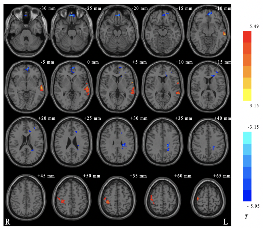
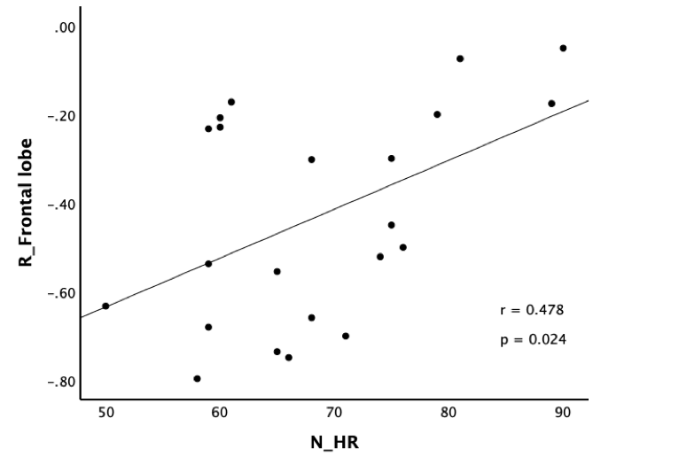
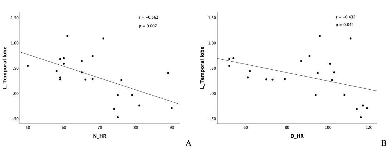
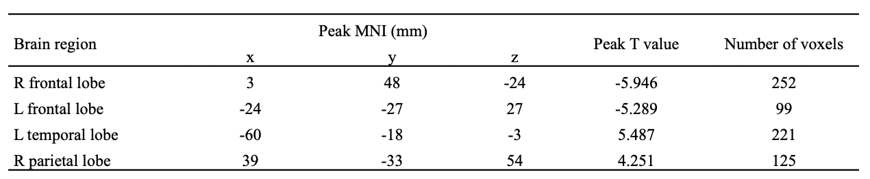
Compared with normal resting state, significantly decreased ReHo was found after dobutamine infusion in bilateral frontal lobe, while increased in left temporal lobe and right parietal lobe (GRF correction, voxel P value < 0.005, cluster P value < 0.05, two tailed) (Table 1 and Figure 1). The difference of ReHo value of right frontal lobe was positively correlated with the value of normal state heart rate (r = 0.478, P = 0.024) (Figure 2), while negative correlation was found between the abnormal ReHo of left temporal lobe and heart rate in two states (r = -0.562, P = 0.007, and r = -0.432, P = 0.044, respectively) (Figure 3).Discussion
A higher ReHo indicates enhanced coordination of the corresponding brain regions, while a lower ReHo reflects the decrease in neuronal activity in these regions or the consistency of tending to be disordered, which indicates that there may be brain dysfunction. The results showed lower ReHo values in bilateral frontal lobe after dobutamine infusion, and it is mainly located in the orbital gyrus. Physiologically, orbitofrontal cortex is responsible for the regulation of behavior and emotions, which displays decreased activity during performance of attention-demanding tasks 5. These results indicate that the dobutamine infusion may influence the mood, thereby affecting the neural activity of frontal lobe. The temporal lobe, as an auditory and visual related brain region, is involved in the processing of emotions and working memory 6. Increased ReHo value of the temporal gyrus after dobutamine infusion reminds us to pay attention to its impact on hearing and vision. Another brain region where ReHo increased is mainly located in the right postcentral gyrus in parietal lobe, which indicate that dobutamine may increase emotional changes in normal subjects, because it is the main sensory receptive area for somatosensory information 7. Pearson correlation analysis revealed that the heart rate is associated with interregional synchronized activity, which may due to neurovascular protection mechanisms.Conclusion
By applying rs-fmri studies based on ReHo analysis, we identified brain regions that underwent abnormal changes after dobutamine infusion, and the change values showed correlation with a variety of physiological parameters. This study provides preliminary insight into the effects of dobutamine on normal subjects brain function and provide new ideas for further understanding of the pharmacological mechanisms of dobutamine.Acknowledgements
This work was supported by Beijing Scholar 2015, Space Medical Experiment Project of China Manned Space Program (No. HYZHXM01012) and National Natural Science Foundation of China (No. 61931013).References
1. Ogoh S, Moralez G, Washio T, et al. Effect of increases in cardiac contractility on cerebral blood flow in humans. Am J Physiol - Hear Circ Physiol. 2017;313(6):H1155-H1161. doi:10.1152/ajpheart.00287.2017
2. Meier M, Bettschart-Wolfensberger R, Schwarzwald CC, Portier K, Gysler A, Ringer SK. Effects of dobutamine on cardiovascular function and oxygen delivery in standing horses. J Vet Pharmacol Ther. 2020;43(5):470-476. doi:10.1111/jvp.12869
3. Dubin A, Lattanzio B, Gatti L. The spectrum of cardiovascular effects of dobutamine - from healthy subjects to septic shock patients. Rev Bras Ter Intensiva. 2017;29(4):490-498. doi:10.5935/0103-507X.20170068
4. Mutoh T, Mutoh T, Sasaki K, et al. Isoflurane postconditioning with cardiac support promotes recovery from early brain injury in mice after severe subarachnoid hemorrhage. Life Sci. 2016;153:35-40. doi:10.1016/j.lfs.2016.04.020
5. Wu K, Liu M, Zhao G, He L, Tan Y. Altered regional homogeneity in delayed encephalopathy after carbon monoxide poisoning: A resting-state fMRI study. Neurosci Lett. 2020;729:135002. doi:10.1016/j.neulet.2020.135002
6. Shan X, Qiu Y, Pan P, et al. Disrupted Regional Homogeneity in Drug-Naive Patients With Bipolar Disorder. Front Psychiatry. 2020;11(August):1-10. doi:10.3389/fpsyt.2020.00825
7. Liu Y, Zhao X, Cheng Z, et al. Regional homogeneity associated with overgeneral autobiographical memory of first-episode treatment-naive patients with major depressive disorder in the orbitofrontal cortex: A resting-state fMRI study. J Affect Disord. 2017;209(November 2016):163-168. doi:10.1016/j.jad.2016.11.044
Figures