3024
Evaluation of banding artifacts suppression for inner ear MR imaging by 3D b-FFE-XD technique1Department of Radiology, Peking Union Medical College Hospital,Chinese Academy of Medical Sciences, Beijing, China, 2Philips Healthcare, Beijing, China
Synopsis
High resolution inner ear MR imaging plays an important role for preoperative plan of hearing system, such as cochlear implant surgery etc. Nowadays, 3D balanced fast-field echo sequence (3D b-FFE) is an effective technique to deliver high-resolution images up to a spatial resolution 0.5 0.5 0.5mm. However, black banding artifacts are frequently observed in the images of inner ear anatomic structures at 3.0T due to magnetic field inhomogeneity. In this study, the 3D b-FFE-XD technique which combines a cycling RF phase with the conventional b-FFE sequence to suppress the banding artifacts and improve the image quality.
Synopsis
High resolution inner ear MR imaging plays an important role for preoperative plan of hearing system, such as cochlear implant surgery etc. Nowadays, 3D balanced fast-field echo sequence (3D b-FFE) is an effective technique to deliver high-resolution images up to a spatial resolution 0.5 0.5 0.5mm. However, black banding artifacts are frequently observed in the images of inner ear anatomic structures at 3.0T due to magnetic field inhomogeneity. In this study, the 3D b-FFE-XD technique which combines a cycling RF phase with the conventional b-FFE sequence to suppress the banding artifacts and improve the image quality.Summary of main findings
The 3D b-FFE-XD technique can effectively reduce banding artifacts in inner ear imaging at 3T compared with the conventional 3D b-FFE sequence.Introduction
It remains clinically challenging to diagnose diseases in inner ear due to the complicated mixture of soft tissues, dense bones, and fluid-filled compartments [1]. Despite the use of 3D balanced fast field echo (3D b-FFE) technique, this sequence is prone to magnetic field inhomogeneity and is frequently disturbed by banding artifacts with increased severity at 3.0T. This study proposed a RF phase-cycling technique as an addition to the traditional b-FFE sequence, namely b-FFE-XD, to suppress the potential banding artifacts in inner ear imaging.Method
Ten healthy volunteers (4 women, 6 men), ranging in age range from 31 to 64 years (mean age, 53.30±8.73 years) were recruited and underwent 3D b-FFE-XD and 3D b-FFE scans at 3.0T (Ingenia Elition, Philips healthcare, Best, the Netherlands) on the inner ear in coronal orientation with a 32-channal head coil for RF transmitting and receiving. The detailed acquisition parameters for both sequences were kept the same, listed in Table1 as below. b-FFE-XD adopts a specially designed excitation RF pulse, the phase of which equals to [0, 180+1*X, 2*X, 180+3*X, 4*X,…], where X=n*360/NSA, and n represents the nth average. Two radiologists (both over 3 years radiology experience) who were blind to all the clinical and sequence information evaluated and scored all the images of 3D b-FFE and 3D b-FFE-XD using 5-point scale criteria (5 for the best result), the score items include overall image quality, Diagnostic certainty, and black band artifact. Statistical analyses were performed by intra-class correlation coefficients (ICC) in SPSS (IBM). If the consistency of the scores between the two observers was good, then the scores were averaged and performed a Wilcoxon signed-rank test with a significance threshold of P < 0.05.Results
The representative images obtained from both sequences and the corresponding post-processed images of the dedicated inner ear structure were shown in Figure1(A,B). Fewer banding artifacts could be observed in the images from the 3D b-FFE-XD sequence, shown in Figure1(C,D). Good interobserver agreements for the image scoring using the 5-point scale criteria (shown as Table2) was achieved. The scores on diagnostic certainty, banding reduction are significantly higher in b-FFE-XD than in b-FFE (4.55 0.60v.s. 3.70 0.47; 4.85±0.49v.s. 3.40±0.50, respectively, both P<0.05), and the scores on overall image quality show no significant difference (4.80 0.41v.s. 4.70 0.57), as shown in Table3.Discussion and Conclusion
This study demonstrated the black banding artifact suppression of the proposed b-FFE-XD was superior to that of the conventional b-FFE without extending the scan time at 3T. High resolution of inner ear structures.Acknowledgements
NoReferences
[1] lmari Pyykko ̈,Jing Zou, MD,Dennis Poe, Tsutomu Nakashima, Shinji Naganawa, Magnetic resonance imaging of the inner ear in Meniere’s disease. Otolaryngologic clinics of north America, 2010, 43(5): 1059-1080.
[2] M.A. van der Jagt, W.M. Brink, M.J. Versluis, S.C.A. etc. Visualization of Human Inner Ear Anatomy with High-Resolution MR Imaging at 7T: Initial Clinical Assessment, 2015, 36 (2) 378-38
Figures
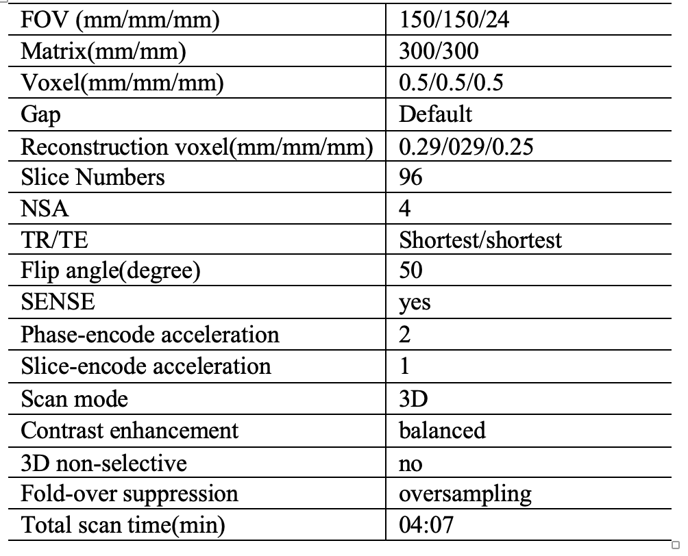
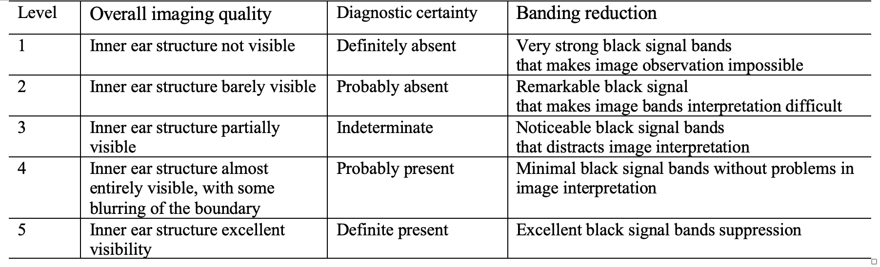

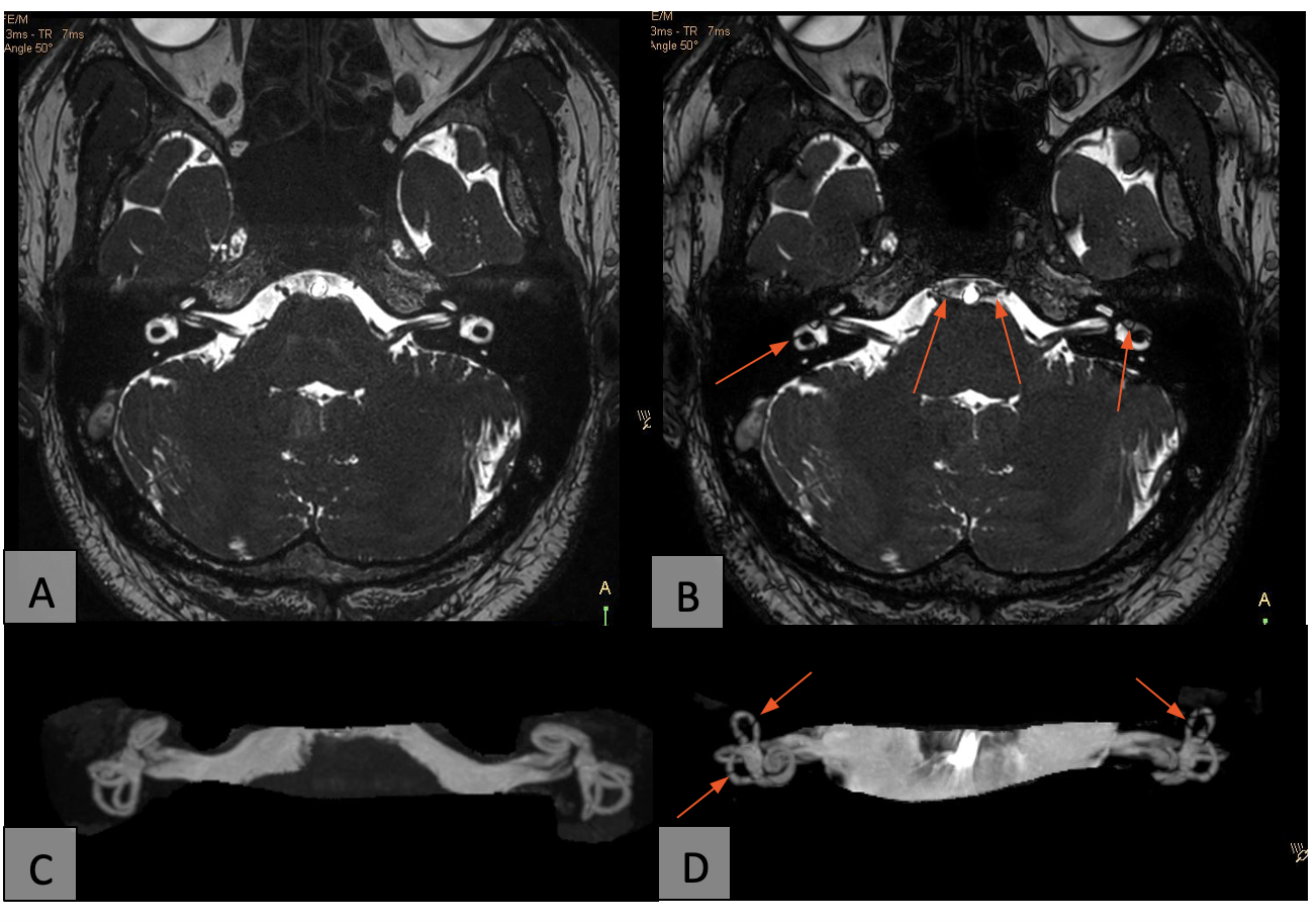
Figure 1.A healthy volunteer, 31-year-old male. The black banding artifacts were indicated by the red arrows in B and D. A and C can be observed with much fewer black banding artifacts.
A: Coronal 3D b-FFE-XD image of inner ear.
B: Coronal 3D b-FFE image of inner ear
C: 3D b-FFE-XD image of inner ear post-processing
D: 3D b-FFE image of inner ear post-processing