3021
3D NerveView Neurography of the lumbosacral plexus with a deep learning constrained Compressed SENSE Reconstruction1Radiology, First Hospital of Jilin University, changchun, China, 2Philips Heathcare, Beijing, China
Synopsis
High resolution three dimensional (3D) sequences of MR Neurography always takes a very long scan time. However, using very high acceleration factors leads to degradation of image quality due to insufficient noise removal. In this study, we used Compressed-SENSE Artificial Intelligence (CS-AI) framework to acquire highly accelerated 3D NerveView imaging of lumbosacral plexus. The result showed that CS-AI reconstruction can significantly decrease scan time with sufficient image quality compared to Compressed SENSE(CS) and might be clinically useful in assessment of lumbosacral plexus.
Introduction
Advancements in MRI scanner technology and imaging protocols nowadays enable the visualization of the anatomic origin of the lumbosacral plexus nerves, their trunks and small branches, and their path towards the lower limbs. Recently, magnetic resonance neurography (MRN) is increasingly applied to identify and characterize changes of lumbosacral plexus nerves. A successful MRN imaging depends on high resolution three-dimensional (3D) sequences 1-2. However, this always takes a very long scan time and cause patient discomfort. Although compressed sensing (Compressed SENSE: CS) was proven to achieve scan time reduction significantly especially with 3D sequence, using very high acceleration factors leads to degradation of image quality due to insufficient noise removal 3-4. More recently, deep learning-based algorithms have been combined with CS to overcome these challenges by learning optimal reconstruction parameters from the data itself and showed superior performance for reconstructing the knee image from highly undersampled k-space data 5-8.Purpose
The purpose of this study was to acquire highly accelerated 3D NerveView imaging of lumbosacral plexus using the Compressed-SENSE Artificial Intelligence (CS-AI) framework reconstruction and to evaluate the impact of different acceleration factors on the image quality of conventional CS and CS-AI.Methods
A total of 19 volunteers were examined on a 3.0 T system (Ingenia Elition X, Philips Healthcare) , including 5 men and 14 women(mean age 48.65±17.35 years, range: 21-79 years). This study was approved by the local IRB, and written informed consent was obtained from all subjects. The 3D NerveView sequence was acquired with four acceleration factors of 5, 8, 12, 16 (CS 5, CS 8, CS 12, CS 16) as well as time-equivalent CS-AI reconstruction (CS-AI 5, CS-AI 8, CS-AI 12, CS-AI 16). Among them, CS 5 is a commercially available sequence. Following parameters were common to all examinations: voxel size=1.25*1.25*2.1 mm3, FOV=280*360*100 mm, 95 slices, TR=2200 ms, TE=170 ms, 1 echo, TSE factor=51, NSA = 2. The scan results were exported to Philips IntelliSpace Portal (ISP, Philips Healthcare). Quantitative image analysis was performed by a radiologist with more than 4 years of experience. Signal to noise ratio (SNR) and contrast to noise ratio (CNR) were calculated by setting the nerve root as the region of interest (ROI) and setting the psoas muscle as a contrast. The image quality of CS and CS-AI was visually compared, including anatomical structures, diagnostic certainty and artifacts scored on a five-point-scale.The paired Wilcoxon test was performed to test for an influence of the different sequences. P values< 0.05 were considered significant.Results
Scan time decreased with increasing acceleration factors (CS 5: 5min4s, CS 8/CS-AI 8: 3min14s, CS 12/CS-AI 12: 2min8s, CS 16/CS-AI 16: 1min37s). For quantitative analysis of image quality, the SNR and CNR values of CS 12, CS-AI 12, CS 16 and CS-AI 16 were all significantly lower than CS 5 (P<0.05), and the CS-AI 5 sequence was rated highest for SNR and CNR value with an average of 11.96±4.11 and 11.2±3.71. With the increase of acceleration factor, the mean SNR and CNR values decreased (P<0.05). On the other hands, all the CS-AI sequences had significantly higher CNR values than the CS sequences with the same acceleration factor ( P<0.05).For subjective analysis, CS-AI 5 and CS-AI 8 sequences had higher scores for artifacts compared to CS 5 (P<0.05), without difference for the scores of anatomical structures and diagnostic certainty. With the increase of acceleration factor, all the subject analysis mean values decreased (P<0.05). However, all the subject scores of CS-AI sequences were significantly higher than time-equivalent CS (all P<0.05), expect for anatomical structures and diagnostic certainty for CS-AI 5 (p>0.05). The mean SNR,CNR and subject scores for all sequences were listed in the Table 1. The representative high resolution 3D NerveView images using the CS and CS-AI reconstructions with different acceleration factor were shown in Figure 1 and Figure 2.Conclusion
CS-AI reconstruction can significantly decrease scan time in 3D MR Neurography of the lumbosacral plexus, with better image quality compared with conventional CS with same acceleration factors. CS-AI 8 provided sufficient quality with scan time reduction (36.18%). Although further clinical investigation is needed, high-resolution 3D NerveView images with CS-AI reconstruction might be clinically useful in assessment of lumbosacral plexus.Acknowledgements
No acknowledgement found.References
1.Sollmann N, Cervantes B, Klupp E, et al. Magnetic resonance neurography of the lumbosacral plexus at 3 Tesla - CSF-suppressed imaging with submillimeter resolution by a three-dimensional turbo spin echo sequence. Magn Reson Imaging 2020; 71: 132-9.
2.De Paepe KN, Higgins DM, Ball I, Morgan VA, Barton DP, deSouza NM. Visualizing the autonomic and somatic innervation of the female pelvis with 3D MR neurography: a feasibility study. Acta Radiol 2020; 61(12): 1668-76.
3.Gao T, Lu Z, Wang F, Zhao H, Wang J, Pan S. Using the Compressed Sensing Technique for Lumbar Vertebrae Imaging: Comparison with Conventional Parallel Imaging. Curr Med Imaging 2021; 17(8): 1010-7.
4.Iuga AI, Abdullayev N, Weiss K, et al. Accelerated MRI of the knee. Quality and efficiency of compressed sensing. Eur J Radiol 2020; 132: 109273.
5.Knoll F, Zbontar J, Sriram A, et al. fastMRI: A Publicly Available Raw k-Space and DICOM Dataset of Knee Images for Accelerated MR Image Reconstruction Using Machine Learning. Radiol Artif Intell. 2020;2(1):e190007.
6.Knoll F, Murrell T, Sriram A, et al. Advancing machine learning for MR image reconstruction with an open competition: Overview of the 2019 fastMRI challenge. Magn Reson Med. 2020;(January):mrm.28338.
7.Pezzotti N, Yousefi S, Elmahdy MS, et al. An Adaptive Intelligence Algorithm for Undersampled Knee MRI Reconstruction. IEEE Access. 2020;8:204825-204838.
8.Pezzotti N, de Weerdt E,Yousefi S, et al. Adaptive-CS-Net: FastMRI with Adaptive Intelligence. arxiv. 2019;(NeurIPS).
Figures
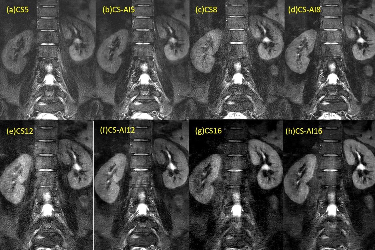
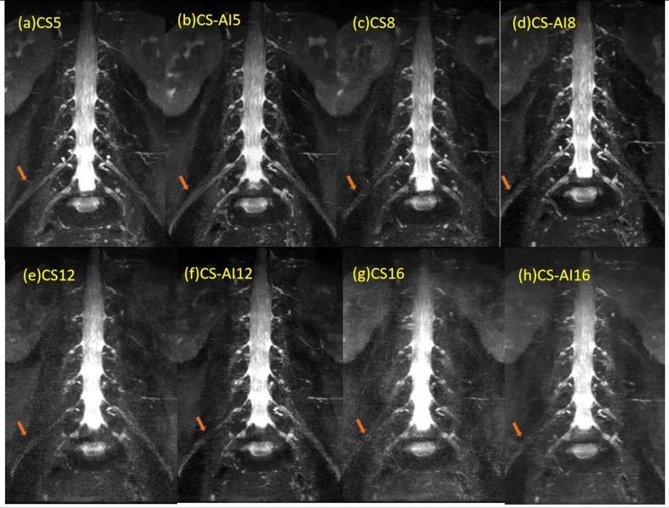

Table 1
Ana: anatomical structures, Dia: diagnostic certainty, Art:artifacts
* denotes statistically significant differences (p < 0.05) compared to the CS 5 sequences.