3020
Performance of single-shot echo-planar imaging DTI in rat sciatic nerve compared with readout-segmented echo-planar imaging1Department of Radiology, Shenzhen Traditional Chinese Medicine Hospital (The Fourth Clinical Medical College of Guangzhou University of Chinese Medicine), Shenzhen, China, 2MR Collaboration, Siemens Healthineers Ltd., Guangzhou, China
Synopsis
This study compared single-shot echo-planar imaging (SS-EPI) with readout-segmented echo-planar imaging (RS-EPI) diffusion tensor imaging (DTI) in vivo anesthetized rat sciatic nerve at 3T. The result showed that the image quality scores of SS-EPI were higher than those of RS-EPI in terms of the nerve morphology and homogeneity of the neuromuscular region. The coefficients of FA and RD obtained with SS-EPI were higher than those obtained with RS-EPI SS-EPI. This suggests that in rat sciatic nerve DTI, SS-EPI showed higher image quality and offered more sensitive and stable parameters to detect the histopathological change in the rat sciatic nerve.
Introduction/Purpose
DTI is the most important MR functional sequence in peripheral nerve study. However, due to the very small size of small animal’s nerve, DTI imaging is a difficulty in nerve MR study. Although readout-segmented echo planar imaging DTI (RS-DTI) was considered to correct artifacts and improve resolution clinically1, its performance in the peripheral nerves of small animals was unknown. Our study compared single-shot echo-planar imaging DTI (SS-DTI) in rat sciatic nerve with RS-DTI to correlate between DTI parameters and fiber histopathology, to clarify which DTI is more superior for small animal peripheral nerve study and to provide reference values for the DTI study of tiny nerves of the human body.Materials and Methods
Eight healthy adult male Sprague-Dawley rats were anaesthetized to deep sleep (7% chloralhydrate, 5mL/kg, intra-peritoneal injection) and scanned at 3T (MAGNETOM Prisma, Siemens Healthcare, Erlangen, Germany) with SS-EPI and RS-EPI (seven segments) DTI (resolution: 0.7×0.7×1.5 mm3; b-values: 0, 800 s/mm²; and diffusion directions: 20), as shown in Table 1. During the examination, rat limbs were attached to the animal coil (8-ch animal coil, Suzhou Medcoil Healthcare Co.,Ltd.) by proof fabric to further prevent movement.Image quality were evaluated by two independent readers (C.Y.Y., radiologist with 10 years of experience in DTI of the nerve; P.Z.X., with 3 years of experience in DTI of the nerve) on DWI b=800 s/mm² image. Morphology of the sciatic nerve, distortions of the nearby femur, muscles, and homogeneous of neuromuscular were evaluated and scored, good performance with 3 points and poor performance with 1 point. Standard DTI parameters of FA (Fractional Anisotropy), AD (Axial Diffusivity), RD(Radial Diffusivity), and ADC(Apparent Diffusion Coefficient) values were acquired from the workstation singo.via.
The rats were sacrificed after MR imaging. The middle stumps of the nerves were harvested and stained with toluidine blue. The toluidine blue slices were analyzed morphometrically by ImageJ software (https://imagej.net) for histological quantification. The degree of correlation between the DTI parameters and histopathological parameters (the percentage of axon area, percentage of myelin area, thickness of myelin, diameter of myelinated fiber) was calculated by using the Pearson correlation coefficient and compared by the modified Fisher Z-transform respectively.
Results
The image quality scores of SS-EPI were higher than those of RS-EPI in the blurring of the nerve margin (P= 0.008), artifacts of the nerve (P= 0.008), and homogeneity of the neuromuscular region (P= 0.007) (Figure 1, Figure 2). A higher FA (0.661 ± 0.010 vs. 0.607 ± 0.009, P<0.001) and lower RD (0.593 um2/ms ± 0.020 vs. 0.693 um2/ms ± 0.018, P<0.001) were found for SS-EPI compared to RS-EPI. As shown in Table 2 and Figure 3, for both sequences, FA was correlated with all of the histopathological parameters (r=0.913 to 0.963 for SS-EPI, and r=0.741 to 0.897 for RS-EPI, P=<0.001~0.036), except the diameter of myelinated fibers for RS-EPI (r=0.623, P= 0.099); and RD was correlated with all of the histopathological parameters (r= -0.886 to -0.948 for SS-EPI, and r= -0.709 to -0.812 for RS-EP, P=<0.001~0.049), except the percentage of the axonal area for RS-EPI (r= -0.691, P=0.058). The overall correlation coefficients of FA and RD obtained with SS-EPI were numerically higher than those obtained with RS-EPI, and the coefficients between FA and myelin thickness (P=0.027), FA and myelinated fiber diameter (P=0.036), and RD and myelin thickness (P=0.05) for SS-EPI were statistically higher than those for RS-EPI.Discussion and Conclusion
We found significantly higher image quality of rat sciatic nerve DW imaging based on single-shot echo-planar imaging (SS-EPI) compared with readout-segmented echo-planar imaging (RS-EPI). The SS-EPI derived DTI showed a stronger linear correlation with histopathological parameters than RS-EPI derived DTI parameters. This may be because RS-EPI is extremely sensitive to motion artifacts2. Even the invisible trembling after anesthesia can lead to motion artifacts, especially in tiny structures like sciatic nerve. What’s more, SS-EPI has a much shorter scanning time than RS-EPI in our study (4:06min VS 14:12min), reducing the risk of motion artifacts. Secondly, the main advantage of RS-EPI compared to SS-EPI is the significant reduction of geometric distortion caused by susceptibility artifacts3, 4, especially in regions prone to distortion, such as the orbit, skull base, and posterior fossa. However, in areas with less susceptibility artifacts such as the sciatic nerve (located in the deep surface of the muscle and has a small difference in magnetization rate with the adjacent tissues), the main advantage of RS-EPI is not obvious anymore3. FA and RD value are sensitive biomarkers to detect the minor changes of myelin5, 6. Our study indicated that SS-EPI can not only acquire better image quality, but possess stronger correlation with myelin parameters compared to RS-EPI. However, RS-EPI with various readout segmentations did not investigate in this study. Less readout segmentation with short acquisition time may reduce the motion artifact and shows high performance for the rat sciatic nerve, which need to be investigated in our future study.In conclusion, for the rat sciatic nerve DW imaging, SS-EPI sequence has significantly higher image quality compared with RS-EPI sequence. The FA and RD derived from the SS-EPI sequence might be more sensitive and quantitative biomarkers to detect the histopathological change of rat sciatic nerve.
Acknowledgements
We thank the national application master Gui Jin Li from Siemens HealthCare for his contribution to technical support and sequence optimization on Prisma 3T MR. We thank the SYNGO application support of Asia Pacific Soon Fee Boon from Siemens HealthCare for her excellent assistance in DTI post-processing and analysis. And we thank the Suzhou Medcoil Healthcare Co.,Ltd. for the MRI animal coil supplied for our study.References
1. Porter DA, Heidemann RM. High resolution diffusion weighted imaging using readout segmented echo planar imaging, parallel imaging and a two dimensional navigator based reacquisition. Magn Reson Med. 2009;62:468-75.
2. Friedli I, Crowe LA, de Perrot T, et al. Comparison of readout-segmented and conventional single-shot for echo-planar diffusion-weighted imaging in the assessment of kidney interstitial fibrosis. J Magn Reson Imaging. 2017;46(6):1631-1640.
3. Yeom KW, Holdsworth SJ, Van AT, et al. Comparison of readout-segmented echo-planar imaging (EPI) and single-shot EPI in clinical application of diffusion-weighted imaging of the pediatric brain. AJR Am J Roentgenol. 2013;200(5):W437-43.
4. Bogner W, Pinker-Domenig K, Bickel H, et al. Readout-segmented echo-planar imaging improves the diagnostic performance of diffusion-weighted MR breast examinations at 3.0 T. Radiology. 2012;263(1):64-76.
5. Naraghi AM, Awdeh H, Wadhwa V, et al. Diffusion tensor imaging of peripheral nerves. Semin Musculoskelet Radiol. 2015;19(2):191-200.
6. Chen YY, Zhang X, Lin XF, et al. DTI metrics can be used as biomarkers to determine the therapeutic effect of stem cells in acute peripheral nerve injury. Journal of Magnetic Resonance Imaging. 2017;45(3):855-862.Figures
Figure 1. T2WI coronal view displayed the morphology of bilateral sciatic nerves. The transverse DTI images of the rat sciatic nerve in SS-TI and RS-DTI showing different image quality. Toluidine blue myelin staining of the sciatic nerves (×1000).
Figure 2. Score of image quality in both SS-EPI and RS-EPI.
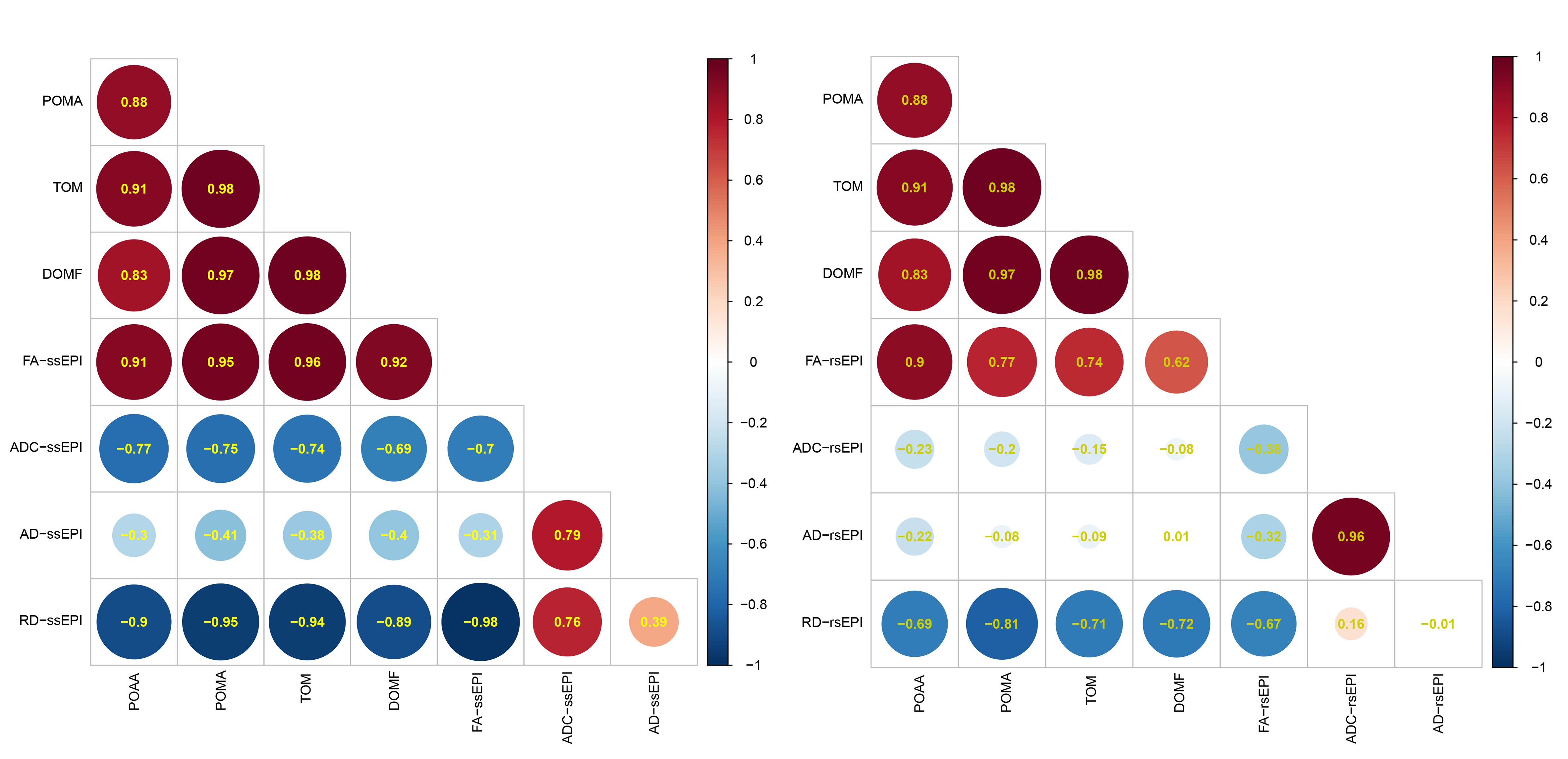
Figure 3. Heatmap of Correlation between DTI parameters and histopathological parameters.