3009
Correction of Gibbs ringing artifact improves visualization of epileptogenic pathology1Florey Institute of Neuroscience & Mental Health, Heidelberg, Australia, 2Florey Department of Neuroscience and Mental Health, The University of Melbourne, Melbourne, Australia, 3Department of Medicine, Austin Health, Heidelberg, Australia, 4Department of Neurology, Austin Health, Heidelberg, Australia
Synopsis
We evaluate the utility of a recently-proposed method tailored for the removal of Gibbs ringing artifacts in 3D MRI data, by applying it to high-resolution isotropic T2-weighted data acquired during the Australian Epilepsy Project (AEP) pilot. The filter is highly effective at removing this known artifact whilst preserving pathological detail. We therefore encourage its uptake in not only the radiological assessment of the Epilepsies, but any 3D imaging context where the manifestation of Gibbs ringing may be detrimental.
Introduction
Efficacy of radiological assessment is predicated on the absence of gross image artifacts, particularly in the pursuit of subtle pathologies of potential epileptogenesis. One particularly intrusive artifact is the presence of Gibbs ringing in high-resolution T2-weighted imaging, where sharp transitions of strong intensity contrast between CSF and parenchyma can result in artificial bands of high and low intensity adjacent to tissue-fluid interfaces. Recently, an algorithm for the estimation and correction of Gibbs ringing in 2D image data based on sub-voxel shifts1 was augmented to enable operation on data acquired by 3D MRI sequences2, such as those used for high-resolution T2-weighted imaging. Here we demonstrate the influence of this novel algorithm on pathological cases acquired during the Australian Epilepsy Project (AEP)3 pilot.Methods
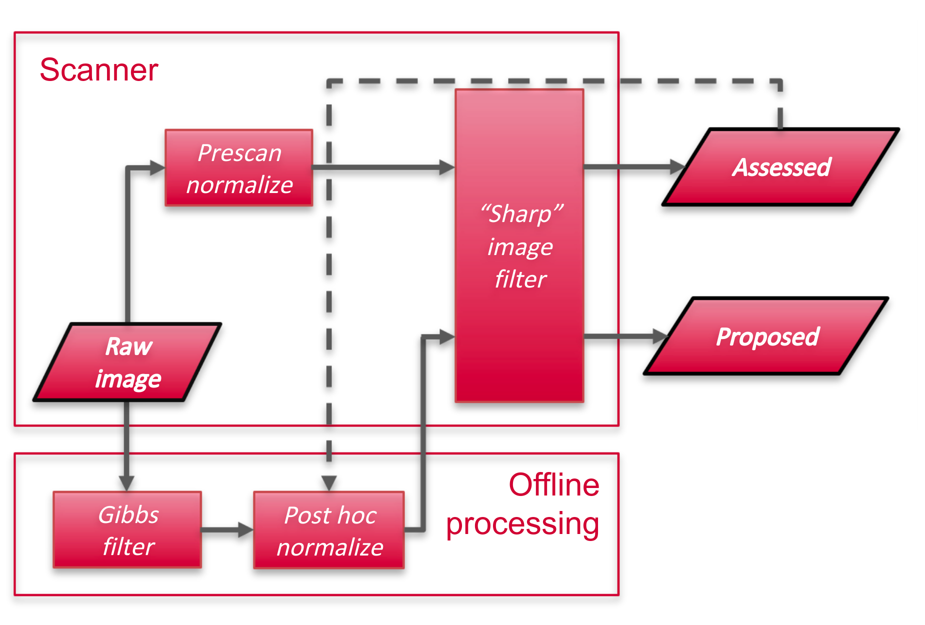
Image data were acquired on a Siemens Prisma 3T scanner. T2-weighted data were obtained at 0.9mm isotropic resolution using a turbo spin-echo sequence (TE=564ms; TR=3200ms; in-plane acceleration factor 2). Image processing prior to radiological assessment included vendor-specific B1- bias field correction (“Prescan normalize”) and inline filtering (“Sharp” filter); raw image data omitting both steps were additionally exported.The 3D Gibbs ringing removal algorithm2 was applied to raw magnitude image data. An approximation of the vendor B1- bias field correction was produced by fitting a polynomial spline to the log-ratio of filtered and raw images4 within a brain mask produced by ROBEX5. These data were reuploaded to the scanner console for application of vendor image filtering. Manually processed images were subjectively compared to those used for radiological assessment. The pipeline is shown diagrammatically in Figure 1.
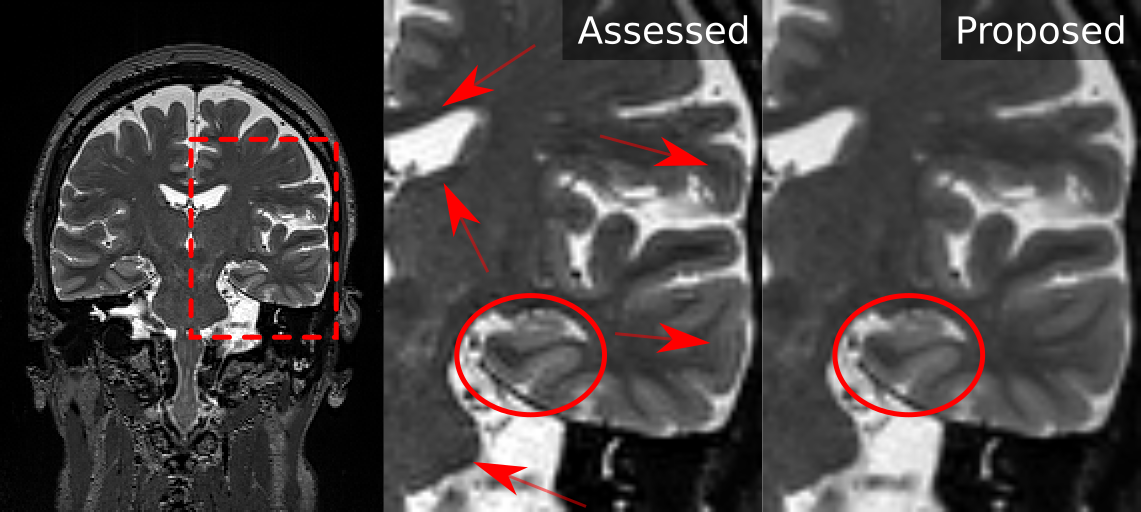
A subset of four exemplar cases from the AEP pilot were selected based on prior radiological confirmation of epileptic pathology; these included oligodendroglioma, polymicrogyria, temporal encephalocele, and hippocampal sclerosis.
Results
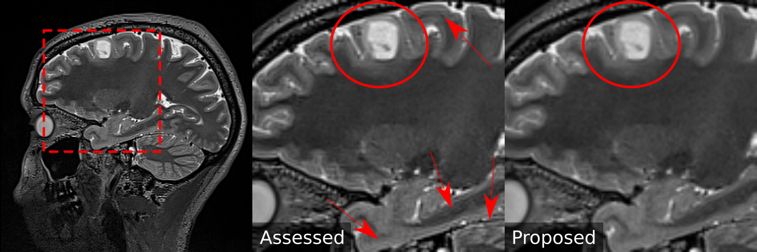
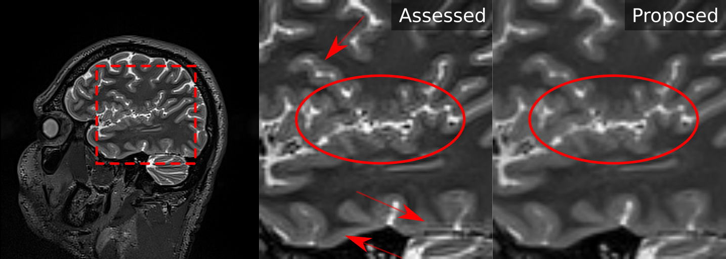
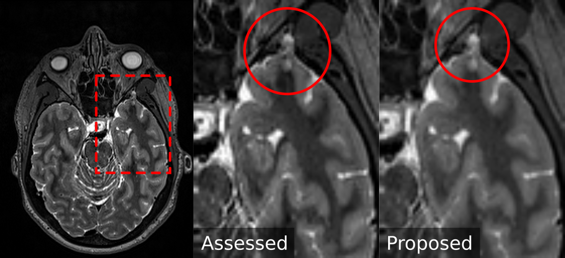
Figures 2-5 contrast image data between the two image processing pipelines across the four exemplar cases. Pathological regions are highlighted, as are regions where manifestation of the Gibbs ringing effect differs considerably between the two images.Discussion
The 3D Gibbs ringing removal filter is highly effective at removing this known image artifact (without introduction of unwanted image blurring as occurs with k-space filtering techniques2). Subsequent filtered images retain similar visual properties to standard clinical images, but with reduced distortions particularly at high-contrast boundaries.Inclusion of the algorithm:
- Improves the delineation of pathological borders and reduces artificial contrast within homogeneous tumours (Figure 2);
- Improves confidence in diagnosis of fine structural features contaminated by Gibbs ringing (Figure 3);
- Does not detrimentally affect diagnosis of fine structural features not contaminated by Gibbs ringing (Figures 4-5);
- Removes visual distractions to radiological assessment both near to, and distant from, pathologies (Figures 2-5).
Conclusion
Given the effectiveness of the 3D Gibbs ringing removal filter, we encourage its widespread adoption; not only in radiological assessment of the Epilepsies, but indeed across qualitative and quantitative applications of 3D MRI where the manifestation of Gibbs ringing is of notable detriment.Acknowledgements
RS is supported by fellowship funding from the National Imaging Facility (NIF), an Australian Government National Collaborative Research Infrastructure Strategy (NCRIS) capability.
We thank J-Donald Tournier for early access to an implementation of the 3D Gibbs ringing removal filter.
References
- Elias Kellner, Bibek Dhital, Valerij G. Kiselev, and Marco Reisert. “Gibbs-Ringing Artifact Removal Based on Local Subvoxel-Shifts.” Magn. Reson. Med. 76, no. 5 (November 2016): 1574–81.
- Thea Bautista, Jonathan O’Muircheartaigh, Joseph V Hajnal, and J-Donald Tournier. “Removal of Gibbs Ringing Artefacts for 3D Acquisitions Using Subvoxel Shifts.” In Proceedings of the ISMRM, 3535, 2021.
- Australian Epilepsy Project | Changing Lives | Supercharging med-tech. Australian Epilepsy Project https://epilepsyproject.org.au/.
- Thijs Dhollander, Rami Tabbara, Jonas Rosnarho-Tornstrand, J-Donald Tournier, David Raffelt, Alan Connelly. Multi-tissue log-domain intensity and inhomogeneity normalisation for quantitative apparent fibre density. In Proc. ISMRM, 2021, 2472
- Juan Eugenio Iglesias, Cheng-Yi Liu, Paul M. Thompson, Zhuowen Tu. Robust Brain Extraction Across Datasets and Comparison With Publicly Available Methods. IEEE TMI, 2011, 30(9), 1617-1634
Figures