3005
Alteration in White Matter Microstructural Integrity and Network Differentiate Drug-naïve Obsessive compulsive disorder and Healthy controls
Suming Zhang 1, Xinyu Hu2, Xuan Bu2, Hailong Li1, LingXiao Cao2, Jing Liu2, KaiLi Liang2, Xue Li3, and Xiaoqi Huang2
1Huaxi MR Research Center (HMRRC), Department of Radiology, West China Hospital of Sichuan University, Chengdu City, China, 2Huaxi MR Research Center (HMRRC), Department of Radiology, West China Hospital of Sichuan University, ChengDu City, China, 3College of Physics, Sichuan University,Chengdu, China, Chengdu City, China
1Huaxi MR Research Center (HMRRC), Department of Radiology, West China Hospital of Sichuan University, Chengdu City, China, 2Huaxi MR Research Center (HMRRC), Department of Radiology, West China Hospital of Sichuan University, ChengDu City, China, 3College of Physics, Sichuan University,Chengdu, China, Chengdu City, China
Synopsis
We investigated the alteration of white matter in obsessive compulsive disorder from voxel-level, tractography-level and connectome-level by combining analysis of TBSS, AFQ and graph theory. TBSS and AFQ revealed widespread changes in WM. Network-based statistic (NBS) analysis demonstrated decreased structural connectivity in patients mainly comprising frontal, temporal, limbic areas, which belong to DMN, FPN, limbic, SMN and VAN network. Along with regions commonly described in the CSTC model of pathophysiology, our results indicate an involvement of white matter in frontal-temporal and limbic regions to differentiate OCD patients and healthy controls, supporting their importance for neurobiological alterations in OCD.
Synopsis
We investigated the alteration of white matter in obsessive compulsive disorder from voxel-level, tractography-level and connectome-level by combining analysis of TBSS, AFQ and graph theory.TBSS and AFQ revealed widespread changes. Network-based statistic (NBS) analysis observed decreased structural connectivity in patients mainly comprising frontal, temporal, limbic areas, which belong to DMN, FPN, limbic, SMN and VAN network. Along with regions commonly described in the CSTC model of pathophysiology, our results indicate an involvement of white matter in frontal-temporal and limbic regions to differentiate OCD patients and healthy controls, supporting their importance for neurobiological alterations in OCD.
Body of the abstracts
BackgroundObsessive-compulsive disorder (OCD) has been conceptualized as a brain dysconnectivity disorder mainly in cortico-striate-thalamo-cortical model. In the past decade, diffusion magnetic resonance imaging (dMRI) studies have demonstrated OCD patients have the widespread alterations either in white matter bundles or in white matter structural connectome and that these alterations are associated with core symptoms.1 2 3 4 However, evidence support that brain regions outside of the classical CSTC model(e.g. hippocampus and amygdala) may play a role in the pathophysiology.5 6 Aiming to comprehensively explore white matter(WM) change in adult OCD patients, we investigated dMRI-based WM microstructure integrity and structural-connectome from a voxel-, tractography-, and network-based perspective. MethodsA total of 71 DSM-IV criteria diagnosed OCD patients and 81age- and gender-matched healthy controls(HC) participated in the study after giving written informed consent. All patients were drug naïve and were scanned using 3-Tesla GE magnetic resonance imaging(MRI) to acquire high-resolution T1 and diffusion MRI data. EPI sequence was acquired with 16 optimized non-colinear diffusion-weighting gradient directions(15 noncollinear diffusion gradients with b=1000 s/mm2 and one reference volumes with b=0 s/mm2) with voxel size 0.935*0.935*3mm3. The diffusion images were preprocessed were performed using the FMRIB Software Library(FSL) 6.0 (https://fsl.fmrib.ox.ac.uk/fsl/). All DTI source data were corrected for eddy current and head motion by “EDDY” and “BET”, respectively. Then we performed voxel-wise statistical analysis of the FA data using TBSS(tract-based spatial statistics). The mean FA skeleton set threshold FA >0.02. Then, permutation-based nonparametric test with threshold-free cluster enhancement and 5000 permutations per analysis to assess group differences. The statistical threshold was set at p <0.05 with family-wise error (FWE) whole-brain multiple comparison correction. Meanwhile, we performed AFQ(automated fiber quantification) analysis based on deterministic tractography. (https://github.com/ yeatmanlab/AFQ) (version 1.2). after getting DTI-induced measures (FA, MD, RD, and AD) , group comparison were compute using two sample T test. Finally, we obtained connectivity matrices composed of nodes parcellation from high resolution brain atlas(BN-246 template, http://www.brainnetome.org/) and edges from deterministic tractography by software PANDA(https://www.nitrc.org/projects/panda). Edges were defined as streamline counts of fibers. We construct structural network and obtained global and local network parameters via GRETNA(https://www.nitrc.org/projects/gretna/). Network-based statistic (NBS) analysis was used to assess differences in the interregional connectivity matrices between the groups. Finally, partial correlation were performed between significant group difference and symptom severity(Yale-Brown Obsessive Compulsive Scale, YBOCS). ResultsThe demographic information and clinical characteristics of the OCD patients and HC were shown in Table1.Voxel level analysis based on TBSSTBSS proved widespread regions with increased FA and decreased RD in OCD patients compared with HC.(Figure1) Those regions include: right posterior limb of internal capsule, bilateral SLF, bilateral cingulum, body and genu of corpus callosum, right fornix and right sagittal stratum.Tractography level analysisAFQ revealed significant increased FA in four bundles (post segment of left thalamic radiation, middle segment of left cingulum cingulate, posterior segment of left ILF, two middle segments of left uncinate fasciculus) in OCD patients compared with HC.(Figure2)Network level analysisBoth OCD and HC exhibited small-worldness (Figure4). No significant differences (p > 0.05) in major network parameters were found except the normalized clustering coefficiency(γ, mean(OCD)=66.9, mean(HC)=69.4, P=0.019) between OCD and HC.Network-based statistic analysis (t-stat = 2.2, P < 0.05, corrected for multiple comparisons) revealed a single network of decreased diminished streamline counts in OCD patients compared to HC. The network comprised a total of thirty six nodes connected by thirty seven edges(Table 2 and Figure 3). The entire network included nodes and edges mainly located in frontal lobe, temporal lobe and limbic system, which is overlapped with default mode network(DMN), frontoparietal network(FPN), Limbic, salience network (SMN), and Visual network(VAN) based on Yeo-7 template. (Figure3)Partial correlation with symptom severityOnly the mean FA value of left UF node 41-77 showed significant negatively correlation with YBOCS(Figure5). There is no significant correlation revealed by TBSS and graph theory. Discussion & Conclusion As far as we know, current study is the first one to investigate OCD white matter structure from three diverse level(voxel-level, tractography-level, connectome-level). We revealed specific white matter abnormalities in four bundles(left thalamic radiation, left cingulum cingulate, left ILF and left UF) and decreased streamline counts mainly connecting frontal, temporal and limbic areas, which belong to DMN, FPN, limbic, SMN and VAN network. Along with regions commonly described in the CSTC model of pathophysiology5, our results indicate an involvement of white matter in frontal-temporal and limbic regions(left thalamic radiation, left cingulum cingulate, left ILF, left UF, WM connecting left MFG and Thalamus) to differentiate OCD patients and healthy controls, supporting their importance for neurobiological alterations in OCD.Acknowledgements
This study was supported by National Nature Science Foundation(Grant No.81671669)References
1.Zhong,Zhao. Zhao Teng. Et al. Abnormal topological organization in white matter structural networks revealed by diffusion tensor tractography in unmedicated patients with obsessive–compulsive disorder. Progress in Neuro-Psychopharmacology & Biological Psychiatry 51 (2014) 39–50.2. Dikmeer, N. Besiroglu,L.et al. White matter microstructure and connectivity in patients with obsessive-compulsive disorder and their unaffected siblings. Acta Psychiatr Scand. 2020;00:1–10.3. Versace A. Graur S.et al. Reduced focal fiber collinearity in the cingulum bundle in adults with obsessive-compulsive disorder. Neuropsychopharmacology (2019) 44:1182–1188.4. Watanabe. Takashi. et al . The detection of white matter alterations in obsessive–compulsive disorder revealed by Tracts constrained by Underlying anatomy (TRACULA). Neuropsychiatric Disease and Treatment 2018:14 1635–1643.5. Milad MR, Rauch SL. Obsessive-compulsive disorder: beyond segregated corticostriatal pathways. Trends Cogn Sci 2012; 16: 43–51.6. Menzies L, Chamberlain SR, Laird AR, Thelen SM, Sahakian BJ, Bullmore ET. Integrating evidence from neuroimaging and neuropsychological studies of obsessive-compulsive disorder: the orbitofronto-striatal model revisited. Neurosci Biobehav Rev 2008; 32: 525–549.Figures
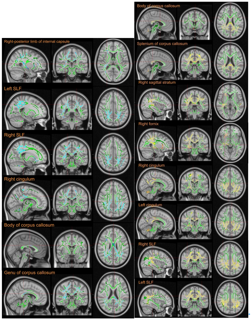
TBSS results. WM structures showed decreased FA (blue) and increased RD( yellow) in OCD compared with HC.
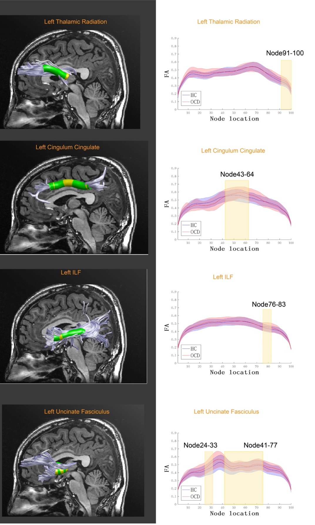
AFQ results. Significantly changed locations of fiber tracts in point-wise comparison of FA profiles and mean FA group comparison results (FDR correction, P< 0.05) in OCD group compared with HC. Abbreviation: FA, Fractional anisotropy; OCD, obsessive compulsive disorder; HC, health control;
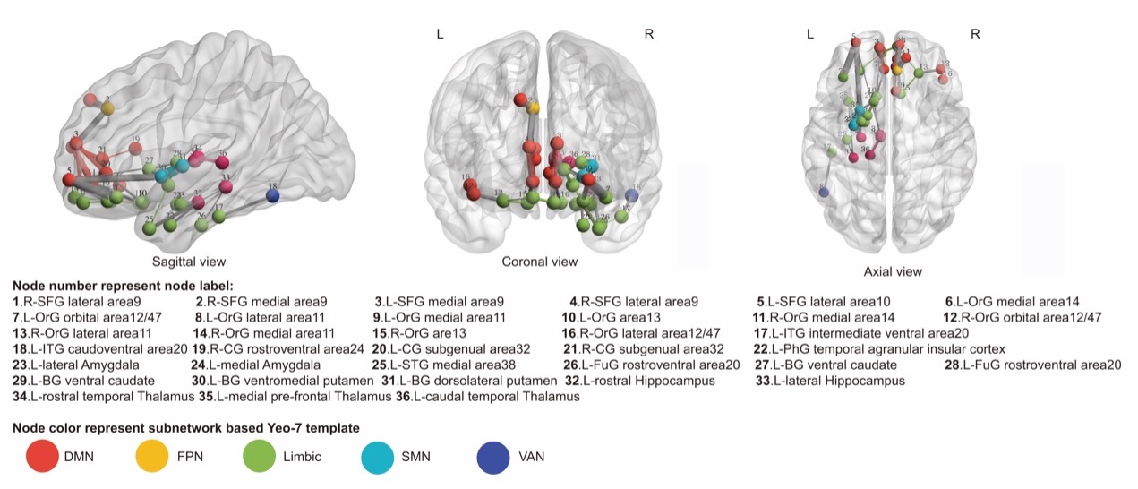
Network-based statistic-derived subnetworks. Significant subnetwork of FN weighted network from comparison between OCD and HC in sagittal, coronal and axial view. The circles represent the nodes and lines represent the connections with a t-stat =2.2 and p < 0.05 (corrected). The color of node show the type of subnetwork composed of all nodes.
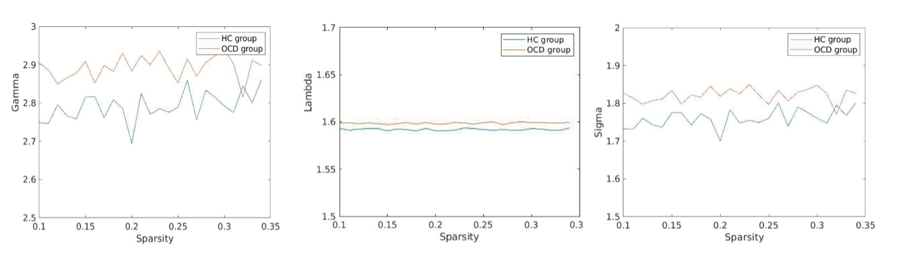
The key small-world parameters of the white matter networks with sparsity threshold. Graphs show that in the defined threshold range, both the OCD group and control groups exhibited Gamma(γ) substantially larger than 1 and Lambda(λ) approximately equal to 1, which indicates that both groups exhibited the typical features of small-world topology. OCD, obsessive-compulsive disorder; γ, normalized clustering coefficient; λ, normalized characteristic path length.
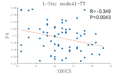
The partial correlation results between mean FA value of left Uncinate fasciculus node41-77 and symptom severity based on AFQ analysis.
DOI: https://doi.org/10.58530/2022/3005