3000
Aberrant cortical gyrification in adolescent major depressive disorder1Huaxi MR Research Center (HMRRC), Department of Radiology, West China Hospital of Sichuan University, Chengdu, China, 2Department of radiology, the Thrid Hospital of Mianyang, Sichuan Mental Health Center, Mianyang, China, Mianyang, China, 3Psychoradiology Research Unit of the Chinese Academy of Medical Sciences (2018RU011), West China Hospital of Sichuan University, Chengdu, Sichuan, China, Chengdu, China
Synopsis
Evidence has emerged to suggest the important role of cortical gyrification morphology in adult major depressive disorder (MDD). However, little is known about how the altered cortical gyrification contribute to adolescent patients who is at a critical period of brain development. We address this gap by examining the difference of local gyrification index (LGI) between adolescent MDD patients and HCs. We found that MDD patients showed significant increased LGI in the left PCC and bilateral lingual gyrus, which characterize the neurobiological mechanisms of adolescent MDD and may be important to understand the developmental effect of this disorder.
Introduction
MDD is characterized by lack of interest, depressed mood, despaired and high suicide rate, which is the most common psychiatric disorder in the world with a high lifetime prevalence of about 16% [1]. Adolescent is especially easily suffered from this disorder, as adolescence is a critical period for brain development, which is sensitive to surrounding impacts [2]. Despite its high prevalence and significant suffering, its neuropathology is still not clear. Accumulated evidence has emerged to suggest the important role of cortical gyrification morphology in adult MDD. However, this level of investigation has been largely absent in adolescent MDD. Therefore, in the current study, we aimed to explore the cortical gyrification as measured by local gyrification index (LGI), which can specifically reflect the early development of our cortical folding [3], in a relatively large sample of adoloscent MDD versus HC.Methods
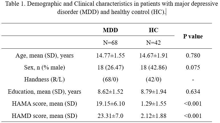
68 drug-naïve MDD patients and 42 age and sex matched HCs were recruited in our study. The Hamilton Anxiety Scale (HAMA) and Hamilton Depression Scale (HAMD) were used to assess the severity of anxiety and depression symptoms seperately in subjects. High resolution T1 weighted images were obtained using a volumetric 3-dimensional Spoiled Gradient Recall (SPGR) sequence (TR/TE = 1900/2.25ms; flip angle = 9du; 176 axial slices with thickness =1mm, field of view = 256×256 mm and data matrix = 256×256) via Siemens Skyra 3.0 T scanner. FreeSurfer 6.0 software was utilized for image processing and reconstruction. Quality control on FreeSurfer output was conducted based on the ENIGMA Cortical Quality Control Protocol. LGI was analyzed by computing the degree of gyrification locally at approximately 150000 vertices across each hemisphere. Whole-brain vertex-wise comparison was conducted by using a general linear model (GLM) analyses on QDEC software with age, sex and ICV as covariates. The Monte Carlo simulation was performed to correct for multiple comparisons (10,000 iterations, cluster-forming p<0.01, cluster-wise corrected p < 0.05).Results
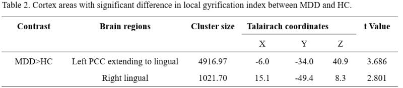
The demographic and clinical information can be seen in Table 1. As shown in Figure 1 and Table 2, compared to HC group, MDD showed increased local gyrification index in the left posterior cingulate cortex (PCC) extending to left lingual gyrus (P<0.01, Monte Carlo-corrected), and also increased in the right lingual gyrus (P<0.01, Monte Carlo-corrected).Discussion & Conclusion
To our knowledge, this is the first study to investigate the alteration of LGI in adolescent depression. Previous studies have identified decreased LGI of left PCC and lingual in adult MDD [4]. Interestingly, we found increased LGI values in the left PCC and bilateral lingual gyrus. PCC has been reported to play a critical role in pre-executive motor production during a motor imagery task [5]. Therefore, we hypothesized that the abnormal LGI in PCC may be associated with the disruption in pre-executive production function in MDD individuals, which may manifest as slow in movement, reduced facial expression symptoms. In addition, some researches have demonstrated that the lingual gyrus is involved in high-level visual processing and visual memory, which is correlated with cognitive function [6-7]. And the altered development of bilateral lingual gyrus may be associated with cognitive impaired in patients. Our findings extended the neurobiological mechanisms to adolescent MDD which need further study identifying the distinctions between adolescent and adult MDD to verify.Summary of Main Findings
MDD patients showed significant increased LGI in the left posterior cingulate cortex and bilateral lingual gyrus, which characterize the neurobiological mechanisms of adolescent MDD and may be important to understand the developmental effect of this disorder.Acknowledgements
This study was supported by grants from 1.3.5 Project for Disciplines of Excellence, West China Hospital, Sichuan University (ZYJC21041) and Clinical and Translational Research Fund of Chinese Academy of Medical Sciences (2021-I2M-C&T-B-097).References
[1]. Kessler RC, Berglund P, Demler O, Jin R, Merikangas KR, Walters EE. Lifetime prevalence and age-of-onset distributions of DSM-IV disorders in the National Comorbidity Survey Replication. Arch GenPsychiatry. 2005; 62(6):593–602.
[2]. Andersen SL, Teicher MH. Stress, sensitive periods and maturational events in adolescent depression. Trends Neurosci.2008;31:183-91.
[3]. Petanjek Z, Judas M, Kostovic I, Uylings HB. Lifespan alterations of basal dendritic trees of pyramidal neurons in the human prefrontal cortex: a layer-specific pattern. Cereb Cortex. (2008) 18:915–29.
[4]. Palaniyappan L, Marques TR, Taylor H, Handley R, Mondelli V, Bonaccorso S, Giordano A, McQueen G, DiForti M, Simmons A, David AS, Pariante CM, Murray RM, Dazzan P. Cortical folding defects as markers of poor treatment response in first-episode psychosis. JAMA Psychiatry. 2013 Oct;70(10):1031-40.
[5]. Liberg B, Adler M, Jonsson T, Landen M, Rahm C, Wahlund L-O, et al. Motor imagery in bipolar depression with slowed movement. J Nerv Ment Dis. (2013) 201:885–93.
[6]. Ancelin ML, Carrière I, Artero S, Maller J, Meslin C, Ritchie K, Ryan J, Chaudieu I. Lifetime major depression and grey-matter volume. J Psychiatry Neurosci. 2019 Jan 1;44(1):45-53.
[7]. Jung J, Kang J, Won E, Nam K, Lee MS, Tae WS, Ham BJ. Impact of lingual gyrus volume on antidepressant response and neurocognitive functions in Major Depressive Disorder: a voxel-based morphometry study. J Affect Disord. 2014 Dec;169: 179-87.
Figures