2995
Use lesion network localization to locate related brain lesions in incidental white matter lesions children with verbal comprehension delay1Radiology, The first afflicted hospital of Xi'an Jiaotong University, Xi'an, China
Synopsis
Incidental white matter lesions (IWMLs) is a common disease in children with an incidence rate between 1.9% - 20%. Locating the brain lesions that might be related to the decline in verbal comprehension by overlapping lesion-associated networks can help define the possible response lesion. Our finding would help pediatricians to recognize the significant IWMLs that could potentially cause language delay in children. Thus, an early intervention can be applied to prevent the occurrence and progression of verbal comprehension decline in children.
Abstract
Introduction:Incidental white matter lesions (IWMLs) in children are the WMLs that are incidentally discovered during magnetic resonance imaging (MRI) examination.1 The incidence rates of IWMLs in children is between 1.9% - 20%,1-2 and it often occurs with diseases such as epilepsy or headache.1 IWMLs are often overlooked by doctors since their clinical effect in children is unclear.1 Only a few studies have mentioned that severe IWMLs can reduce children's information processing speed thus affecting language cognitive function.3 In humans, the language cognitive function, specifically verbal comprehension is majorly processed by the frontal and temporal lobes of the brain.4 Therefore, we suspect that brain lesions incidentally found at these lobes could possibly affect the verbal comprehension ability of children. However, not all children with IWMLs at these brain lobs present with verbal comprehension defects. Thus, we aimed to define the possible brain lesions that might be related to the decline in verbal comprehension by overlapping lesion-associated networks.5
Method:
The institutional review board of the First Affiliated Hospital of Xi’an Jiaotong University approved this prospective study and written informed consent was obtained from all participants. Subjects This study enrolled IWMLs children aged 4-8 years who participated in both 3.0T brain MRI examination and Wechsler intelligence test (WISC) at our hospital from January 2015 to December 2019. According to the verbal comprehension index (VCI) of the Wechsler intelligence test, these participants were divided into normal-VCI group (VCI ≥ 85) and delayed-VCI group (VCI < 85). MR Protocols All subjects were examined by using a 3.0T scanner (Signa HDxt, General Electric Medical System, Milwaukee, WI, USA) with an 8-channel head coil. Data acquisition included three-dimensional fast spoiled gradient-echo T1-weighted sequence (TR/TE:10.2ms/4.6ms; NEX:1; isotropic:1×1×1mm3; FOV:24cm) and T2-weighted fluid-attenuated inversion recovery (TR/TE:9600ms/117ms; NEX:1; matrix:256×256; thickness:4mm; FOV:24cm), followed by diffusion-tensor imaging (DTI:30 directions; b-value:600s/mm; TR/TE:11000ms/67.4ms; NEX:1; thickness:2.5mm; FOV:24cm; matrix:172×172). Data and statistical analysis Based on T2 flair, IWMLs were delineated, and the probability map of lesions was generated. FSL v5.0 (FSL,www.fmrib.ox.au.uk/fsl) was used for whole-brain fiber tracking. The region of interest (ROI) of IWMLs were first transferred onto a reference brain; then the intrinsic functional connectivity of the lesion volume with the rest of the brain was assessed using normative connectome data; last, we overlapped lesion-associated networks to identify regions common to both normal and delayed VCI groups.5 The demographic and clinical data of normal and delayed VCI groups were collected and compared. The Fisher-exact test was used for categoric data, and the Analysis-of-Variance (ANOVA) was used for continuous data. Statistical analysis was performed by using statistical software (SPSS 23.0; SPSS, Chicago, Ill). All P values were considered statistically significant at P<0.05.
Results:
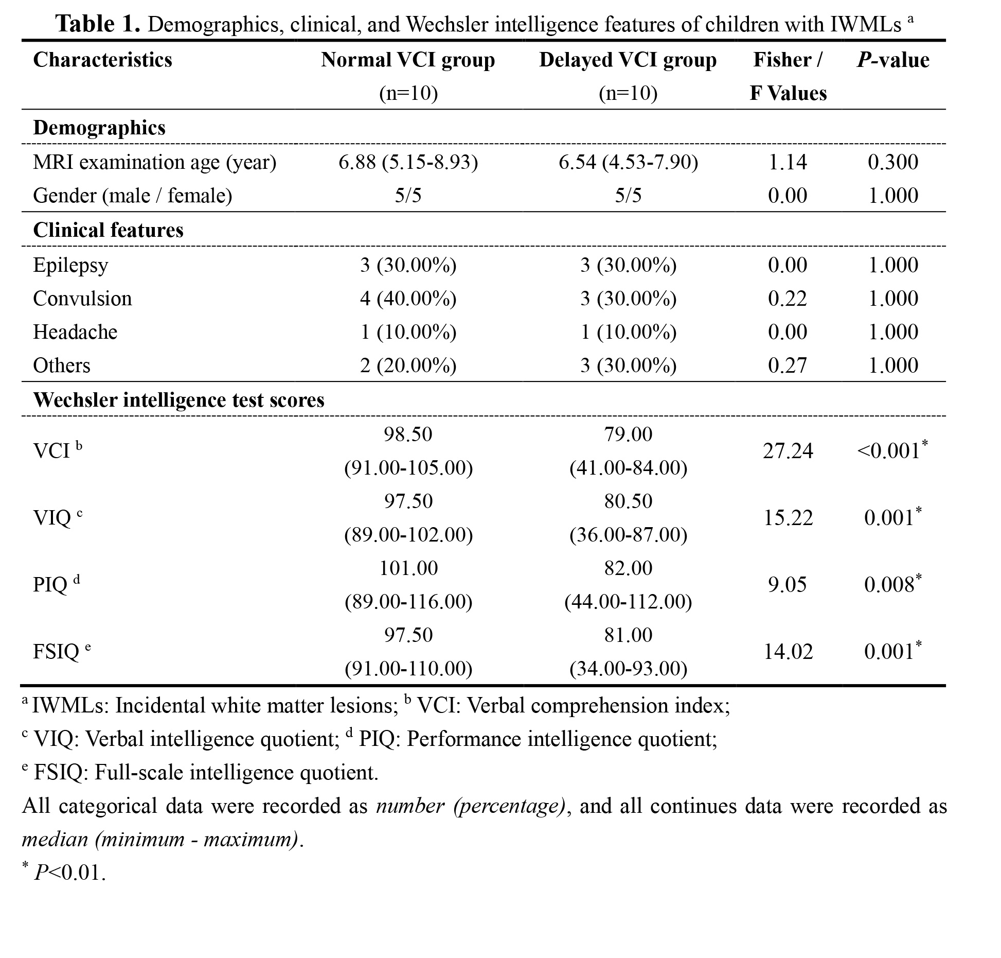
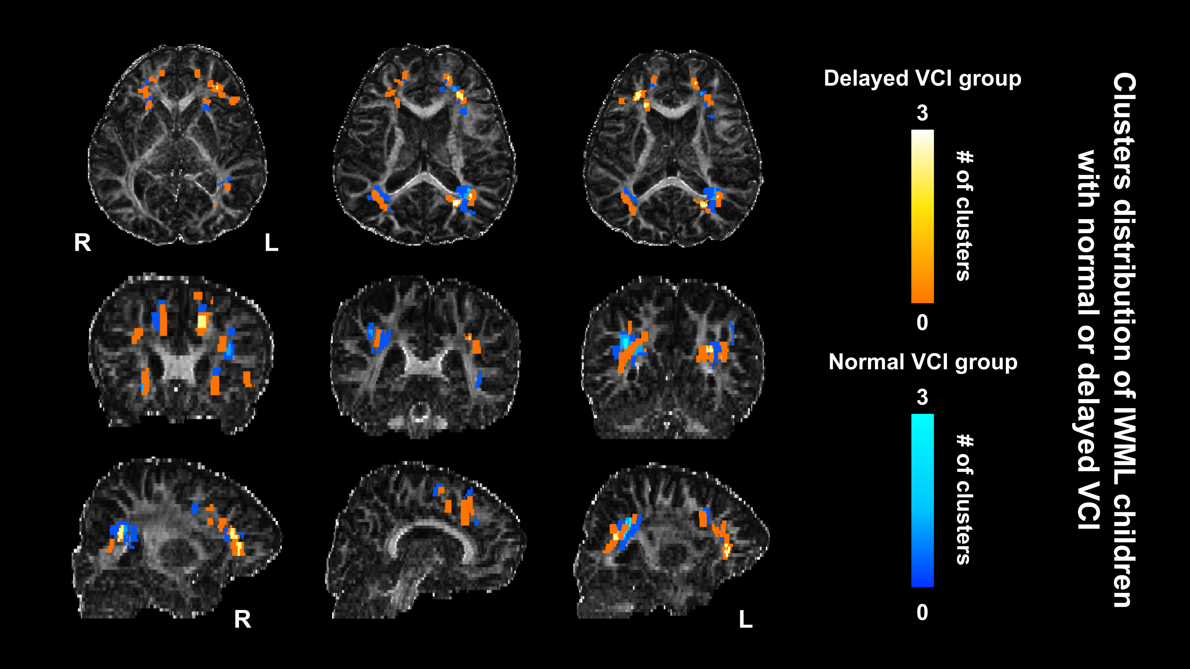
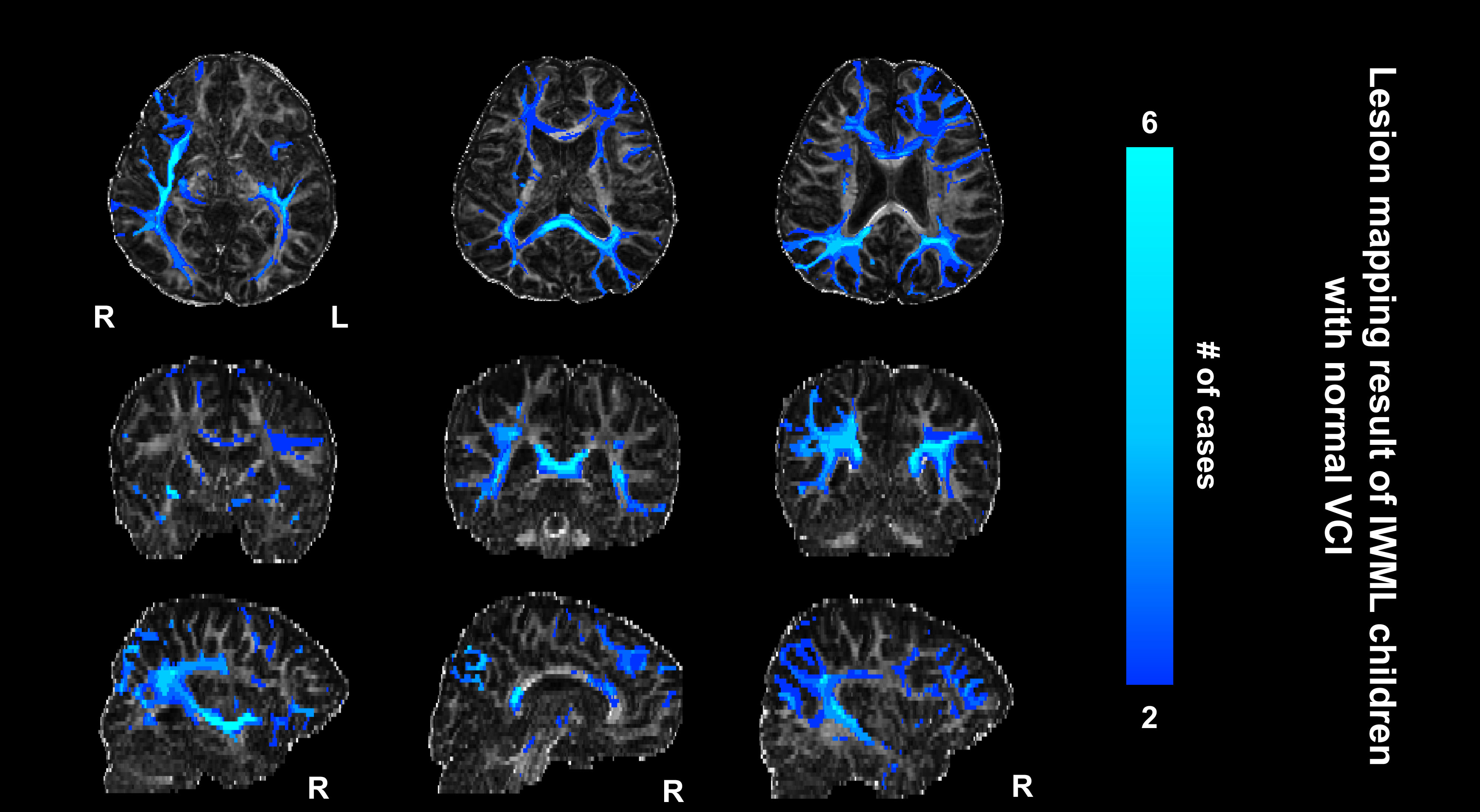
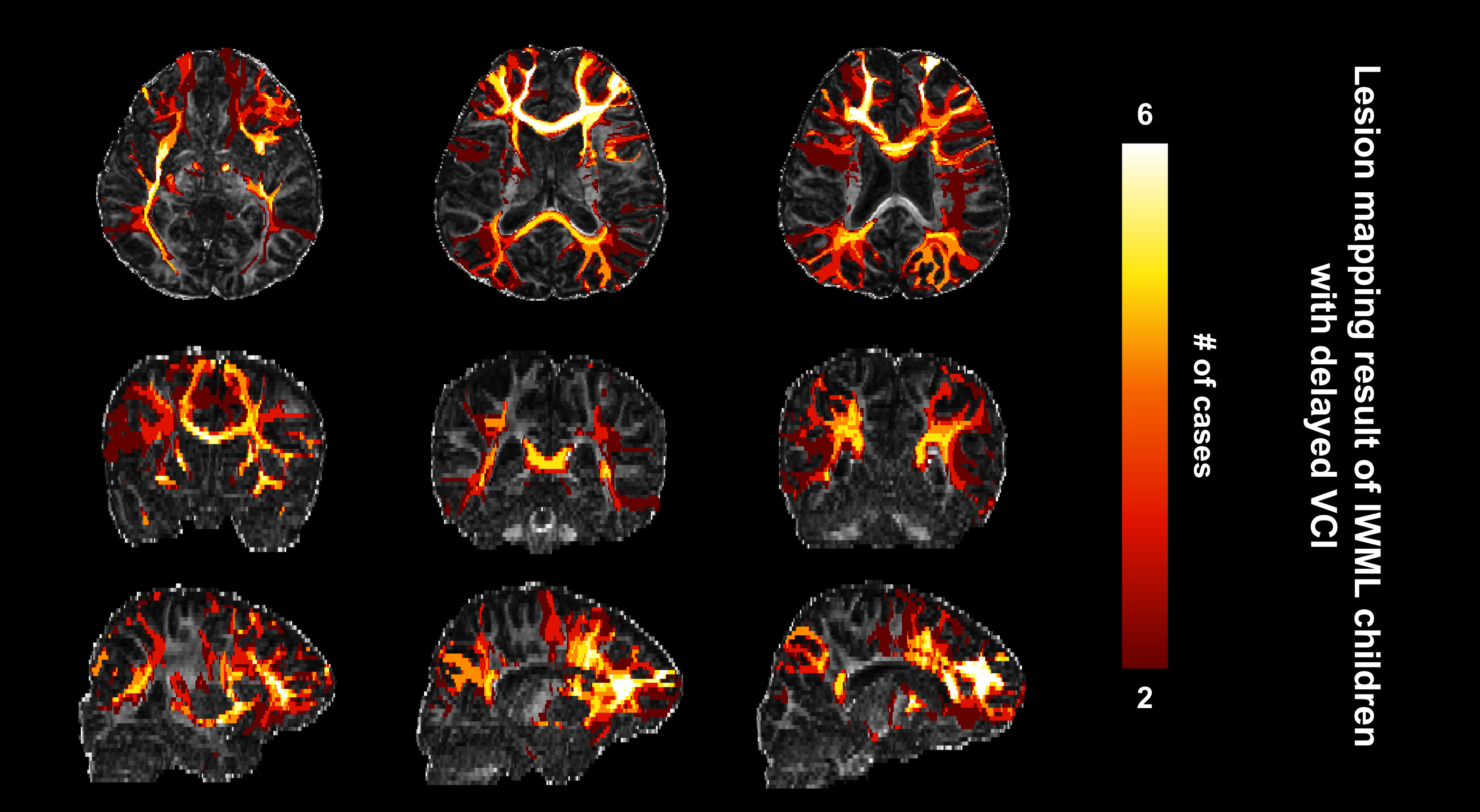
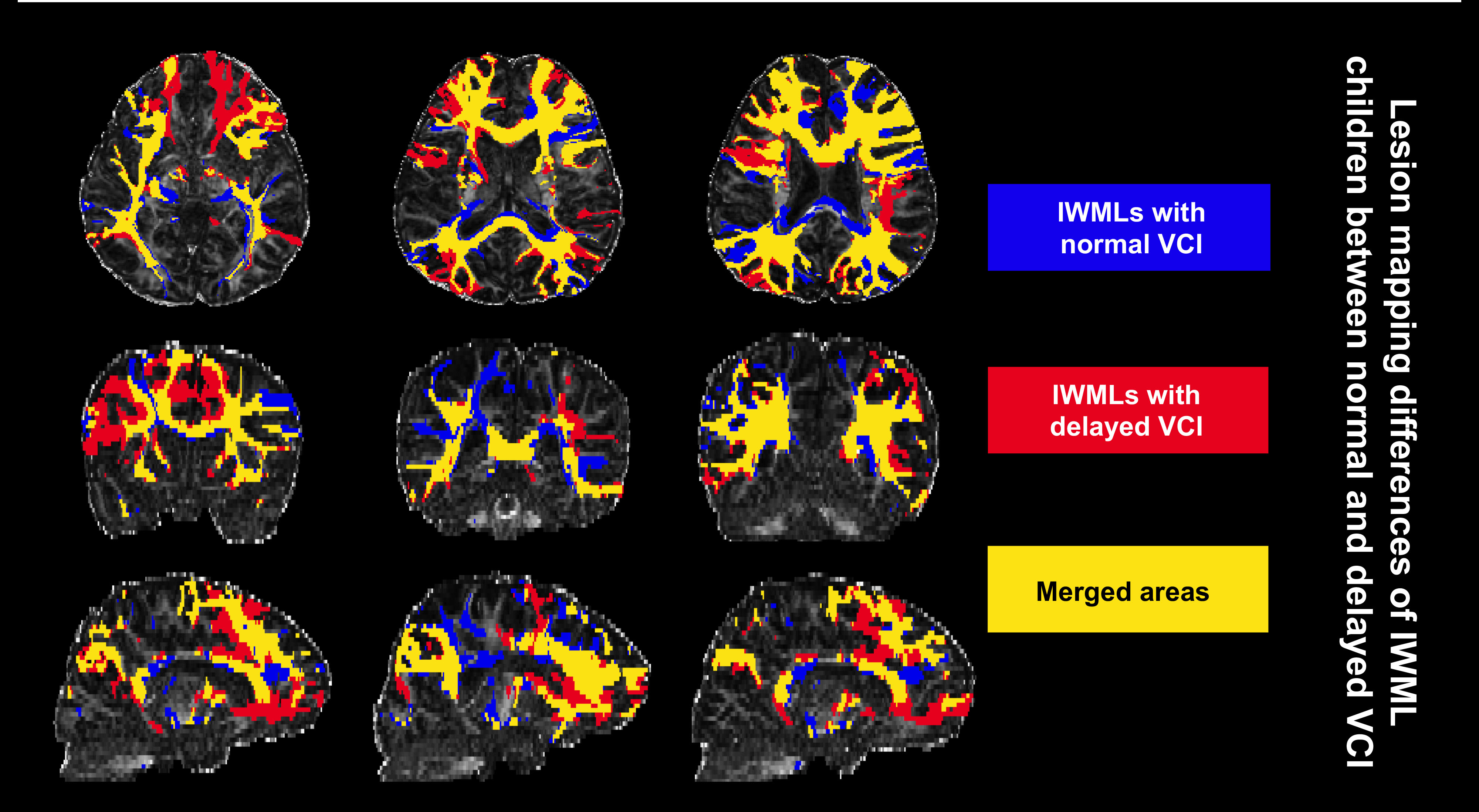
A total of 20 patients (normal-VCI: n=10, delayed-VCI: n=10) were included. No significant difference was found in demographic and clinical features between the two groups (Table 1). A significantly higher score in all subcategories of WISC was found for normal-VCI group (all P<0.01) (Table 1). The probability map of lesions showed that the lesions were mainly located in frontal and parietal lobes for both groups (Figure 1). Specifically, in areas of bilateral inferior fronto-occipital fasciculus (IFOF), bilateral uncinate fasciculus (UF), bilateral anterior thalamic radiation (ATR), and bilateral arcuate fasciculus (AF) (Figure 1). The lesion network map showed the fasciculus tracked by IWMLs’ ROI are partially similar between the normal and delayed-VCI groups, which include bilateral IFOF, corpus callosum, and right UF (Figure 2, Figure 3). However, more fasciculus has been tracked commonly by delayed-VCI groups (Figure 4). The left UF was only found in delayed-VCI groups, and was found in 8/10 participants. Bilateral AF was also found in delayed-VCI groups only, but with just 2/10 participants. Thus, we believe that the left UF and bilateral AF found in delayed-VCI groups might be the response lesion that caused the decline in verbal comprehension.
Discussion:
In this study, we found the normal and delay VCI groups have similar clusters distribution, but the fasciculus tracked by lesion ROIs are partially different between the two groups. The left UF and bilateral AF are only found in delayed-VCI groups, which could be the potential risk that caused IWMLs children with a decline in verbal comprehension. The fasciculus that is only tracked in delayed-VCI groups is mainly located in the left hemisphere, which may be related to the fact that most people’s language dominant hemisphere is located in the left hemisphere.6 The UF is a connection fiber that connects the temporal and frontal lobes, which are the areas of the semantic network. Therefore, the UF (especially left UF) is related to the semantic processing process,7 vocabulary retrieval, working memory, auditory/voice recognition, and fluency of language expression.8-12 As a connecting fiber bundle of Broca and Wernicke area, the AF also played a major role in the language process,4,13 however, more data needs to be analyzed to solidify our findings. Thus, we speculate that the white matter damage of the left frontal lobe might cause the connection disrupter of language-related brain areas and affect language functions.
Conclusion:
In conclusion, the IWMLs located at left UF and bilateral AF areas in children could possibly cause a decline in verbal comprehension in children.
Acknowledgements
This study was supported by the National Natural Science Foundation of China (81901823, 81971581, and 81901516), the Innovation Team Project of Natural Science Fund of Shaanxi Province (2019TD-018).References
1. Fish N, Konen O, Halevy A, et al. Incidental multifocal white matter lesions in pediatric magnetic resonance imaging [J]. Pediatric neurology, 2012, 47(1): 7-12.
2. Dangouloff-Ros V, Roux C, Boulouis G, et al. Incidental Brain MRI Findings in Children: A Systematic Review and Meta-Analysis [J]. AJNR American journal of neuroradiology, 2019, 40(11): 1818-23.
3. Van Der Land V, Hijmans C T, De Ruiter M, et al. Volume of white matter hyperintensities is an independent predictor of intelligence quotient and processing speed in children with sickle cell disease [J]. Br J Haematol, 2015, 168(4): 553-6.
4. Fuji M, Maesawa S, Ishiai S, et al. Neural Basis of Language: An Overview of An Evolving Model [J]. Neurologia medico-chirurgica, 2016, 56(7): 379-86.
5. Aaron D. B, Sashank P, Hesheng L, et al. Network localization of neurological symptoms from focal brain lesions [J]. Brain: a journal of neurology, 2015: 138; 3061–3075.
6. Ocklenburg S, Beste C, Arning L, et al. The ontogenesis of language lateralization and its relation to handedness [J]. Neurosci Biobehav Rev, 2014, 43(191-8.
7. Lu L, Crosson B, Nadeau S, et al. Category-specific naming deficits for objects and actions: semantic attribute and grammatical role hypotheses [J]. Neuropsychologia, 2002, 40(9): 1608-21.
8. Catani M, Jones D K, Ffytche D H. Perisylvian language networks of the human brain [J]. Ann Neurol, 2005, 57(1): 8-16.
9. Frey S, Kostopoulos P, Petrides M. Orbitofrontal contribution to auditory encoding [J]. NeuroImage, 2004, 22(3): 1384-9.
10. Grossman M, Mcmillan C, Moore P, et al. What's in a name: voxel-based morphometric analyses of MRI and naming difficulty in Alzheimer's disease, frontotemporal dementia and corticobasal degeneration [J]. Brain: a journal of neurology, 2004, 127(628-49.
11. Hickok G, Poeppel D. Dorsal and ventral streams: a framework for understanding aspects of the functional anatomy of language [J]. Cognition, 2004, 92(1-2): 67-99.
12. Ahtam B, Link N, Hoff E, et al. Altered structural brain connectivity involving the dorsal and ventral language pathways in 16p11.2 deletion syndrome [J]. Brain Imaging Behav, 2019, 13(2): 430-45.
13. Axer H, Klingner C, Prescher A. Fiber anatomy of dorsal and ventral language streams [J]. Brain and language, 2013, 127(2): 192-204.
Figures