2947
Clinical Feasibility of Compressed SENSE Artificial Intelligence Accelerated SWI in Cerebral Hemorrhage Imaging1Radiology, The First Hospital Of Jilin University, Changchun, China, 2Philips Healthcare, Beijing, China
Synopsis
Susceptibility weighted imaging (SWI) is more sensitive than T2* gradient echo MRI in detecting intracranial hemorrhage, but its acquisition time is too long. The purpose of this study is to prospectively assess the clinical feasibility of Compressed SENSE Artificial Intelligence (CS-AI) SWI by comparing it with SENSE SWI and Compressed SENSE (CS) SWI. The results suggested that the CS-AI SWI with an acceleration factor 8 was able to reduce the acquisition time of SWI by 74.58% while maintaining the image quality and accuracy of the diagnosis of intracerebral hemorrhage, except for the diagnosis of cerebral microbleeds.
Introduction
Susceptibility weighted imaging (SWI) is a technique based on T2* sequences that exploits differences in magnetic susceptibility to paramagnetic material1. It is valuable for detecting micro and massive hemorrhage and demonstrating the cerebral microvascular system and vascular malformation 2,3, which has been proved more sensitive than T2* gradient echo MRI in detecting intracranial hemorrhage 4,5. However, its relative long acquisition time may lead to motion artifacts and anxiety in some patients during examination. Several studies have shown that the Compressed SENSE (CS) reconstruction algorithm can reduce scan time without effecting image quality while providing comparable diagnostic efficiency to that of conventional images,and it has been applied to different sequences at multiple parts of the body. 6-8. Compressed SENSE Artificial Intelligence (CS-AI) is a new reconstruction algorithm developed from the basis of CS. It is assumed that brain SWI can significantly reduce scan time while maintaining image quality by using the CS-AI reconstruction algorithm.The purpose of this study was to compare the image quality of SENSE SWI, CS SWI and CS-AI SWI with different acceleration multipliers, and their diagnostic efficiency on intracerebral hemorrhage (ICH) and cerebral microbleeds (CMBs).Methods
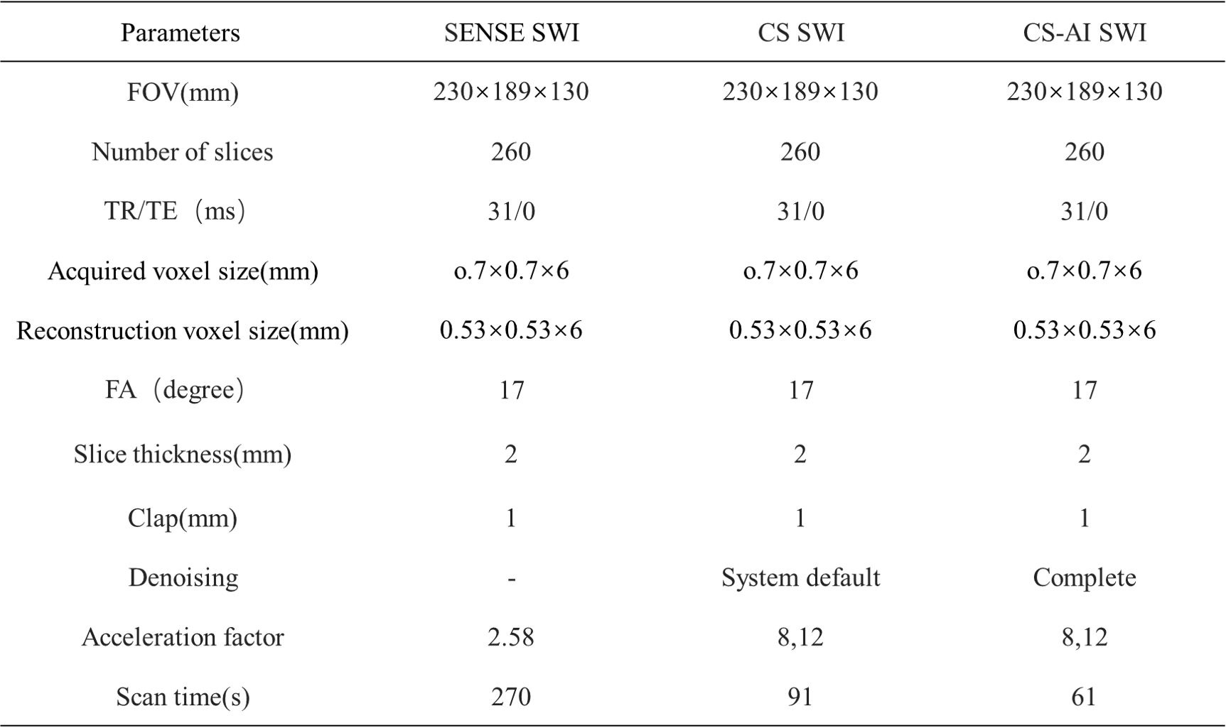
A total of seven patients with ICH confirmed by CT scan were prospectively enrolled. All patients were examined on 3.0T MRI system(Ingenia Elition 3.0T; Philips Healthcare, Best, The Netherlands) and three of them were diagnosed with CMBs. All patients received two different acceleration factors 8 and 12 for CS SWI(CS8, CS12) and CS-AI SWI(CSAI8, CSAI12), as well as SENSE SWI with acceleration factor 2.58 as a reference. The scanning parameters are shown in Table 1. The denoising level of CS group was set to "system default", and the CS-AI group was set to "complete". Two uninformed radiologists identified brain hemorrhage lesions in all patients and counted the number of CMBs in the brains of the three patients. Two radiologists rated the overall image quality of SENSE SWI, CS SWI and CS-AI SWI using a five-level scale based on visual analysis. The ROI of cerebral hemorrhage lesions and the proximity of white matter in all patients were outlined. Five levels were selected for each lesion and the proximity of white matter, and their mean and SD values were recorded, SNR and CNR were calculated. The SNR and CNR of the images were statistically analyzed using the Friedman test and post hoc analysis. Statistical significance was considered at P < 0.05.$$SNR_{lesion}=\frac{SI_{lesion}}{SD_{lesion}}$$,$$CNR_{lesion-white matter}=\frac{|SI_{lesion}-SI_{white matter}|}{\sqrt{SD_{leision}^{2}+SD_{white matter}^{2}}}$$
Results and Discussions
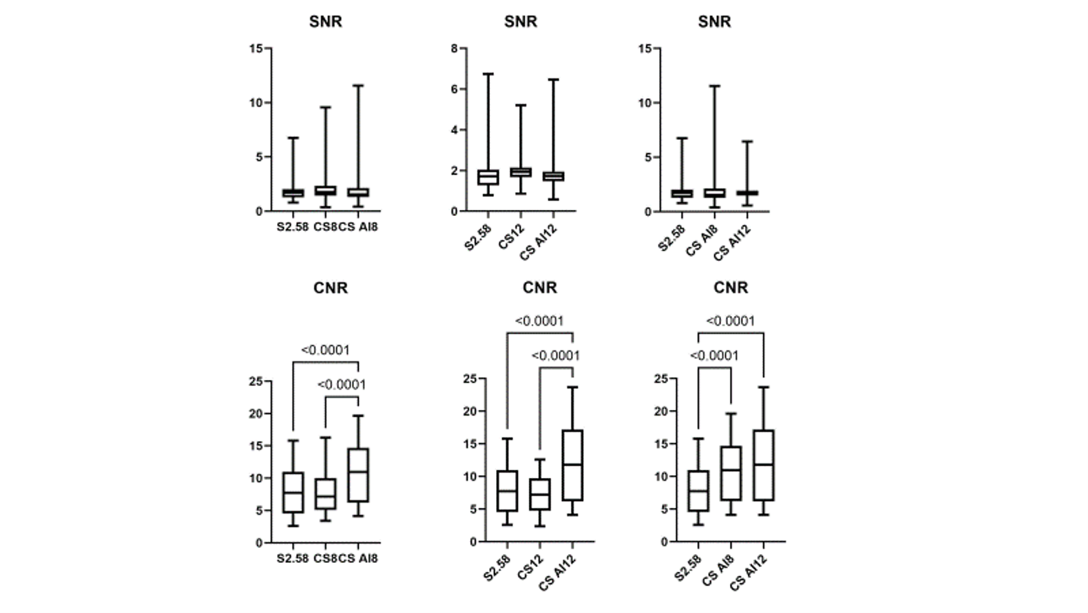
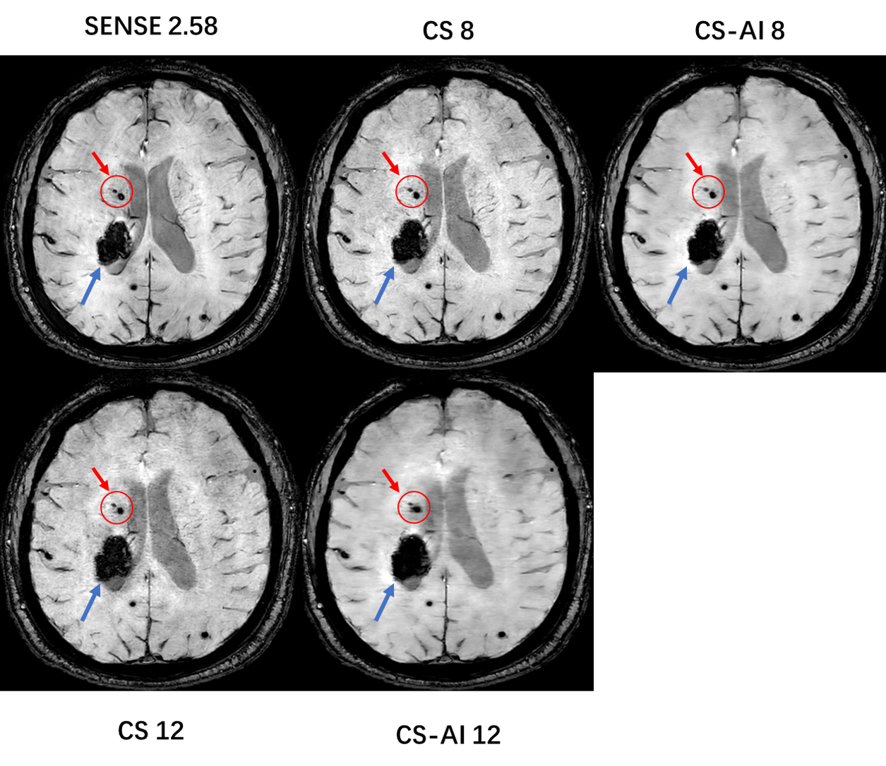
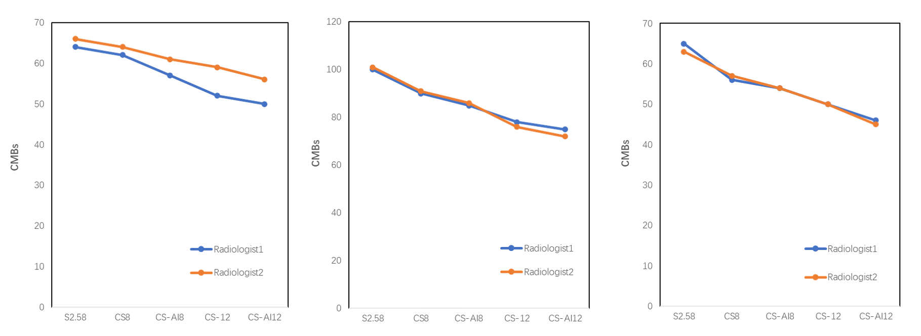
The scan time of CS 8/CS-AI 8 was reduced 74.58% and 77.41% for CS 12/CS-AI 12 compared to the conventional SWI( SENSE 2.58). In the objective evaluation, there was no statistical difference in SNR among conventional SENSE SWI, CS SWI and CS-AI SWI with different acceleration factors, while both the CNR of CS-AI 8 and CS-AI 12 were significantly higher than that of conventional SENSE SWI (P<0.0001), and both the CNR of CS-AI 8 and CS-AI 12 were significantly higher than that of CS sequences with the same acceleration multiplier (P<0.0001)(Figure 1). IIn the subjective evaluation of image quality ,the radiologists considered the overall image quality of SENSE SWI and CS-AI 8 comparable(Figure 2). All of SENSE, CS and CS-AI SWI diagnosed cerebral hemorrhagic lesions in 7 patients. However, in the statistics of the number of CMBs, CS and CS-AI SWI with different accelerating factors showed less CMBs than that of SENSE SWI probably due to the small size of CMBs and some of small CMBs were incorrectly removed as noise during CS and CS-AI reconstruction(Figure 3). Further work should test the detection performance of CMBs using different levels of denoising parameters.Conclusion
Although the diagnosis efficiency of CMBs is slightly reduced, the current findings suggest that CS-AI SWI with an acceleration factor 8 is superior to either SENSE SWI or CS SWI in reducing the scan time significantly while maintaining the considerable diagnostic efficiency of cerebral hemorrhage.Acknowledgements
No acknowledgement found.References
1.Larsen JP,Britt W,Kido D,et al.Susceptibility weighted magnetic resonance imaging in evaluation of dementia.Radiology Case Reports 2007;2:102.
2.Haacke EM, Mittal S, Wu Z, et al.Susceptibility-weighted imaging: technical aspects and clinical applications, Part 1. AJNR Am J Neuroradiol 2009;30:19–30.
3. Mittal S, Wu Z, Neelavalli J, et al.Susceptibility-weighted imaging: technical aspects and clinical applications, Part 2. AJNR Am J Neuroradiol 2009;30:232–52.
4.Nandigam RN, Viswanathan A, Delgado P, et al. MR imaging detection of cerebral microbleeds: effect of susceptibility-weighted imaging, section thickness, and field strength. AJNR Am J Neuroradiol 2009;30(2):338–43.
5.Cheng AL, Batool S, McCreary CR, et al. Susceptibility weighted imaging is more reliable than T2*-weighted gradient-recalled echo MRI for detecting microbleeds. Stroke 2013;44(10):2782–6.
6.Vranic JE, Cross NM, Wang Y, et al. Compressed Sensing-Sensitivity Encoding (CS-SENSE) Accelerated Brain Imaging: Reduced Scan Time without Reduced Image Quality. AJNR Am J Neuroradiol. 2019 Jan;40(1):92-98.
7. Winkel DJ, Heye TJ, Benz MR, et al. Compressed Sensing Radial Sampling MRI of Prostate Perfusion: Utility for Detection of Prostate Cancer. Radiology. 2019;290(3):702-708.
8. Iuga AI, Abdullayev N, Weiss K, et al. Accelerated MRI of the knee. Quality and efficiency of compressed sensing. Eur J Radiol. 2020;132:109273.
Figures