2946
Surface-based Cortical Network Analysis in Post-Stroke Dementia with Subcortical lesions1Key Laboratory for Biomedical Engineering of Ministry of Education, Department of Biomedical Engineering, College of Biomedical Engineering & Instrument Science, Zhejiang University, Hangzhou, China, 2Department of radiology, Sir Run Run Shaw Hospital, School of Medicine, Zhejiang University, Hangzhou, China
Synopsis
This study aimed to investigate alterations of cortical network in post-stroke dementia (PSD) patients with subcortical lesions using a surface-based morphometry analysis. We calculated nine cortical morphometric metrics and constructed the structural covariate network for each participant. Comparisons between groups showed that PSD and post-stroke nondemented (PSND) groups both displayed an increased local efficiency and a similar global efficiency compared with the normal controls group, and the cortical network in three groups all remained a small-world property. Moreover, we found PSD-specific decreases in gray matter volume and surface area in lateral occipital cortex and middle temporal gyrus.
Introduction:
Post-stroke dementia (PSD) is a common neurological complication after stroke [1]. Previous studies demonstrated abnormal small-world properties and network efficiency in cortical regions in the patients with dementia [2], and anomalous structural covariance patterns with more covariant brain regions in stroke patients [3]. However, the alternations in cortical network are still unclear in PSD patients. Therefore, this study used a surface-based morphometry method to construct individual structural covariance network (SCN) and explored how it would be affected by PSD.Methods:
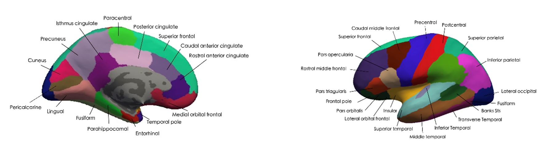
The present study is a cross-sectional experimental design based on high-resolution T1-weighted MR images from 48 stroke patients with subcortical lesions (PSD: n = 20; post-stroke nondemented (PSND): n = 28), and age-matched 21 normal controls (NC). Mini-Mental State Examination (MMSE) and miniCog were used to evaluate the cognitive performance of each participant. We first used FreeSurfer 6.0 to segment the whole brain into thirty-one cortical regions for each hemisphere (three subcortical regions were excluded) (Figure 1) and obtained nine surface-related morphological metrics. Then, we constructed the SCN connectivity matrix at the individual level, and further calculated the network properties for each participant. The network analysis was carried out under a sparsity of 0.05–0.5 with an interval of 0.05. For the graph of each participant, we evaluated the cortical network using this predefined range as the thresholds with the following parameters: small-world coefficient (σ), global efficiency (Eg), and local efficiency (Eloc)) [4]. Finally, we used a linear mixed model, with the age, sex, education, intracranial volume and lesion information as the covariates and the subject factor as a random variable, to compare the difference between groups, and further performed the correlation analysis between MRI measurements and clinical assessments. False discovery rate (FDR) was performed to correct the multiple comparisons for the p value.Results:
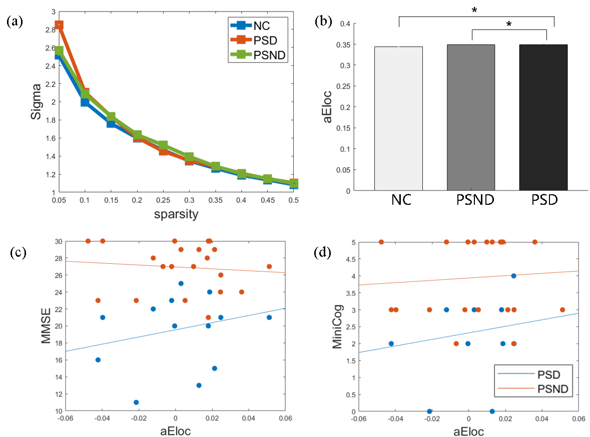
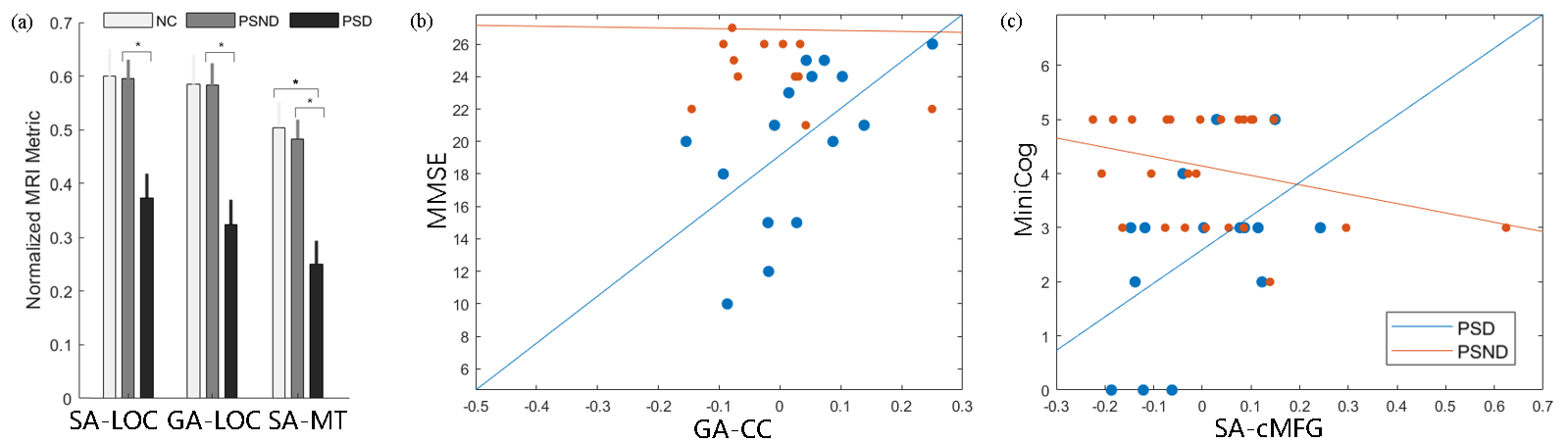
Network analysis showed that PSD and PSND groups both displayed an increased local efficiency compared with the NC group (Figure 2b), which was positively correlated with cognitive function in the PSD but not in the PSND groups (Figure 2c and 2d). The global efficiency did not show significant differences between the three groups, and the cortical network in three groups all remained a small-world property (Figure 2a). Moreover, we found PSD-specific changes (which show significant difference between PSD and PSND while exhibit insignificant differences between PSND and NC) in gray matter volume and surface area of lateral occipital cortex and middle temporal gyrus (Figure 3a). Moreover, the surface area in right caudal middle frontal gyrus and the volume in right cingulate cortex were correlated with the cognitive function (Figure 3b and 3c).Discussion:
The previous studies found that patients with dementia showed lower global and local efficiency of structural networks compared with those without dementia, which was associated with cognitive decline [2]. Given that the local efficiency is a measure of the segregation and specialization within a network [2], the increased local efficiency in the PSD patients in the present study, which was positively correlated with MMSE and MiniCog scores, maybe a reflection of the compensation mechanism after stroke. Moreover, we found PSD-specific decreases in gray matter volume and surface area of lateral occipital cortex and middle temporal gyrus. This was partly consistent with the previous reports in Parkinson’s disease [5] and stroke [6] that the two regions showed reduced gray matter volume/density associated with cognitive impairments.Conclusion:
The increased local efficiency of cortical network may indicate a more modulated brain in the PSD than NC. We also found dementia-specific morphological changes in lateral occipital cortex and middle temporal gyrus, which may serve as the biomarkers of PSD.Acknowledgements
This work is supported by the Youth Program of National Natural Science Foundation of China (82001907), Natural Science Foundation of Zhejiang Province of China (LY19H090027), and China Postdoctoral Science Foundation (2020M671726).References
1. Pasi M, Poggesi A, Salvadori E, et al. Post-stroke dementia and cognitive impairment. Manifestations of Stroke. 2012;30(65-69.
2. Tuladhar A M, van Uden I W, Rutten-Jacobs L C, et al. Structural network efficiency predicts conversion to dementia. Neurology. 2016;86(12): 1112-1119.
3. Wei Y, Wang C, Liu J, et al. Progressive gray matter atrophy and abnormal structural covariance network in ischemic pontine stroke. Neuroscience. 2020;448(255-265.
4. Huang Y, Liu Y, Zhao D, et al. Small-world properties of the whole-brain functional networks in patients with obstructive sleep apnea‐hypopnea syndrome. Sleep medicine. 2019;62(53-58.
5. Chen Y-S, Chen H-L, Lu C-H, et al. Reduced lateral occipital gray matter volume is associated with physical frailty and cognitive impairment in Parkinson’s disease. European radiology. 2019;29(5): 2659-2668.
6. von Deneen K M. Correlations between cognitive function and gray matter alterations in patients with acute lacunar stroke. Brain Science Advances. 2021;7(2): 112-123.
Figures