2939
Visualizing the habenula using high resolution MPRAGE, MP2RAGE and QSM at 3T MRI1The first hospital of Jilin University, changchun, China, 2MR Scientific Marketing, Siemens Healthineers Ltd., Beijing, China, 3MR CS APP, Siemens Healthineers Digital Technology Co., Harbin, China
Synopsis
MPRAGE, MP2RAGE and QSM can clearly visualize the Hb compared to conventional T1WI. Due to its higher CNR, MP2RAGE and QSM could be considered for structural imaging in Hb morphology study.
BACKGROUND AND PURPOSE
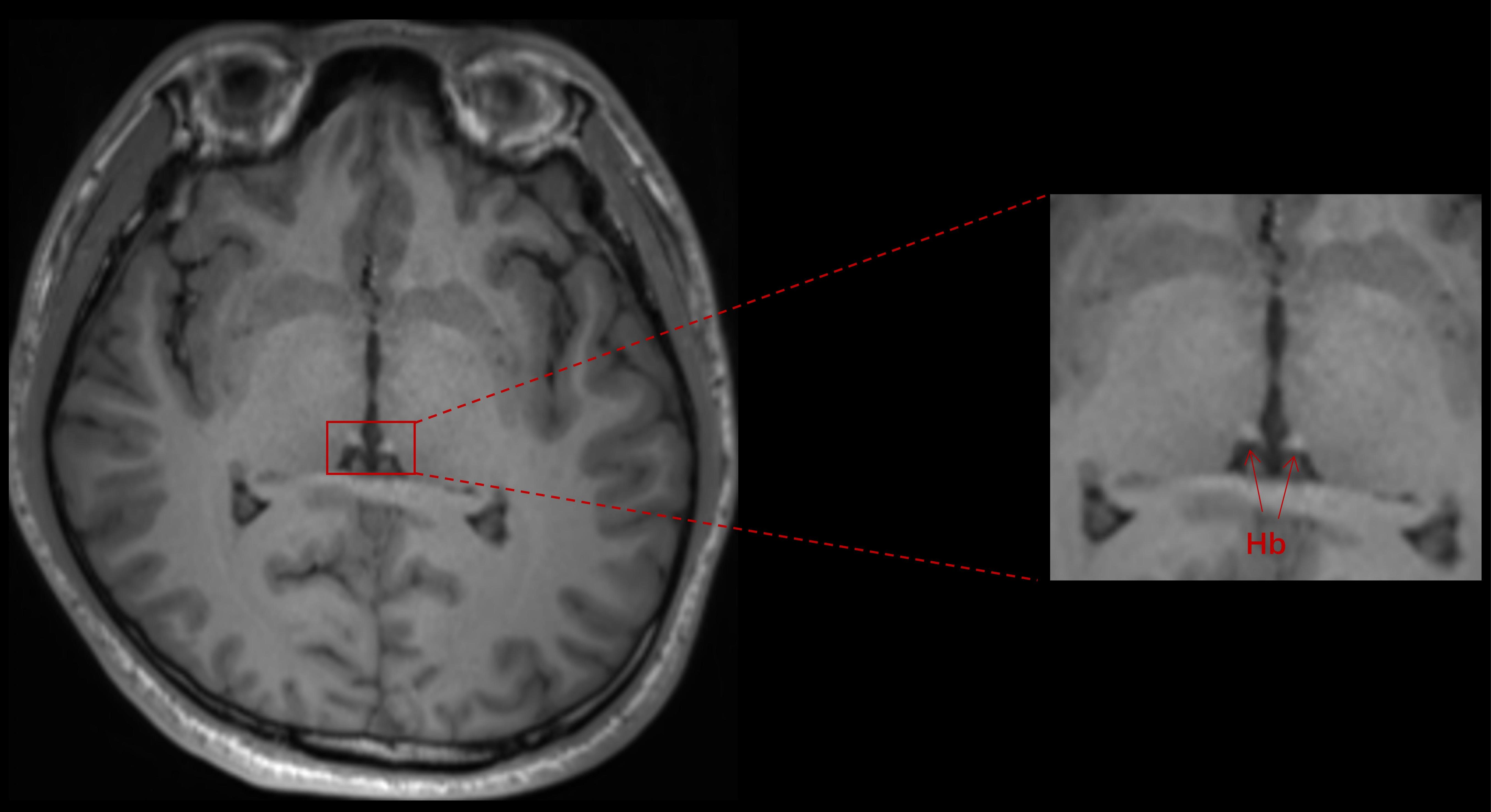
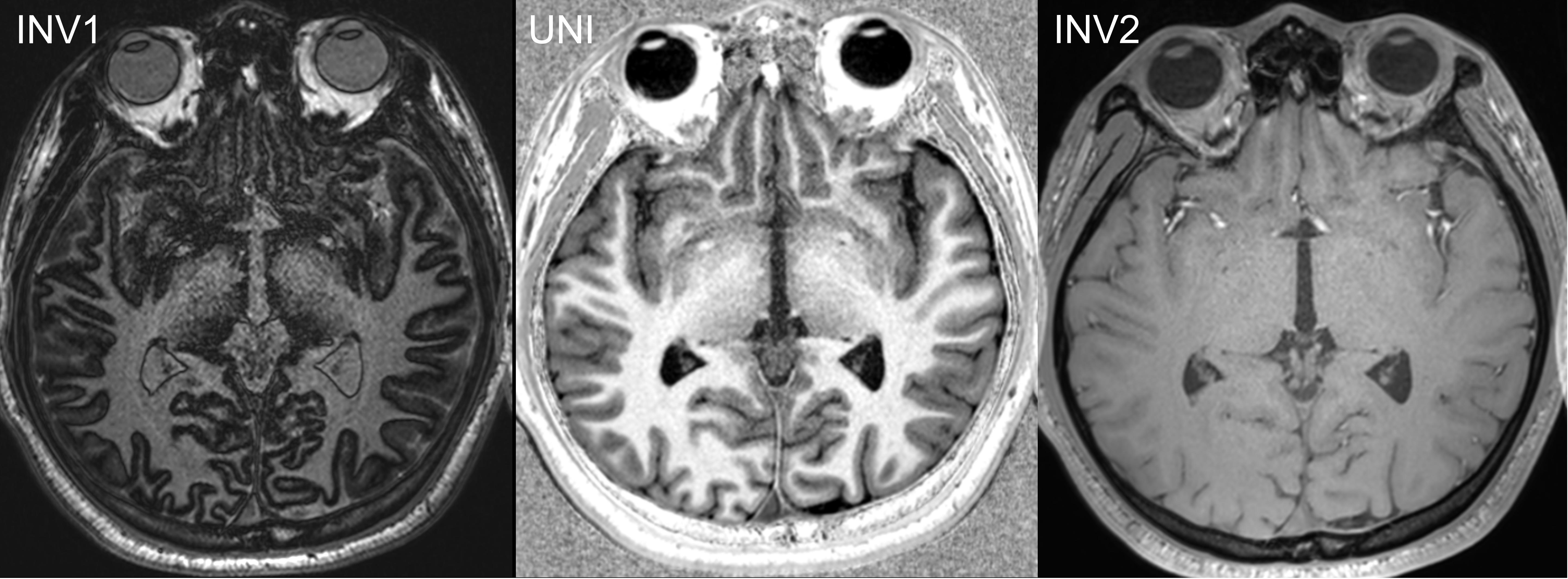
The Habenular(Hb)is a bilateral, paired, and small structure of approximately 5-9 mm in diameter close to the thalamus in human brain1, which plays an important role in brain reward circuitry and psychiatric conditions2. The dysfunction of Hb may underlie several psychiatric disorders, such as major depressive disorder and bipolar disorder3. Conventional T1WI or T2WI of Hb show limited contrast and conspicuity because of its small size and uncertain of its heterogeneous content. Currently, several studies applied MPRAGE and QSM techniques in Hb imaging studies2-3, however, the tissue contrast and resolution of Hb were inconsistent in current literatures and image quality of Hb need to be further improved. Recently, new sequence techniques came into clinical routine with high resolution and multi-contrasts, such as Magnetization prepared 2 rapid gradient echo (MP2RAGE) sequence, which offers superior gray (GM) and white matter (WM) contrast. In MP2RAGE, two gradient echo images are acquired at different inversion times and are combined to yield a T1 contrast enhanced T1W image (UNI) that is free from B1-inhomogeneity. In addition, a quantitative T1 map in high resolution is also obtained4-5[Fig2]. The purpose of this study was to propose high-resolution MR image of Hb with MPRAGE, MP2RAGE and QSM at 3T, and compare the image quality of Hb.METHODS
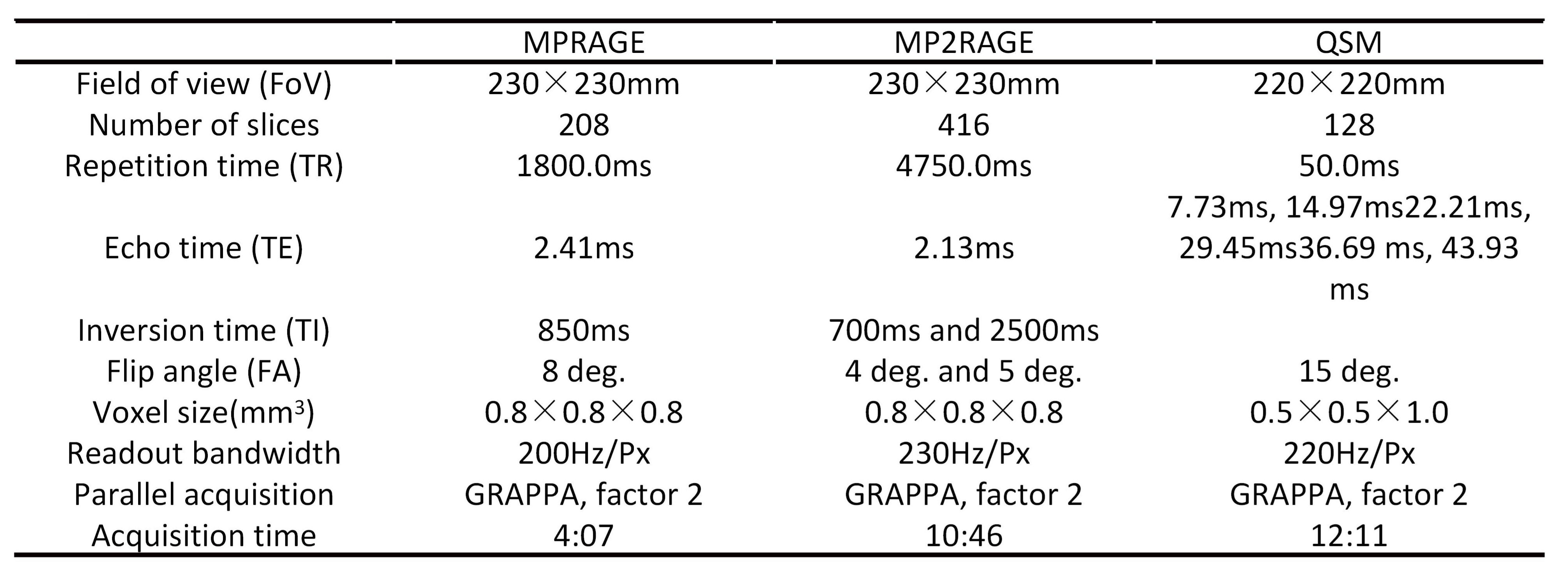
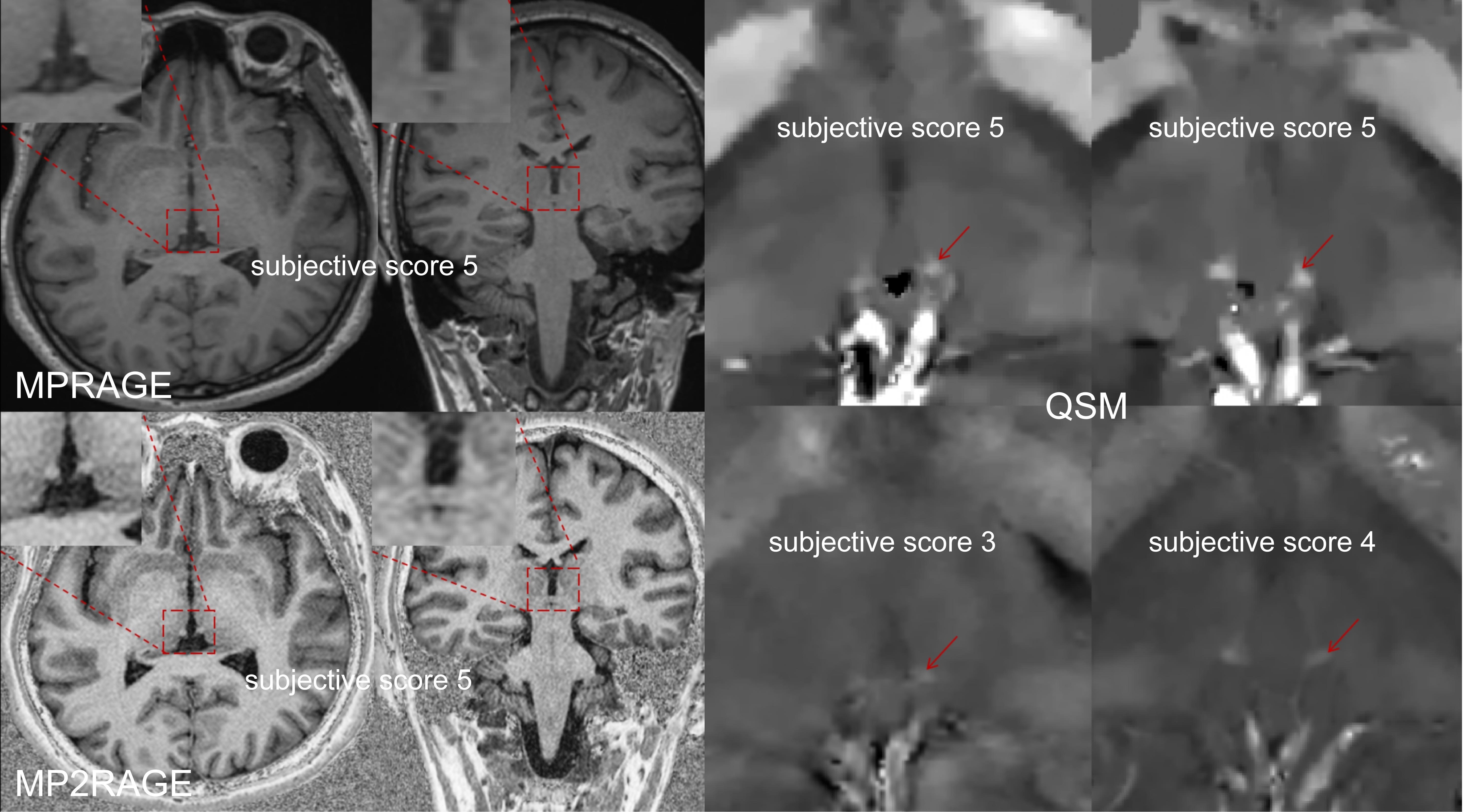
Ten (age 32±4.6; 5 males, 5 females) healthy volunteers were examined on a 3T system (MAGNETOM Vida, Siemens Healthcare, Erlangen, Germany) acquiring MP2RAGE, MPRAGE and QSM images. To show the outline of the Hb, we imaged the Hb and thalamus to highlight the contrast between Hb and thalamus. The detailed imaging parameters are listed in Fig3. Two radiologists with 25 and 2 years of experience independently evaluated Hb MR images in random order. Qualitative assessment of MPRAGE and MP2RAGE sequences was performed based on the following 5-point criteria:1: rarely seen, 2: vaguely seen, 3: identifiable, 4: blurry borders, 5: sharp borders1, markedly reduced SNR; 2, moderately reduced SNR; 3, diminished SNR; 4, mildly diminished SNR; and 5, optimal SNR. Quantitative assessments of T1WI MPRAGE, T1WI MP2RAGE and UNI MP2RAGE were performed on a workstation (Syngo MR, Siemens Healthcare, Erlangen, Germany). The slice in which the Hb showed the largest cross-sectional area was chosen for quantitative analysis. ROIs were drawn independently by two neuroradiologists. Signal to noise ratio (SNR) of Hb and contrast-to-noise ratio (CNR) of Hb and thalamus were calculated based on MPRAGE and MP2RAGE by the following equations: $$$ SNRHb=SIHb/SDHb $$$and $$$ CNRHb-thalamus=|SIHb−SIthalamus|/√SDHb2+SDthalamus2 $$$
where SI is signal intensity and SD is standard deviation. Two radiologists performed the qualitative and quantitative analysis independently. The mean values of two readers were applied to further statistical analyses.
RESULT and DISCUSSION
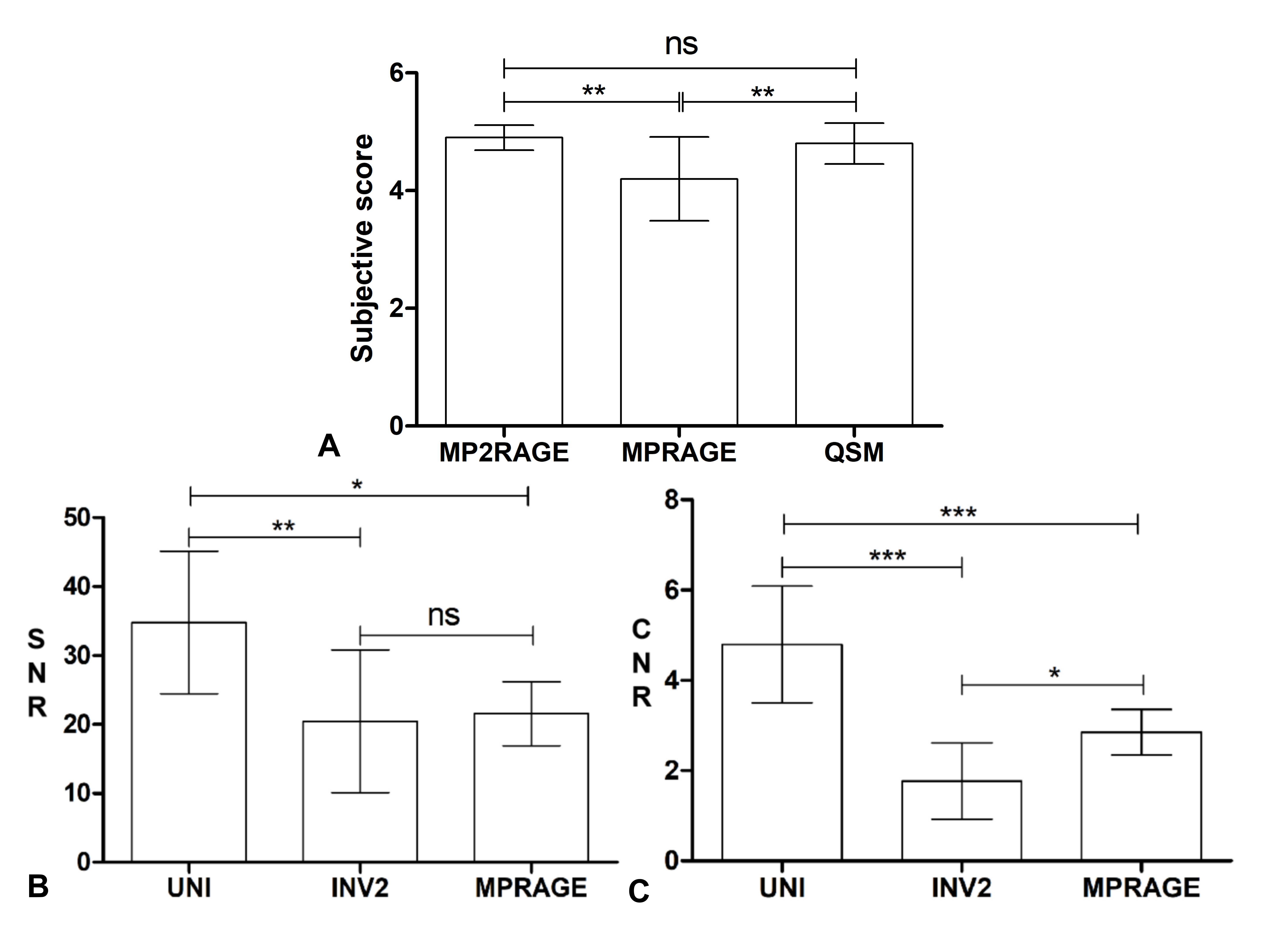
Figure 4 and figure 5A shows that subjective scores of Hb image in MP2RAGE and QSM are significantly higher than MPRAGE (p<0.05). Compared with MPRAGE T1WI, significantly higher SNRHb and CNRHb-thalamus was detected in MP2RAGE UNI (p<0.05) as shown in figure 5B-C. The CNR variation range (5-9%) in UNI images of MP2RAGE was reduced compared with that of T1WI MPRAGE (10-40%). Based on the above results, high-resolution MP2RAGE UNI and QSM images shows higher potentials in visualizing Hb than MPRAGE. MP2RAGE UNI yielded higher CNR and clearer signal intense compared to MPRAGE in Hb, and consequently, provides a high-quality scanning sequence for more accurate segmentation of Hb in the future.CONCLUSION
MP2RAGE and QSM can clearly display the Hb compared to MPRAGE. Due to its higher CNR, MP2RAGE could be considered for structural imaging in Hb morphology study. Meanwhile, QSM might similarly provide a novel, conspicuous anatomical contrast for human habenula imaging.Acknowledgements
No acknowledgement found.References
1. Strotmann B, et al. High-resolution MRI and diffusion-weighted imaging of the human habenula at 7 tesla. J Magn Reson Imaging. 2014, 39(4): 1018-26.
2. Yoo, S., Kim, Jw., Schenck, J.F. et al. Magnetic susceptibility imaging of human habenula at 3 T. Sci Rep 10, 19357 (2020). https://doi.org/10.1038/s41598-020-75733-y.
3. Germann J, et al. Fully Automated Habenula Segmentation Provides Robust and Reliable Volume Estimation Across Large Magnetic Resonance Imaging Datasets, Suggesting Intriguing Developmental Trajectories in Psychiatric Disease. Biol Psychiatry Cogn Neurosci Neuroimaging. 2020, 5(9): 923-929.
4. He N, et al. Visualizing the lateral habenula using susceptibility weighted imaging and quantitative susceptibility mapping. Magn Reson Imaging. 2020, 65(undefined): 55-61.
5. Okubo G, et al. MP2RAGE for deep gray matter measurement of the brain: A comparative study with MPRAGE. Magn Reson Imaging. 2016, 43(1): 55-62.
Figures