2934
Simultaneous Multislice Imaging Using Multiphoton MRI1Electrical Engineering and Computer Sciences, University of California, Berkeley, Berkeley, CA, United States, 2Helen Wills Neuroscience Institute, University of California, Berkeley, Berkeley, CA, United States
Synopsis
Simultaneous multislice (SMS) imaging accelerates data acquisition by exciting several slices simultaneously but has a higher specific absorption rate (SAR) due to additional radiofrequency (RF) pulses required to excite multiple frequencies. Multiphoton excitation achieved by oscillating gradient fields provides frequency sidebands, eliminating the need for additional excitation pulses. We have demonstrated SMS imaging for multiphoton MRI and used SENSE reconstruction to separate simultaneously acquired slices with the help of coil sensitivity profiles. Multiphoton SMS images achieve a similar quality to standard one-photon SMS images but offer the benefit of reduced SAR as they do not require extra RF power.
Introduction
Simultaneous multislice (SMS) imaging is a parallel imaging technique commonly used for accelerating image acquisition by exciting multiple slices at the same time1-3. SMS is advantageous because it reduces scan time without a significant signal-to-noise ratio (SNR) penalty1. A major difficulty, however, is the increased specific absorption rate (SAR) from multislice excitation pulses. We aim to address the SAR issue by utilizing multiphoton excitation which is a novel technique in MRI that uses multiple magnetic field frequencies different from the Larmor frequency to excite multiphoton resonances4. In this work, multiphoton excitation is obtained by oscillating the gradient fields, and these oscillating magnetic fields provide frequency sidebands which can easily be used for SMS imaging. As additional radiofrequency (RF) power is not required for the extra frequency bands, multiphoton excitation offers a reduced SAR benefit for SMS imaging.Theory
RF fields, $$$B_{1}$$$, polarized in the xy-plane result in standard one-photon resonances. Applying additional z-axis RF fields excites multiphoton resonances4. For any integer $$$n$$$, the resonance condition can be expressed as $$$\omega_{xy}=\gamma B_0+n\omega_{z}$$$, where $$$B_{0}$$$ is the main magnetic field, $$$\omega_{xy}$$$ and $$$\omega_{z}$$$ refer to the frequencies of xy-plane and z-axis $$$B_{1}$$$ fields respectively, and $$$n$$$ is the number of z-axis photons in the resonance4. If $$$n=0$$$, then we have one-photon resonance where $$$\omega_{xy}=\gamma B_0$$$.Gradients have z-axis magnetic fields, hence, when they are oscillating, they provide z-axis RF which can be used to excite multiphoton resonances. In our case, frequency sidebands at $$$\pm\omega_ {z}$$$ provide two-photon excitation and are utilized for SMS imaging. Effective $$$B_{1}$$$ fields can be written as $$B_{1, eff}\left(t\right)=B_{1, xy}\left(t\right)J_1\left(\frac{\gamma B_{1,z}}{\omega_z}\right),$$ where $$$J_1$$$ is the Bessel function of the first kind of order 1, $$$B_{1, xy}$$$ is the amplitude of the xy-RF field, and $$$B_{1, z}$$$ is the amplitude of the z-axis RF field produced by the oscillating gradients. As the effective $$$B_{1}$$$ field is spatially varying due to the spatially varying $$$B_{1, z}$$$, flip angles are also spatially varying. This will introduce a contrast difference between images of different slices. For efficiency, $$$\frac{\gamma B_{1,z}}{\omega_z}$$$ is chosen to be 1.5, which is the point where the Bessel function achieves roughly its peak amplitude.
Methods
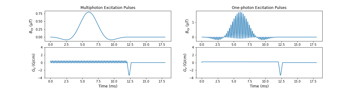
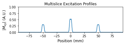
Custom gradient-echo sequences with multiphoton or one-photon excitation pulses (Figure 1) were written using HeartVista (HeartVista, Inc., Los Altos, CA) and implemented on a 3T GE MR750w scanner equipped with a 22 channel head-and-neck receiver array. The multiphoton excitation pulse had a duration of 12 ms and is a combination of a 20$$$^{\circ}$$$ SLR RF pulse with a 0.293 G/cm sinusoidal gradient oscillating at 4.167 kHz superimposed on a 0.196 G/cm DC gradient. The SLR pulse had a time-bandwidth product of 5. Since flip angle is proportional to the magnitude of the effective $$$B_{1}$$$ field, slices at $$$\pm$$$50 mm had a flip angle of 11.16$$$^{\circ}$$$, whereas the center slice had a flip angle of 20$$$^{\circ}$$$. The sinusoidal gradient frequency was calculated to excite two-photon resonances at 5 cm away from the isocenter based on the DC gradient amplitude. The sinusoidal gradient amplitude was calculated to give $$$\frac{\gamma B_{1,z}}{\omega_z}=$$$ 1.5 at 5 cm away from the isocenter. Figure 2 shows the excitation profiles. The one-photon excitation pulse was a combination of three 12 ms SLR pulses with time-bandwidth products of 5. The center pulse had a 20$$$^{\circ}$$$ flip angle, and the two side pulses had 11.16$$$^{\circ}$$$ flip angles to match with the multiphoton pulse. Other parameters are: TE/TR = 20/45 ms, 5.0 mm slice thickness, in-plane resolution = 1.25x1.25 mm2, FOV = 32x32 cm2, 62.5 kHz readout bandwidth. A coronal orientation was chosen for greater coil sensitivity differences.To estimate the sensitivity maps of each coil, a calibration scan was taken immediately before the SMS scan. Each slice was excited individually during the calibration scan and sensitivity maps were estimated using BART5. Using these coil sensitivity maps, SENSE6 reconstruction was implemented to disentangle the simultaneously acquired slices.
Results
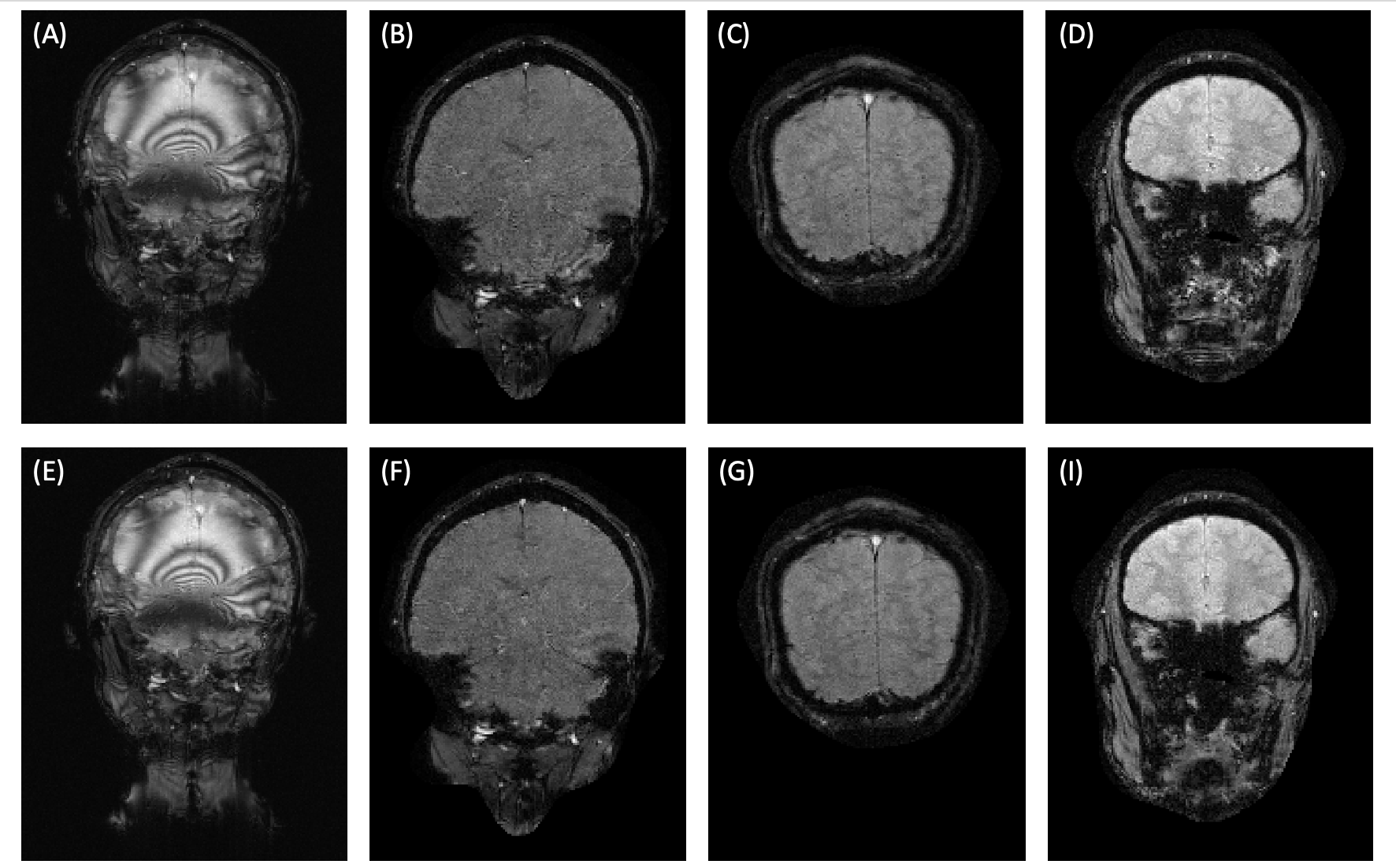
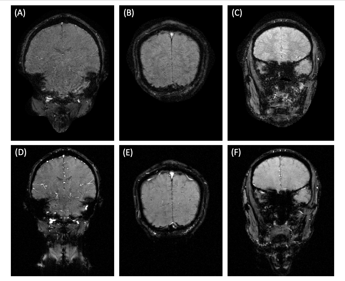
Figure 3 shows the overlapping images of three simultaneously excited slices and their disentangled versions obtained by SENSE reconstruction for both multiphoton and one-photon SMS. Quality of reconstructed multiphoton and one-photon SMS images are seen to be very similar. Figure 4 shows that reconstructed multiphoton SMS images do not have significant aliasing artifacts or SNR reduction, hence, they are comparable in quality to their individually acquired counterparts.Discussion and Conclusion
We have demonstrated SMS imaging for multiphoton MRI at 3T. Disentangled SMS images for multiphoton and one-photon excitation were found to be very similar in quality. However, one-photon SMS requires additional high power RF pulses to excite multiple slices, whereas frequency sidebands are provided by oscillating gradients in multiphoton MRI. Hence, multiphoton SMS provides a significant SAR reduction by making it possible to acquire multiple slices using the same RF power required by one-photon single slice excitation. More frequency bands can be easily produced with three-photon and higher order resonances without any increase in SAR.Although here we demonstrate unequal flip angles among the slices, equal flip angles can be achieved with adiabatic pulses7 or with a dedicated coil providing uniform $$$B_{1,z}$$$ fields8. Further, parallel transmit can be used to achieve equal flip angles as well, since transmit arrays have spatially varying sensitivities.
Acknowledgements
This work was supported in part by NIH grant R21EB030157. The authors thank Ekin Karasan, Shreya Ramachandran and Alfredo De Goyeneche of UC Berkeley for discussions and assistance with coil sensitivity profile estimation.References
1. Larkman DJ, Hajnal JV, Herlihy AH, Coutts GA, Young IR, Ehnholm G. Use of multicoil arrays for separation of signal from multiple slices simultaneously excited. J Magn Reson Imaging. 2001; Feb;13(2):313-7. doi:10.1002/1522-2586(200102)13:2<313::aid-jmri1045>3.0.co;2-w.
2. Setsompop K, Gagoski BA, Polimeni JR, Witzel T, Wedeen VJ, Wald LL. Blipped-controlled aliasing in parallel imaging for simultaneous multislice echo planar imaging with reduced g-factor penalty. Magn Reson Med. 2012 May;67(5):1210-24. doi: 10.1002/mrm.23097.
3. Barth M, Breuer F, Koopmans PJ, Norris DG, Poser BA. Simultaneous multislice (SMS) imaging techniques. Magnetic Resonance in Medicine. 2016; 75: 63-81. doi:https://doi.org/10.1002/mrm.25897
4. Han V, Liu C. Multiphoton magnetic resonance in imaging: A classical description and implementation. Magnetic Resonance in Medicine. 2020;84(3):1184-1197. doi:https://doi.org/10.1002/mrm.28186
5. Uecker M, Ong F, Tamir JI, Bahri D, Virtue P, Cheng JY, Zhang T, Lustig M. Berkeley advanced reconstruction toolbox. Proceedings of the International Society for Magnetic Resonance in Medicine, Toronto; 2015;23:2486.
6. Pruessmann KP, Weiger M, Scheidegger MB, Boesiger P. SENSE: sensitivity encoding for fast MRI. Magn Reson Med. 1999 Nov;42(5):952-62.
7. Han V, Liu C. Multiband Adiabatic Inversion Using Multiphoton Excitation. Proceedings of the International Society for Magnetic Resonance in Medicine; 2020;28:0605.
8. Chi J, Han V, Liu C. In vivo Two-photon Magnetic Resonance Imaging of Human Brain at 3T. Proceedings of the International Society for Magnetic Resonance in Medicine; 2021;29:0564.
Figures