2930
Making Reduced FOV Imaging Applicable on Low-Cost MRI Systems: A Sheared 2DRF Excitation Approach
Bahadir Alp Barlas1,2 and Emine Ulku Saritas1,2,3
1Department of Electrical and Electronics Engineering, Bilkent University, Ankara, Turkey, 2National Magnetic Resonance Research Center (UMRAM), Bilkent University, Ankara, Turkey, 3Neuroscience Graduate Program, Bilkent University, Ankara, Turkey
1Department of Electrical and Electronics Engineering, Bilkent University, Ankara, Turkey, 2National Magnetic Resonance Research Center (UMRAM), Bilkent University, Ankara, Turkey, 3Neuroscience Graduate Program, Bilkent University, Ankara, Turkey
Synopsis
The utilization of low-cost low-field MRI systems has been rising due to the recent image quality improvements. However, these systems cannot incorporate numerous popular MRI techniques due to their hardware limitations. This work proposes a sheared 2DRF pulse design to make reduced FOV imaging applicable for low-cost MRI systems. The proposed sheared 2DRF pulse design provides significant reduction in pulse duration together with improved signal under B0 field inhomogeneities, while ensuring robust sidelobe suppression and unlimited slice coverage. We demonstrate the proof-of-concept applicability of the proposed approach for a 0.35T and a 1.5T MRI scanner.
Introduction
Although the advances in high-field MRI systems achieved high-quality images, installation and maintenance of these systems can be costly and complex, preventing wide accessibility to these systems1-5. Recently, relatively low-cost low-field MRI systems with laudable image quality have been introduced as cheaper alternatives to the high-field systems1,2,6. However, the hardware limitations of these low-cost systems continue to be a major inconvenience, preventing the applicability of numerous popular MRI techniques1-3. Previously, we proposed sheared 2DRF excitation to enhance off-resonance robustness of reduced field-of-view (FOV) imaging and demonstrated this technique on a 3T MRI environment7. In this work, we propose sheared 2DRF excitation to make reduced FOV imaging applicable for low-cost MRI systems restricted by hardware limitations. We demonstrate the proof-of-concept applicability of the proposed design for 0.35T and 1.5T MRI scanners.Methods
2DRF Pulse DesignStandard 2DRF pulses with blipped axis along the slice-select (SS) direction were designed with the following parameters: Time-bandwidth products in SS and phase-encoding (PE) directions of TBWSlice=3 and TBWSlab=8, FOVSlab=40mm slab thickness, and Δz=4mm slice thicknesses. The hardware limitations were set as GMAX=25mT/m and 24mT/m for maximum gradient amplitude; and SMAX=90T/m/s and 55T/m/s for maximum slew rate for the low-cost 1.5T and 0.35T8 MRI systems, respectively. The standard 2DRF pulses had 25.6ms and 20.24ms pulse durations for 0.35T and 1.5T MRI systems, respectively, exceeding the applicable duration limits of MRI scanners (16ms for Siemens MRI scanner in this work). For each MRI system, sheared 2DRF pulses with shear angles (θ) between 30°-85° were generated, ensuring unlimited slice coverage. The pulse durations were minimized within the hardware limits using variable-rate selective excitation (VERSE)9.
Simulations
The 2D excitation profiles for the 2DRF pulses were simulated using Bloch simulations. The primary effects of shearing are shortening the pulse duration and repositioning the sidelobes on the sheared axis. After applying a subsequent 180° refocusing RF pulse with 2.34ms pulse duration, the sidelobes are suppressed, together with some of the fat signal. The effective mainlobe, sidelobe and fat signals as a function of θ were computed after the 180° RF pulse. Mainlobe signals of the best performing sheared 2DRF pulses were compared with their standard counterparts under off-resonance field.
Phantom Experiments
In this work, the above-mentioned hardware limitations were emulated by enforcing them on a 3T MRI scanner (Siemens Magnetom Trio). The phantom experiments were performed using a custom 1:1 scale agar-agar brain and cervical spine phantom10. Reduced FOV imaging was performed in the cervical spinal cord region of the phantom. T2-weighted reduced-FOV EPI images were acquired using the sheared 2DRF pulses with 200X50mm2 reduced-FOV, 128X32 acquisition matrix, and TE ranging between 25-31ms depending on θ, GMAX and SMAX. For comparison, T2-weighted full-FOV EPI images were acquired using a standard 3ms 1DRF pulse with 200X162.5mm2 full-FOV, 128X104 acquisition matrix, and TE ranging between 60-77 ms depending on θ, GMAX and SMAX. The common parameters were Δz=4mm, 6/8 partial Fourier acquisition, TR=6500ms, BW=1563Hz/pixel and BW=1628Hz/pixel for 0.35T and 1.5T, respectively. Off-resonance maps were obtained for the chosen ROI.
Results and Discussion
In Fig.1, 2D excitation profiles, k-space trajectories and pulse waveforms of the standard and 75° sheared 2DRF pulses for 1.5T hardware limitations are plotted. The sheared 2DRF pulse has significantly shorter duration than its standard counterpart for the same k-space coverage.Fig.2-3 show the effective mainlobe, sidelobe, and fat signals together with pulse durations as a function of θ under 0.35T and 1.5T hardware limits, respectively. The standard 2DRF pulses exceed the applicable duration limit. The sheared 2DF pulse duration decreases with increasing θ. The sidelobe signal demonstrates reliable suppression until θ=79°. The increase in the fat signal with θ indicates that additional fat suppression may be required at high θ. To minimize the pulse duration while preserving robust sidelobe suppression, the optimal sheared 2DRF pulses are chosen as θ=75° for 0.35T and θ=75° for 1.5T. θ=56° for 1.5T was chosen as an alternative design with favorable fat suppression. As shown in Fig.2c-3c, the sheared 2DRF pulses achieve improved signal levels under off-resonance compared to their standard counterparts, advantageous for high B0 inhomogeneity in low-cost MRI systems.
Fig.4-5 show phantom experiment results at 0.35T and 1.5T hardware limitations, respectively. Full-FOV images suffer severely from in-plane off-resonance effects especially for 0.35T, while reduced-FOV images with the sheared 2DRF pulses successfully alleviate these artifacts. For 1.5T, 75° sheared 2DRF pulse further improves the signal level with respect to 56° sheared 2DRF pulse. This result is verified in Fig.5f by pixel-wise signal comparison of the reduced-FOV images for 75° and 56°, where the experimental and theoretical signal ratios closely follows each other. The signal improvement with respect to the standard 2DRF pulses could not be verified experimentally, as the standard 2DRF pulses were not applicable. However, the results in Fig.5f indicate the validity of the signal improvements predicted in Fig.2c-3c.
Conclusion
We have proposed a sheared 2DRF design to make reduced FOV imaging applicable on low-cost MRI systems, where the standard 2DRF pulses are not applicable due to hardware limitations. The sheared design achieves 2DRF pulse duration reduction and enhanced signal levels under B0 inhomogeneities while offering unlimited slice coverage and robust sidelobe suppression.Acknowledgements
This work was supported by the Scientific and Technological Research Council of Turkey (TUBITAK 117E116).References
- Marques JP, Simonis FFJ, Webb AG. Low‐field MRI: An MR physics perspective. J Magn Reson Imaging 2019;49(6):1528-1542.
- Bhat SS, Fernandes TT, Poojar P, da Silva Ferreira M, Rao PC, Hanumantharaju MC, Ogbole G, Nunes RG, Geethanath S. Low‐field MRI of stroke: Challenges and opportunities. J Magn Reson Imaging 2021;54(2):372–390.
- Wald LL, McDaniel PC, Witzel T, Stockmann JP, Cooley CZ. Low-cost and portable MRI. J Magn Reson Imaging 2020;52(3):686–696.
- World Health Organization. (2017). Global atlas of medical devices. World Health Organization. https://apps.who.int/iris/handle/10665/255181. License: CC BY-NC-SA 3.0 IGO
- Health equipment - Magnetic resonance imaging (MRI) units - OECD Data. The OECD. http://data.oecd.org/healtheqt/magnetic-resonance-imaging-mri-units.htm. Accessed October 3, 2021
- Campbell-Washburn AE, et al. Opportunities in interventional and diagnostic imaging by using high-performance low-field-strength MRI. Radiology 2019;293(2):384-393
- Barlas BA, Bahadir CD, Kafali SG, Yilmaz U, Saritas EU. Off-resonance robustness in reduced FOV imaging using sheared 2DRF excitation. In Proceedings of the 2021 Annual Meeting of ISMRM, 2021
- Siemens Healthineers Turkey - Magnetom C! 0.35T. Siemens Healtineers. https://www.siemens-healthineers.com/tr/manyetik-rezonans-goruntuleme/0-35-to-1-5t-mri-scanner/magnetom-c. Accessed October 5, 2021
- Lee D, Lustig M, Grissom WA, Pauly JM. Time‐optimal design for multidimensional and parallel transmit variable‐rate selective excitation. Magn Reson Med. 2009;61(6):1471-1479
- Aygun E, Cagil R, Saritas EU. 1-1 Scale agar-agar paramagnetic phantom for brain and cervical spine MRI. In Proceedings of the 2021 Annual Meeting of ISMRM, 2021
Figures
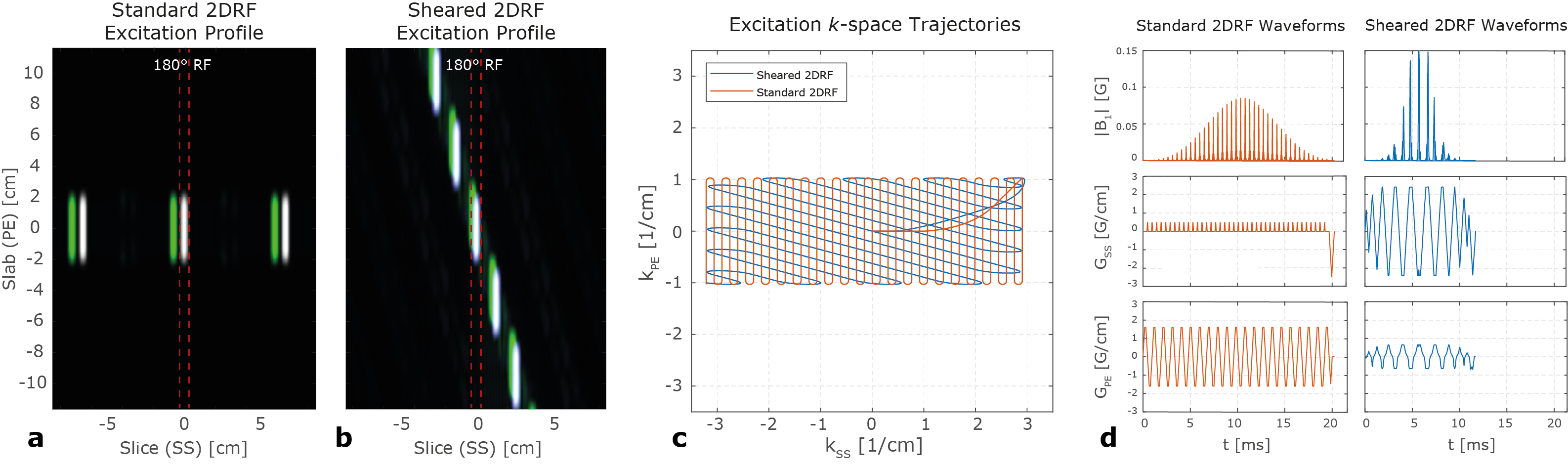
FIG. 1. 2D excitation profiles (white for water and green for fat) for (a) the standard 2DRF pulse and (b) 75° sheared 2DRF pulse under 1.5T hardware limitations. (c) Excitation k-space trajectories and (d) RF and gradient waveforms for the standard and sheared 2DRF pulses. The sheared 2DRF pulse achieves identical k-space coverage with considerably reduced pulse duration.
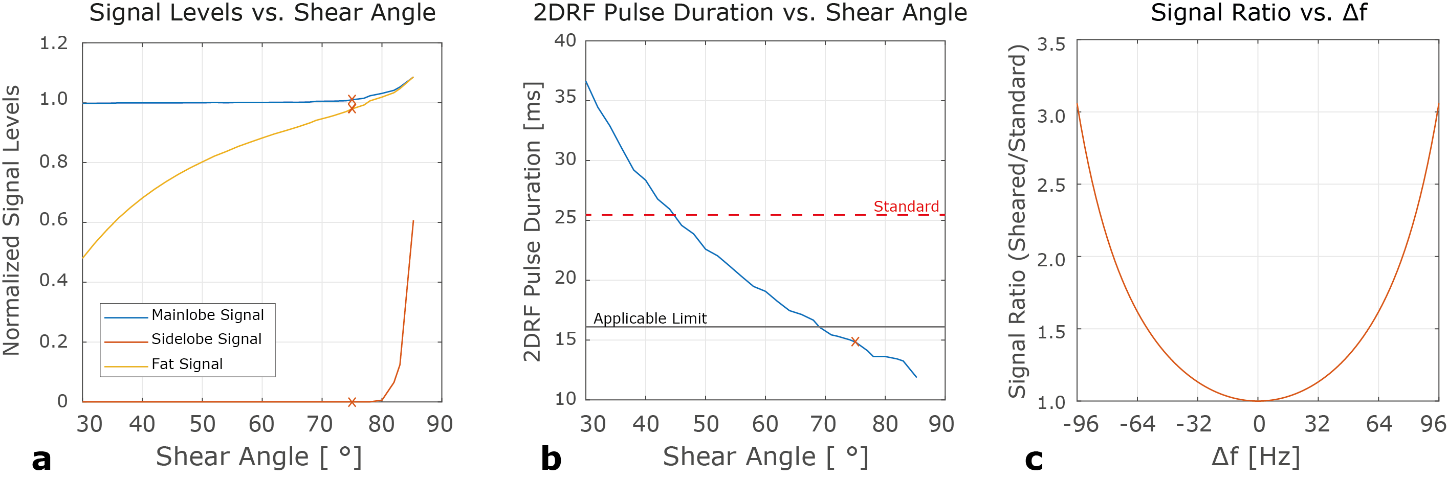
FIG. 2. Simulation results for 0.35T MRI system. (a) Effective mainlobe, sidelobe and fat signals as a function of θ. (b) The standard and sheared 2DRF pulse durations as a function of θ. θ=75° (red crosses) provides the shortest pulse duration with robust sidelobe suppression. (c) Signal ratio of the standard and 75° sheared 2DRF pulses under off-resonance in the range of -96Hz to 96Hz. The sheared 2DRF pulse achieves increased signal improvement under increased off-resonance, showing the improvements of shearing for low-cost MRI systems with high B0 inhomogeneity.
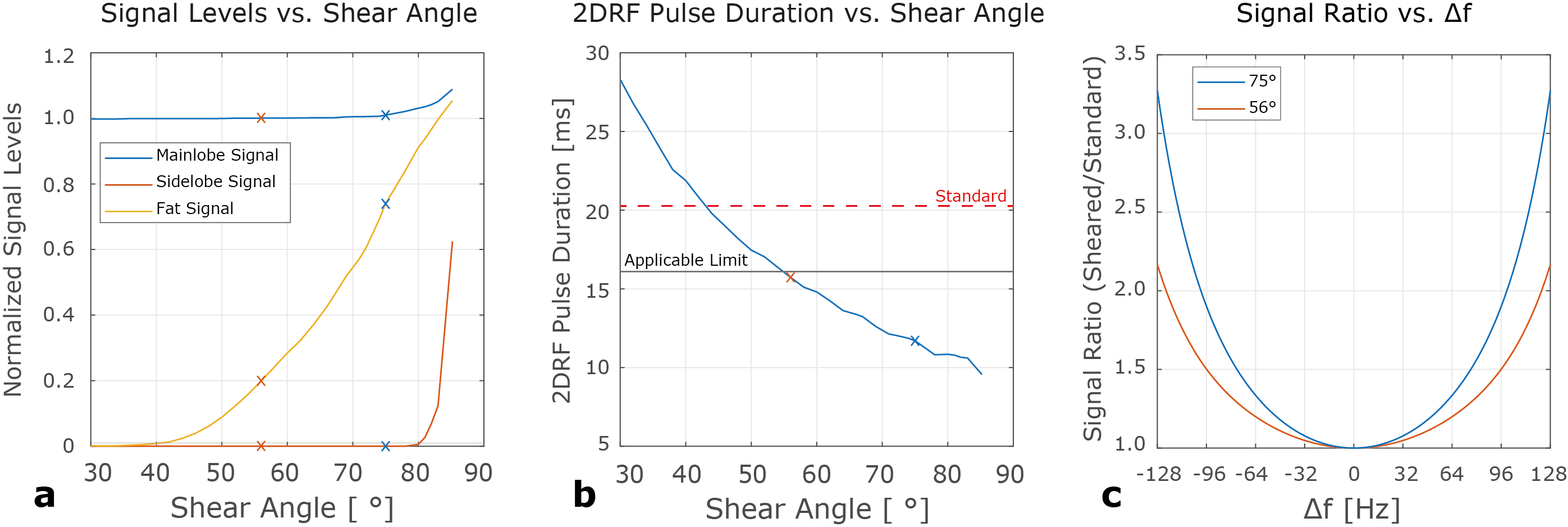
FIG. 3. Simulation results for 1.5T MRI system. (a) Effective mainlobe, sidelobe and fat signals as a function of θ. (b) The standard and sheared 2DRF pulse durations as a function of θ. Again, θ=75° (blue crosses) provides the shortest pulse duration with robust sidelobe suppression while θ=56° (red crosses) is an alternative with favorable fat suppression performance at the expense of increased pulse duration. (c) Signal ratio of the standard and the sheared 2DRF pulses under off-resonance in the range of -128Hz to 128Hz.
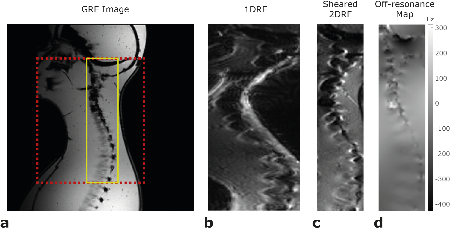
FIG. 4. Phantom imaging results at 0.35T hardware limitations. (a) GRE image indicating full-FOV ROI (red dashed line) with 200X162.5mm2 and reduced-FOV ROI (yellow line) with 200X50mm2. (b) Full-FOV image suffers severely from in-plane off-resonance effects, while (c) the reduced FOV image using sheared 2DRF pulse successfully alleviates these artifacts. (d) Off-resonance map for the reduced-FOV ROI.
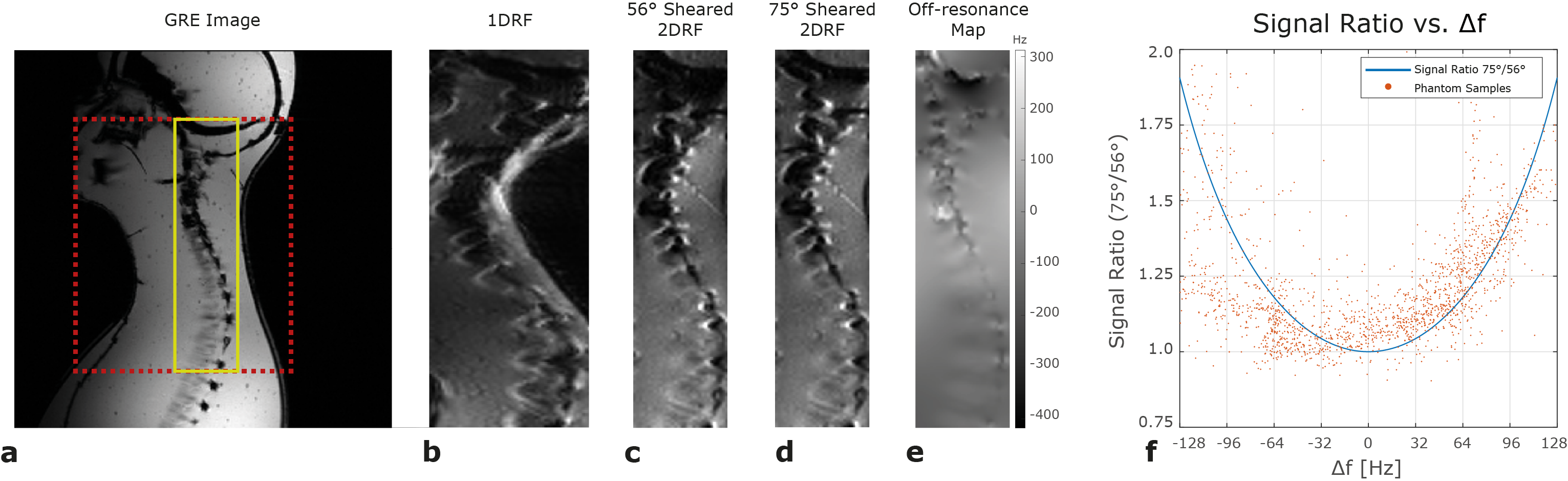
FIG. 5. Phantom imaging results at 1.5T hardware limits. (a) GRE image showing full-FOV ROI (red dashed line) with 200X162.5mm2 and reduced FOV ROI (yellow line) with 200X50mm2. (b) Full-FOV image suffers from in-plane off-resonance effects, while (c-d) the reduced FOV images obtained using sheared 2DRF pulses with θ=56° and θ=75° remove these artifacts. (e) Off-resonance map for the reduced FOV ROI. (f) Pixel-wise signal ratio of the reduced FOV images. The experimental signal ratio closely follows the theoretical signal ratio curve.
DOI: https://doi.org/10.58530/2022/2930