2926
Design of Novel RF Pulse for Fetal MRI Refocusing Trains using Rank Factorization (SLfRank) to Reduce SAR and Improve Image Acquisition Efficiency
Sebastian Diaz1, Yamin Arefeen2, Borjan Gagoski3,4, Ellen Grant3,4, and Elfar Adalsteinsson2,5,6
1Department of Biomedical Engineering, University of Arizona, Tucson, AZ, United States, 2Department of Electrical Engineering and Computer Science, Massachusetts Institute of Technology, Cambridge, MA, United States, 3Department of Radiology, Harvard Medical School, Boston, MA, United States, 4Fetal-Neonatal Neuroimaging and Developmental Science Center, Boston Children's Hospital, Boston, MA, United States, 5Harvard-MIT Health Sciences and Technology, Massachusetts Institute of Technology, Cambridge, MA, United States, 6Institute for Medical Engineering and Science, Massachusetts Institute of Technology, Cambridge, MA, United States
1Department of Biomedical Engineering, University of Arizona, Tucson, AZ, United States, 2Department of Electrical Engineering and Computer Science, Massachusetts Institute of Technology, Cambridge, MA, United States, 3Department of Radiology, Harvard Medical School, Boston, MA, United States, 4Fetal-Neonatal Neuroimaging and Developmental Science Center, Boston Children's Hospital, Boston, MA, United States, 5Harvard-MIT Health Sciences and Technology, Massachusetts Institute of Technology, Cambridge, MA, United States, 6Institute for Medical Engineering and Science, Massachusetts Institute of Technology, Cambridge, MA, United States
Synopsis
Fetal MRI is inherently time-constrained due to unpredictable motion and comfort concerns. Single-shot spin-echo sequences address these concerns by rapidly acquiring images with desired tissue contrast. However, these sequences typically operate at specific absorption rate (SAR) limitations due to a train of refocusing pulses, which impose long TR times and reduce acquisition efficiency. This study leverages SLfRank, the use of ranked factorization to jointly solve for SLR bi-coupled spin parameters, to design refocusing profiles that reduce SAR while maintaining slice-profile performance. The proposed refocusing pulse reduced SAR by over 20% while producing similar image quality to the standard clinical sequence.
Introduction
Fetal MRI is becoming increasingly more important for imaging evaluation of maternal and fetal health but suffers from unpredictable fetal motion in the presence of long acquisition times1,2 Single-shot, fast-spin-echo T2-weighted (SS-FSE) sequences partially mitigate motion artifacts by acquiring all of k-space in roughly ~500 ms after a single excitation with an echo train of large-flip angle refocusing pulses to maximize signal-to-noise and provide T2-weighted contrast3,4. However, tissue heating induced by the refocusing pulses–denoted by the signal absorption rate (SAR)–increases repetition time (TR) between consecutive slices in order to meet safety limits5,6,7. Although the RF refocusing pulses are designed to reduce energy, SAR limits still arise. Therefore, there is a clinical need to design refocusing pulses that decrease SAR while maintaining diagnostic quality.Methods
The traditional Shinnar-Le Roux (SLR) algorithm8 designs high-flip angle refocusing pulses using a spin-domain representation of the Bloch equations to simplify the pulse design problem into finding a 2x2 unitary matrix of two polynomials, ⍺ and β. Since these parameters are bi-coupled, SLR first determines β to approximate the ideal slice profile, and then solves for ⍺ to minimize pulse energy.SLfRank9, a recently proposed amendment to SLR, jointly solves for a global minimum of the parameters through ranked factorization and convex optimization by relaxing the rank constraint to a semidefinite constraint. The subsequent joint optimization yields pulses with less energy.
We applied SLfRank to design refocusing pulses for fetal SS-FSE sequences, evaluated the SAR and slice-profile of the proposed pulses, and demonstrated initial proof-of-concept in in-vivo experiments.
The target design parameters were a pulse time length of 2 milliseconds and 3 millimeter slice thickness. We implemented these SLfRank designs for HASTE on a Siemens Skyra 3T clinical scanner.
The RF design utilized the SigPy.RF10 and the SLfRank Python packages to design and simulate the proposed 64 time-point pulses. Design using SLfRank code took under one minute on a standard PC laptop for a 64 time-point pulse. Using the same setup, a design of a 128 time-point pulse takes approximately four minutes.
Experiments
For RF pulse design, we simulated varying options using constant values of 1% for the pass-band and stop-band, pulse size of 64 time-points, and linear phase. Time-bandwidth product (TBW) was varied from 1.95 to 4.00 in increments of 0.01 for a total of 205 simulations. The energy of each simulated pulse was compared to the product refocusing pulse through the sum-of-squares of their respective envelope. The pulse with the most power reduction and closest match to the desired transverse magnetization slice profile was chosen, see Figure 1.Simulated signal evolutions for a standard SS-FSE sequence using the product and proposed pulse were nearly identical, with a normalized root-mean-square-difference of 5 x 10⁻⁴. In the transverse magnetization profiles, the proposed pulse produces a stop-band ripple peak of 1.4% of the max amplitude of the magnetization.The best performing pulse was a refocusing pulse with a TBW of 2.21. This pulse, along with a gradient strength of 4.25 mT/m, was tested in-vivo with a human head and a fetus.
Results
In the simulations, the refocusing pulse indicated a SAR reduction of 23%.With IRB approval, an adult brain was scanned with a SS-FSE sequence using the product and proposed pulse on a 3T Siemens Skyra with 300 mm x 300 mm field-of-view, 1.2 mm x 1.2 mm resolution, and 2x in-plane acceleration. The proposed refocusing envelope achieved 17% SAR reduction. Images reconstructed by the scanner are depicted in Figure 2.
Second, a fetal MRI scan was acquired with the proposed and product refocusing pulses on a 3T Siemens Skyra scanner with IRB approval, 330 x 300 mm field-of-view, 1.3 x 1.3 mm resolution, and 2x in-plane acceleration. The proposed envelope achieved 22% SAR reduction; Figure 3 depicts comparison images reconstructed by the scanner.
Discussion
In fetal MRI, the SS-FSE sequences utilize a single 90° pulse in combination with many more 180° pulses to produce a 2D image as quickly as possible in a single TR. Accordingly, the significance of proposed pulses are mainly highlighted through the power reduction of the refocusing pulse.Since fetal MRI often hits the FDA limitation in regard to SAR, our proposed, reduced power refocusing pulse enables decreased TR which will reduce imaging time by approximately 20%.
Future Directions
In future work, we will assess diagnostic imaging performance with the standard and proposed refocusing pulses across a cohort of subjects.The tested proposed refocusing pulse is a designed 180º spin-echo pulse, but scaled to 160º for fetal imaging. To directly design a 160º pulse, we modified the β SLR spin-parameter, in SLfRank’s code. The newly designed pulse achieved a 27% reduction, as seen in Figure 4. Furthermore, if the pulse is scaled to a flip angle of 180º, it achieves a 7% reduction when compared to the product at 160º.
Although we examined refocusing pulses here, SLfRank provides additional opportunities of RF pulse design that can be incorporated into other SAR-demanding MRI-sequences, including SMS acquisitions.
Acknowledgements
MIT Summer Research Program (MSRP), NIH Fetal Grant: NIH R01 EB017337, U01 HD087211, R01HD100009References
- References:Gholipour, A., Estroff, J.A., Barnewolt, C.E., Robertson, R.L., Grant, P.E., Gagoski, B., Warfield, S.K., Afacan, O., Connolly, S.A., Neil, J.J., Wolfberg, A. and Mulkern, R.V. (2014), Fetal MRI: A technical update with educational aspirations. Concepts Magn. Reson., 43: 237-266. https://doi.org/10.1002/cmr.a.21321
- Platt LD, Barth RA, Pugash D. (2018), Current controversies in prenatal diagnosis 3: fetal MRI should be performed in all prenatally detected fetuses with a major structural abnormality. Prenat Diagn 38:166–172
- Bernstein MA, King KF. Handbook of MRI pulse sequences. Elsevier: Burlington, MA, 2004.
- Rahel A. KubikHuch et al. “Ultrafast MR Imaging of the Fetus”. In: American Journal of Roentgenology 174.6 (2000). PMID: 10845491, pp. 1599–1606.
- Victoria, Teresa et al. “Comparison Between 1.5-T and 3-T MRI for Fetal Imaging: Is There an Advantage to Imaging With a Higher Field Strength?.” AJR. American journal of roentgenology vol. 206,1 (2016): 195-201. doi:10.2214/AJR.14.14205
- Weisstanner, C., Gruber, G. M., Brugger, P. C., Mitter, C., Diogo, M. C., Kasprian, G., & Prayer, D. (2017). Fetal MRI at 3T-ready for routine use?. The British journal of radiology, 90(1069), 20160362. https://doi.org/10.1259/bjr.20160362
- Victoria T, Jaramillo D, Roberts TPL et al (2014) Fetal magnetic resonance imaging: jumping from 1.5 to 3 tesla (preliminary experience). Pediatr Radiol 44:376–386
- Pauly, J et al. “Parameter relations for the Shinnar-Le Roux selective excitation pulse design algorithm [NMR imaging].” IEEE transactions on medical imaging vol. 10,1 (1991): 53-65. doi:10.1109/42.75611
- Ong, Frank et al. “SLfRank: Shinnar-Le-Roux Pulse Design with Reduced Energy and Accurate Phase Profiles using Rank Factorization.” (2021).
- J. Martin et al. “SigPy.RF: Comprehensive Open-Source RF Pulse Design Tools for Reproducible Research.” in Proceedings of the ISMRM Annual Meeting and Exhibition, 2020, p. 1045.
Figures
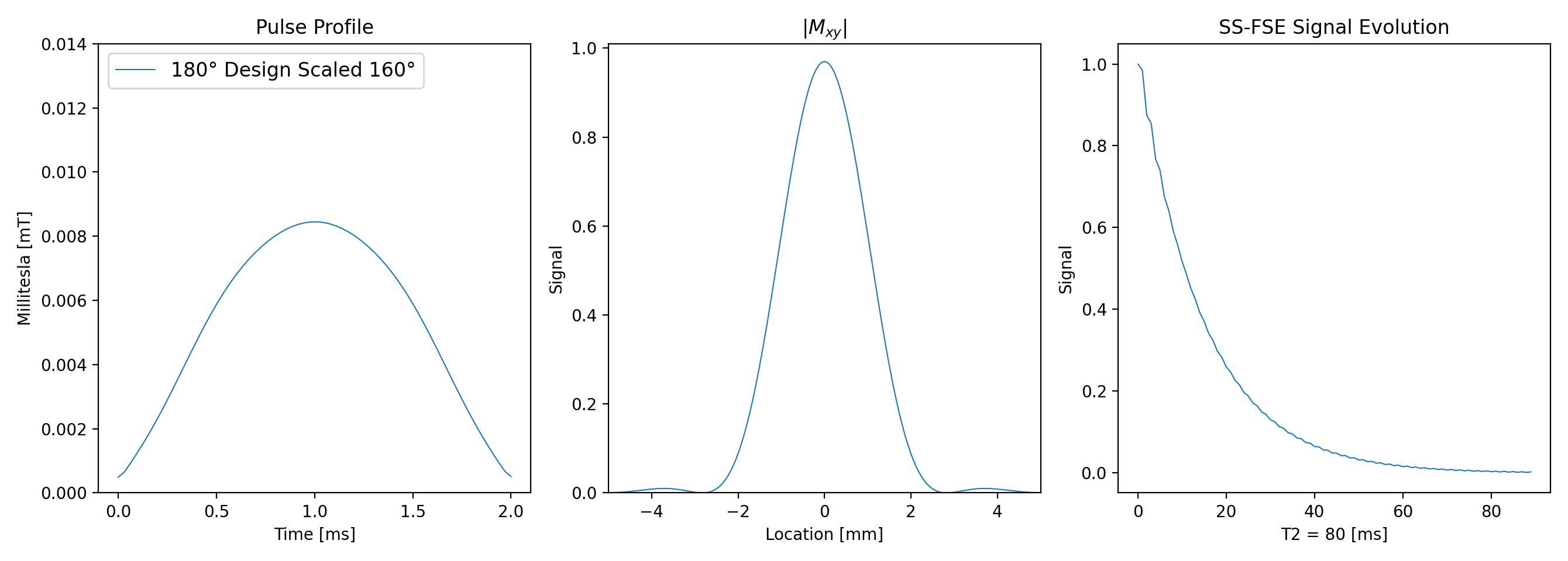
Figure 1. The pulse profile, transverse magnetization, and signal evolutions are displayed for the proposed pulse used in the subsequent in-vivo experiments. In comparison to the product, signal evolution RMS-difference is 0.0004557. The FWHM difference of the transverse magnetization is -0.0547 mm between the proposed and the product, respectively. Simulation slice-selective gradient strength for the proposed was 5.8 mT/m to adequately compare to product parameters.
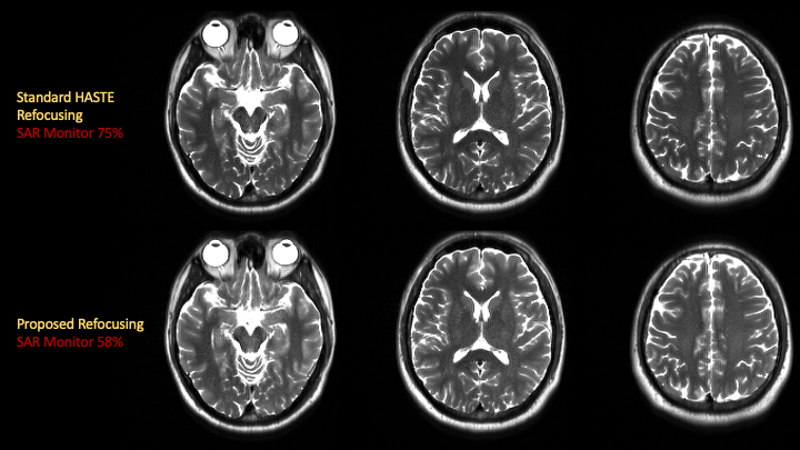
Figure 2. An axial view of the adult brain run with the HASTE sequence at TR = 550 milliseconds on a 3.0T Siemens Skyra scanner is shown. The scanner’s reconstruction of the k-space data is displayed as well as the SAR calculations. The monitor showed a 17% reduction in SAR with the proposed pulse.
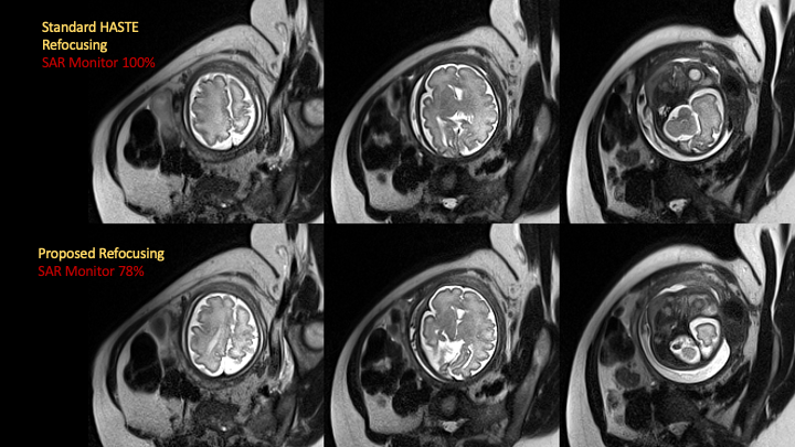
Figure 3. Fetal scans are shown on a 3.0T Siemens Skyra scanner run with HASTE at TR = 1340 milliseconds for both pulses. The SAR monitor calculated a 22% reduction in SAR between the proposed and product refocusing pulses.
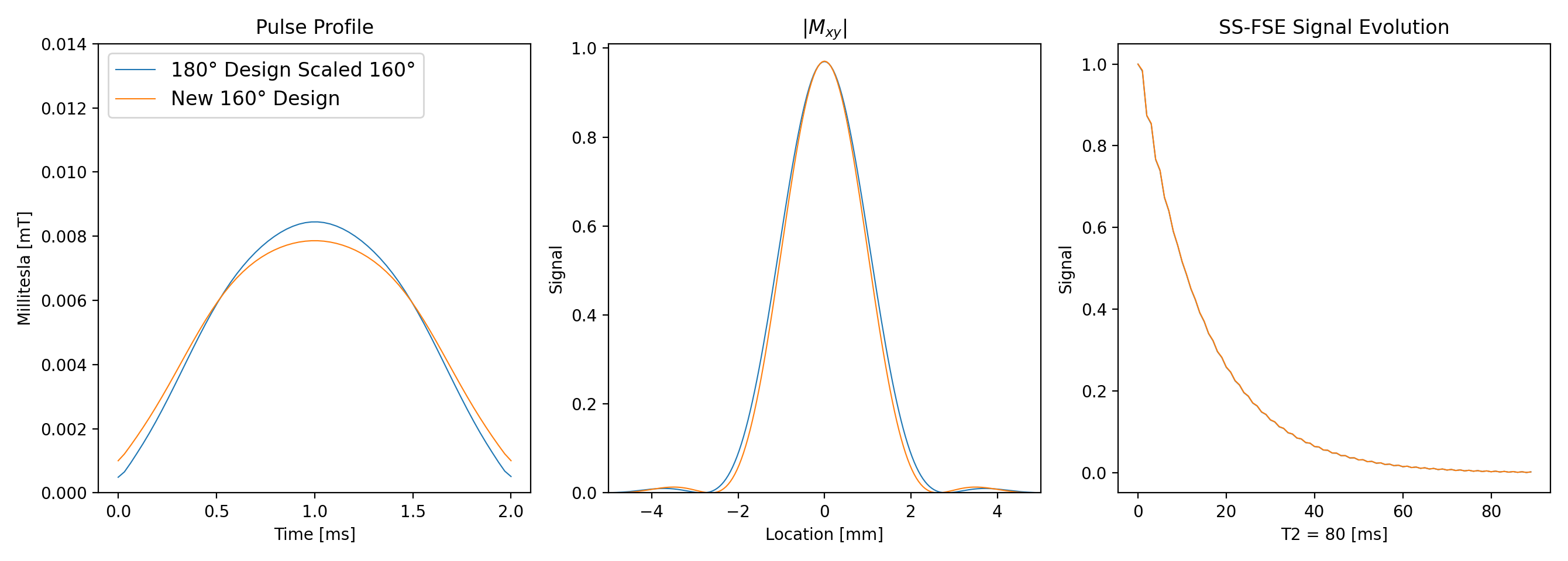
Figure 4. Pulse profile and associated signals are displayed for the utilized 180º scaled to 160º and newly designed 160º pulse. Modifications to the β spin-parameter of SLR in SLfRank’s code enabled the design of a 160º spin-echo pulse as opposed to designing a 180°. Specific flip angle solving resulted in an energy reduction of 26% when compared to the product 160º. Slice-selective gradient strength for each pulse was 5.8 mT/m.
DOI: https://doi.org/10.58530/2022/2926