2841
Low-grade glioma resting-state fMRI pre-operative maps: a comparison with functional atlas from direct electrode stimulation.1CIMeC Center for Mind/Brain Sciences, Trento, Italy, 2Department of Neuroscience, Division of Neurosurgery, S.Chiara Hospital, APSS Trento, Trento, Italy, 3Structural and Functional Connectivity Lab, S.Chiara Hospital, APSS Trento, Trento, Italy
Synopsis
A cortical-subcortical functional atlas has been recently proposed from direct-electrode stimulation on low grade glioma patients. The agreement between that atlas and non-invasive pre-operative resting-state functional MRI (rs-fMRI) data has not yet been investigated. In this study, we use pre-operative rs-fMRI data and the RestNeuMap tool to investigate the agreement between language and sensory networks in a group of low grade glioma patients. We find that rs-fMRI networks of language and speech arrest show greater overlap with DES-derived functional atlas than the sensorimotor network.
Introduction
Intraoperative Direct Electrical Stimulation (DES) remains the elective method to identify eloquent areas of the cortex, to prevent brain surgery from damaging functional regions underlying major cognitive functions1,2,3. This procedure, in conjunction or not with presurgical mapping, can guide the intraoperative localization of functional networks4. One of the main issues with DES is that only a few cortical sites can be tested due to time constraints; this may lead to a small number of electrically stimulated regions, in addition to the potential inaccurate localization given by remote areas’ stimulation due to corticocortical connections5,6. In this framework, ReStNeuMap offers an open-access tool that helps clinicians with the preoperative mapping of functional networks relevant for post-surgical cognitive recovery, automatically selecting resting-state networks in glioma patients7,8. Recently, a new functional atlas obtained by DES coordinates from Low-Grade Gliomas (LGG) patients has been developed. In this study, we evaluate the agreement between single-subject resting-state networks automatically identified by ReStNeuMap in a group of low-grade glioma patients and an atlas of brain functional networks derived from DES mapping during real-time neuropsychological testing9.Methods
Ten LGG patients (Table 1) underwent pre-surgical T1-weighted and resting-state functional MRI (rs-fMRI) on a 1.5 T GE Healthcare MRI scanner at the Department of Radiology of Santa Chiara Hospital, Trento (Italy). Full-brain rs-fMRI data were acquired with a 2D T2*-weighted gradient-echo echo-planar imaging (EPI) sequence (TR = 2600 ms, voxel resolution = 4mm3 isotropic, TE = 45 ms, FA = 87o, FOV = 256×256 mm2, acceleration factor ASSET = 2)7,8.The pre-processing pipeline, probabilistic ICA and template matching procedure implemented in the ReStNeuMap followed the steps described in Gift Cabalo et al. 20218. Individual ReStNeuMap-derived networks as well as DES atlas functional networks were then masked for grey matter.
With respect to the DES atlas (Table 1)9, we included anomia, phonological and semantic, speech articulation (SAN)10, motor and sensorial (SMN) networks. The overlap between the ReStNeuMap8-derived language, SAN and SMN networks and the corresponding functional maps of the DES atlas was computed at subject level. Two metrics were derived: (i) the percentage of overlapping voxels between the ReStNeuMap-derived functional networks and corresponding DES atlas networks relative to the total number of voxels of the DES atlas, as a sensitivity measure; and (ii) the percentage of non-overlapping voxels with respect to the total number of voxels of ReStNeuMap-derived networks, as a specificity measure. The overlap was computed on un-thresholded DES atlas maps (CI00), along with different confidence intervals CI (0.2, 0.6, 0.8).
Results
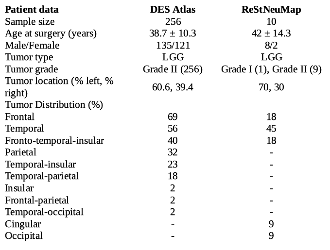
The sensitivity of ReStNeuMap for detecting regions in common to language components (anomia ((CI00) 28.2±10.6), phonemic ((CI00) 31±11), semantic ((CI00) 31.3±11.7,) and SAN ((CI00) 28.6±11.2) components of the DES atlas was in the range of 30-35% (Figure 1). A slight sensitivity increase was seen as the CI of the DES atlas increased. However, the sensitivity of the motor ((CI00) 3.6±7.5) and sensorial ((CI00) 8.8±12.5) components of the DES atlas was lower <10% (Figure 2). Regarding the specificity of the ReStNeuMap language and sensory networks, the voxels overlapping the DES atlas are only few with respect to the whole functional networks detected with this tool, with non-overlapping voxels being >70% (Figure 3). Specifically, for the language and SAN, by increasing CI, the specificity of ReStNeuMap increases for the language network (with respect to anomia ((CI00) 91.1±6.8), phonological ((CI00) 91.4±6.8) and semantic ((CI00) 91.4±6.8) DES atlas) and SAN ((CI00) 89.9±7.6), the SMN (motor ((CI00) 91.4±7.4) and sensorial ((CI00) 90.8±7) (Figure 3). Figure 4 shows a probabilistic map of the overlapping voxels among the LGG patients.Discussion
The agreement between single-subject pre-operative rs-fMRI networks and those defined by the DES atlas depended on whether networks were sensory or associative. We found a greater sensitivity (overlap 30-40%) for detecting the language and speech articulation10 networks, while lower accuracy to detect the sensory motor network (overlap <10%). One possible explanation is that the SMN is a highly plastic functional network in glioma patients, increasing to a great extent the variability across subjects11. Moreover, from a clinical perspective, what ReStNeuMap offers is the presurgical mapping of two functional networks (i.e. language and SAN) non-invasively and with minimal patient cooperation, as opposed to the needs of task-based fMRI or DES. On the other hand, ReStNeuMap showed low levels of sensitivity for all networks.Conclusion
In this study we demonstrated that ReStNeuMap is a promising tool for detecting functional networks pre-surgery, in gliomas patients, with greater sensitivity for the language and speech articulation functions, in relation to invasive DES atlas mapping networks. However, the combination with intraoperative DES is still crucial for mapping primary cortices networks (i.e. SMN)12.Acknowledgements
Fondazione Paolina Lucarelli Irion, Rovereto (Trento), Italy.
Dipartimento di Eccellenza project 2018-2022 (Italian Ministry of Education, University and Research).
References
Duffau H, Capelle L, Denvil D, Sichez N, Gatignol P, Taillandier L, van Effenterre R. Usefulness of intraoperative electrical subcortical mapping during surgery for low-grade gliomas located within eloquent brain regions: functional results in a consecutive series of 103 patients. Journal of neurosurgery, 2003; 98(4), 764-778.
Pendleton C, Zaidi HA, Chaichana KL, Raza SM, Carson BS, Cohen-Gadol AA, Quinones-Hinojosa A. Harvey Cushing’s contributions to motor mapping: 1902–1912. 2012; Cortex, 48(1), 7–14.
Sarubbo S, Le Bars E, Moritz-Gasser S, Duffau H. Complete recovery after surgical resection of left Wernicke's area in awake patient: a brain stimulation and functional MRI study. Neurosurgical review, 2012; 35(2), 287-292.
Qiu TM, Gong FY, Gong X, Wu JS, Lin CP, Biswal BB, Zhuang DX, Yao CJ, Zhang XL, Lu JF, Zhu FP, Mao Y, Zhou LF. Real-Time Motor Cortex Mapping for the Safe Resection of Glioma: An Intraoperative Resting-State fMRI Study. American Journal of Neuroradiology, 2017; 38(11), 2146–2152.
Castellanos FX, Di Martino A, Craddock RC, Mehta AD, Milham MP. Clinical applications of the functional connectome. NeuroImage, 2013; 80, 527–540.
Sinai A, Bowers CW, Crainiceanu CM, Boatman D, Gordon B, Lesser RP, Lenz FA, Crone NE. Electrocorticographic high gamma activity versus electrical cortical stimulation mapping of naming. Brain, 2015; 128(7), 1556–1570.
Zacà D, Jovicich J, Corsini F, Rozzanigo U, Chioffi F, Sarubbo S. ReStNeuMap: a tool for automatic extraction of resting-state functional MRI networks in neurosurgical practice, Journal of Neurosurgery JNS, 2019;131(3), 764-771.
Cabalo D, Saviola F, Tambalo S, Luciani B., Zacà D, Zigiotto L, Sarubbo S, Jovicich J. Automated identification of key resting-state fMRI networks in preoperative glioma patients. Organization of Human Brain Mapping - Annual 2021 Meeting, Abstract 2673
Sarubbo S, Tate M, De Benedictis A, Merler S, Moritz-Gasser S, Herbet G, Duffau H. Mapping critical cortical hubs and white matter pathways by direct electrical stimulation: an original functional atlas of the human brain. Neuroimage, 2020; Jan 15;205:116237.
Zacà D, Corsini F, Rozzanigo U, Dallabona M, Avesani P, Annicchiarico L, Sarubbo S. Whole-brain network connectivity underlying the human speech articulation as emerged integrating direct electric stimulation, resting state fMRI and tractography. Frontiers in human neuroscience, 2018; 12, 405.
Duffau H. Lessons from brain mapping in surgery for low-grade glioma: insights into associations between tumour and brain plasticity. The Lancet Neurology, 2005; 4(8), 476-486.
Sarubbo S, Annicchiarico L, Corsini F, Zigiotto L, Herbet G, Moritz-Gasser S, Avesani P. Planning brain tumor resection using a probabilistic atlas of cortical and subcortical structures critical for functional processing: a proof of concept. Operative Neurosurgery, 2021; 20(3), E175-E183.
Figures
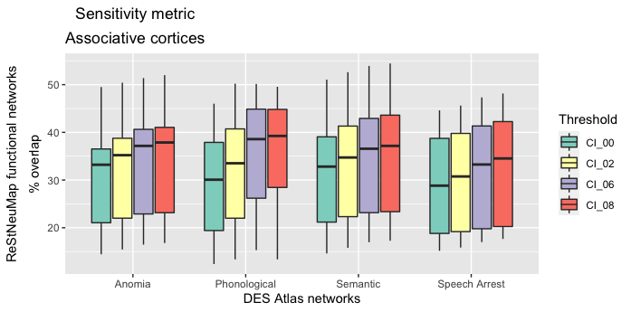
Spatial overlap of functional networks belonging to associative cortices is shown as the percentage of voxels from ReStNeuMap functional networks (y-axis) falling within the corresponding DES Atlas Network (x-axis). Each box represents the % overlap at different Confidence Intervals (CI) of DES atlas.
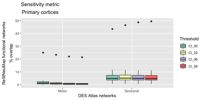
Percentage of rs-fMRI networks that do not overlap with the corresponding DES atlas networks, computed in % relative to the size of the rs-fMRI networks.
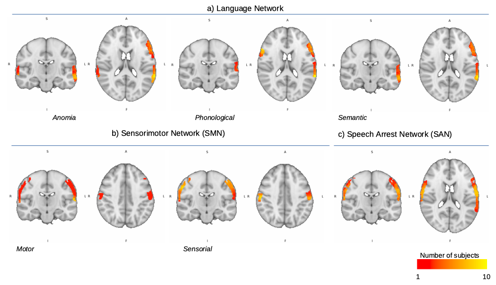
Probabilistic maps for each functional domain derived from the overlap between the DES atlas and the ReStNeuMap-detected networks: (a) language networks, (b) sensory motor network (SMN), and (c) speech articulation network (SAN). Red areas represent overlapping voxels for 1/10 subjects, while yellow regions correspond to voxels where the overlap is found for all 10 LGG patients.